Yellow and Pustular Lesions
Libby Edwards
Yellow lesions primarily result from purulent material. These can occur as intact pustules, or as yellow crusting, when pustules rupture. On occasion, desquamated, hydrated stratum corneum that obstructs or distends follicles form can appear somewhat yellow and mimic pustules.
TRUE PUSTULAR DISEASES
Folliculitis
Folliculitis is a common pustular eruption resulting from the inflammation or infection of the superficial portion of hair follicles (Figs. 8-1, 8-2, 8-3 and 8-4). This can be bacterial, fungal, or irritant.
Clinical Presentation
Folliculitis occurs in all age groups, and the affected population depends on the underlying etiology. Fungal folliculitis occurs primarily in middle-aged or older men, especially those with fungal toenail infection. Irritant folliculitis occurs in women who shave the area and in overweight individuals at risk for prominent friction and chronic moisture. There is no particular patient at risk for Staphylococcal folliculitis, whereas hot tub folliculitis is found in individuals exposed to inadequately treated hot tubs or those who use washcloths or sponges that do not completely dry from day-to-day.
Staphylococcus aureus folliculitis usually presents with symptoms of mild tenderness and pruritus. On examination, there are red papules and variable numbers of small yellow pustules 1 to 5 mm in size with a surrounding red flare. The pustule is short-lived and leaves a residual red papule with crust or a fine collarette of scale. Individual lesions heal within 7 to 10 days. Many patients have associated folliculitis with furunculosis, representing a deeper infection of the follicle. These tender, red nodules often suppurate and drain. Frequent sites include the mons pubis, buttocks, and thighs.
Pseudomonas folliculitis presents as tender, red, nonscaling papules and occasional pustules concentrated in areas where contaminated water has been held against the skin, such as intertriginous skin and under wet swimsuits. Sometimes, patients exhibit constitutional symptoms such as fever, malaise, ear pain, and sore throat.
Follicular pustules within scaly, annular, pink-red plaques of tinea cruris characterize fungal folliculitis (Fig. 8-5). However, women with tinea pedis sometimes develop scattered single papules or pustules of fungal folliculitis on their lower legs and thighs by inoculating fungi into follicles when they shave their legs.
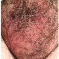
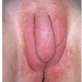
Noninfectious, irritant folliculitis is most often asymptomatic. Shaving folliculitis is a very common form of irritant folliculitis, as increasing numbers of women shave the vulva. Either the shaved, short, curly hair curves back and pierces the skin, producing an inflammatory reaction (Fig. 8-6) or the prominent tips of follicles are removed by the razor, again causing an irritant reaction. In addition, follicular occlusion from pressure, friction, and moisture often produces folliculitis that is often indistinguishable from Staphylococcal folliculitis. Small pustules surrounded by a thin rim of erythema characterize these almost ubiquitous lesions. These occur primarily on those areas of friction and pressure, such as the medial thighs and the buttocks.
Diagnosis
The diagnosis of folliculitis is usually made clinically. A bacterial or fungal culture is often helpful to identify any causative organisms and to guide the treatment course. A potassium hydroxide preparation sometimes reveals hyphae in dermatophyte folliculitis. Histopathology shows neutrophils within the hair follicle with abscess formation. Special stains may reveal the causative organism.
Folliculitis can be confused with scabies, pustular miliaria, Candida infection, eosinophilic folliculitis of human immunodeficiency virus (HIV) infection, arthropod bites, keratosis pilaris, and herpes simplex virus (HSV) infection.
FOLLICULITIS: Diagnosis
Morphology of red papules, pustules, and/or crusts, often with a hair piercing some lesions
Culture showing causative organism, or fungal preparation showing branching hyphae should reveal infectious causes
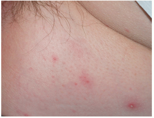 FIG. 8-1. Folliculitis is common on the buttocks, where chronic pressure and occlusion promote follicular obstruction. |
Pathophysiology
Bacterial folliculitis is most often caused by S. aureus, but it can be caused by gram-negative bacteria, especially Pseudomonas aeruginosa, the cause of hot tub folliculitis. Folliculitis may also be caused by dermatophytes and is most often seen in men in association with tinea cruris. Chemical, mechanical, or frictional irritation are also very common causes of superficial folliculitis.
Management
The treatment of folliculitis depends on the etiology. Staphylococcal folliculitis can be treated with oral and topical antibiotics. Oral antibiotics with past predictable coverage include dicloxacillin and cephalexin at 500 mg twice a day for 7 to 10 days. Recently, a large proportion of community-acquired S. aureus is resistant to methicillin, dicloxacillin, and cephalexin. From 4% (France) to 76% (North Carolina) of community-acquired S. aureus is resistant to methicillin (1,2). Clindamycin at 150 mg twice daily is somewhat more likely to be effective, and trimethoprim-sulfamethoxazole double strength is nearly always efficacious. Linezolid, daptomycin, and tigecycline are advanced agents available for the rare infection resistant to the older oral medications (3). Treatment with clindamycin 1% topical solution twice a day in conjunction with antibacterial soaps is also useful. Mupirocin cream and ointment are extremely effective for the first week of use but resistance occurs quickly.
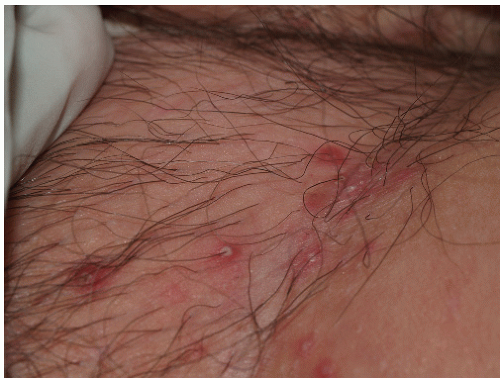 FIG. 8-2. Although the pustule of folliculitis is classically pierced by a hair as seen here, this is often not seen. |
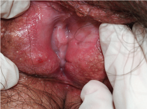 FIG. 8-3. Although devoid of clinical hair, hair follicles are present on the modified mucous membranes of the vulva. This patient exhibited the red papules and pustules of irritant folliculitis on both the clinically hair-bearing skin and the medial aspects of the labia minora. |
Recurrence of Staphylococcal folliculitis is common. Nasal staphylococcal carriage is common in affected individuals with chronic and recurrent folliculitis. These patients benefit by the application of mupirocin ointment to the anterior nares twice a day for 5 days. Because carriers often have recurrent colonization of S. aureus within their
nares, some clinicians recommend that these patients apply the mupirocin treatment for 5 days every 1 to 6 months indefinitely to prevent recurrent folliculitis.
nares, some clinicians recommend that these patients apply the mupirocin treatment for 5 days every 1 to 6 months indefinitely to prevent recurrent folliculitis.
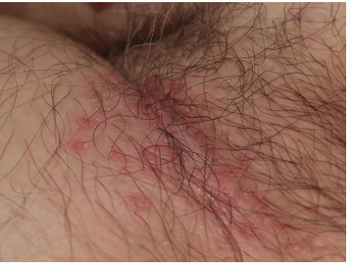 FIG. 8-4. The pustule of folliculitis is very fragile, so that peripheral collarettes, the remaining, circular edge of a ruptured pustule roof, and desquamation are often the predominant lesions. |
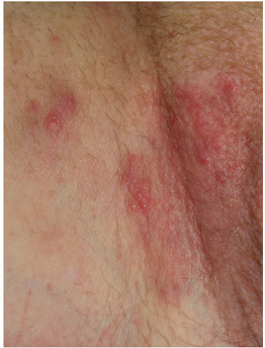 FIG. 8-5. Fungal folliculitis gnereally does not present as isolated, scattered pustules, but rather occurs within plaques of tinea cruris, in which infection extends down follicles to produce pustules or red papules. Often, this occurs in a setting of topical corticosteroids, so that scale is sometimes subtle. |
Pseudomonas folliculitis (hot tub folliculitis) requires no treatment because it resolves when exposure ceases. Therefore, identification of the source of the organism and avoidance of reinoculation are most important. Emptying and cleaning the spa or hot tub and proper maintenance of bactericidal chemicals is also important. However, those patients who exhibit constitutional symptoms may improve more rapidly if they receive oral ciprofloxacin at 500 mg twice a day or levofloxacin 500 mg per day.
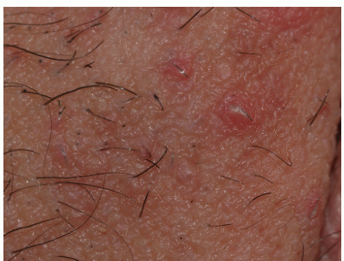 FIG. 8-6. Irritant folliculitis can occur from shaving and is characterized by red papules and pustules limited to the area of shaving, and produced by stiff, curled hairs piercing the skin. |
Fungal folliculitis requires oral therapy because the organism extends into the follicle where topical antifungal creams cannot penetrate. Therapies include oral griseofulvin 500 mg twice daily until the eruption resolves. Alternatively, oral fluconazole or itraconazole may also be prescribed at doses of 100 mg/day orally for 15 days or until the skin is clear. Finally, oral terbinafine at 250 mg/day until the skin is clear is effective, and it is the least expensive solution because this medication is available generically. After treatment, the daily use of an antifungal cream or powder to the groin area helps to prevent recurrences.
Irritant folliculitis can be improved by minimizing irritants. Avoiding shaving clears shaving folliculitis; laser hair removal may be preferable. Cool, nonocclusive clothing, weight loss, and powders may prevent chronic moisture, pressure, and friction. Otherwise, the chronic administration of anti-inflammatory antibiotics such as doxycycline or minocycline 100 mg b.i.d. or clindamycin 150 mg b.i.d. can improve irritant folliculitis, although improvement generally requires about a month and entails ongoing administration.
FOLLICULITIS: Management
Depends on the underlying cause:
Bacterial folliculitis: treat according to culture results. For suspected Staphylococcal folliculitis, oral clindamycin 150 mg b.i.d., trimethoprim-sulfamethoxazole double strength b.i.d.
Fungal folliculitis: terbinafine 250 mg daily, griseofulvin 500 mg b.i.d., fluconazole 100 to 200 mg daily, or itraconazole 200 mg a day until clear
Irritant folliculitis: minimize irritants. Discontinue shaving, add powders to reduce friction and moisture. When required, chronic anti-inflammatory antibiotics such as doxycycline 100 mg b.i.d. can be taken chronically
Furunculosis
While folliculitis is characterized by inflammation of the superficial portion of the hair follicle, furunculosis involves the deeper follicle, producing a red “boil.”
Clinical Presentation
Furunculosis occurs in all patient populations. It is somewhat more common in patients who are immunosuppressed, diabetic, or who exhibit scaling dermatoses that are likely to be colonized with S. aureus.
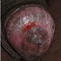
A furuncle is a red, usually painful nodule that typically suppurates and drains (Figs. 8-7 and 8-8). Patients experience evolving lesions, with some appearing as others heal. Often, there is associated staphylococcal folliculitis. Uncommonly, patients develop associated fever and malaise.
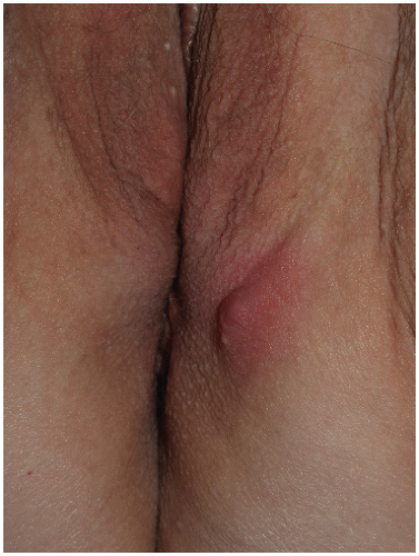 FIG. 8-7. Furunculosis is a deeper and larger infection than folliculitis and exhibits associated induration. |
Diagnosis
The diagnosis is made on clinical grounds, and it is confirmed by a culture that usually yields S. aureus. A biopsy is generally not performed, but it reveals a dermal and subcutaneous abscess with surrounding cellulitis. Special stains show organisms consistent with S. aureus.
A furuncle occurring in the genital area is indistinguishable from an inflamed cyst of hidradenitis suppurativa (Fig. 8-8), which is a form of deep cystic acne. However, furuncles are not confined to the genital and axillary area as is hidradenitis, and comedones and scarring from recurrent draining abscesses are characteristic only of hidradenitis suppurativa. Cultures of hidradenitis suppurativa usually show normal skin organisms or a multitude of bacteria rather than a pure growth of a pathogen. Hidradenitis suppurativa occasionally is secondarily infected by S. aureus, but oral antistaphylococcal agents at most improve, but never eliminate, hidradenitis. Other possible diagnostic challenges include other inflamed cysts, especially an epidermal cyst, but inflamed Bartholin’s gland duct cysts or vestibular cysts can also mimic a furuncle. Occasionally, inflamed tumors such as a basal cell carcinoma can mimic a furuncle.
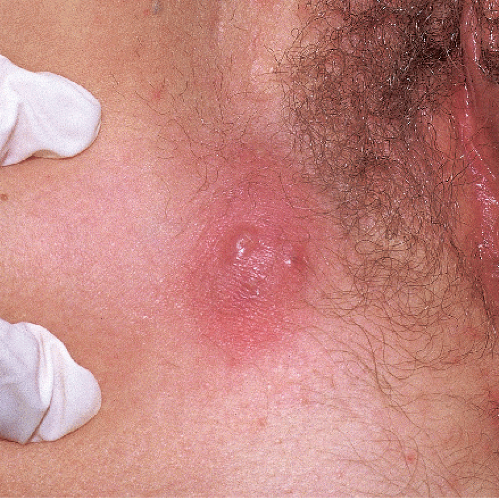 FIG. 8-8. This large, suppurative nodule represents a furuncle, but it is clinically indistinguishable from a ruptured, inflamed epidermal (“sebaceous”) cyst. |
FURUNCULOSIS: Diagnosis
By morphology and acute onset of red painful nodules, often with rupture and draining
Confirmed by culture and complete response to therapy
Pathophysiology
Furunculosis represents a bacterial infection, usually with S. aureus, of the deep portion of the follicle, with a resulting inflammatory nodule.
Management
Oral antistaphylococcal antibiotics are effective in clearing furunculosis. Previous first-line medications such as cephalexin, dicloxacillin, and methicillin are often ineffective due to the increase in community-acquired methicillin S. aureus (see above). Trimethoprim-sulfamethoxazole double strength or antibiotics according to culture results are preferable. Unfortunately, recurrent disease after discontinuation of medication is common. Some patients require prolonged oral therapy of weeks to months, in addition to the insertion of mupirocin ointment to the anterior nares twice a day for the first 5 days of therapy to reduce the carrier state.
FURUNCULOSIS: Management
Oral antibiotics, choice by culture and sensitivities. Initial choices include clindamycin 300 mg b.i.d., trimethoprim-sulfamethoxazole double strength b.i.d.; cephalexin/dicloxacillin/methicillin 500 mg b.i.d. is no longer a good choice in most areas because of high resistance
Warm soaks, incision and draining if fluctuant
For recurrent disease, apply mupirocin ointment or cream in the nose two times a day for 5 days each month for 3 to 6 months to eliminate the carrier state.
Hidradenitis Suppurativa (Acne Inversa) (see also Chapter 7)
This relatively common eruption is often misdiagnosed as furunculosis, but actually represents cystic acne of skin folds rather than an infectious process.
Clinical Presentation
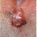
Hidradenitis suppurativa is an inflammatory condition of skin folds that presents with a remarkable variability of severity. Hidradenitis suppurativa generally occurs after puberty, and this condition is worse in overweight individuals (4). There is a strong association with smoking as well (4). Painful, firm, red nodules occur in the axillae and/or the genital area, including the crural crease, proximal inner thighs, scrotum, and vulva (Fig. 8-9). Occasionally, the buttocks and lower abdomen can be involved. These nodules become fluctuant and drain. Some draining tracts become chronic. Although others heal, reaccumulation of keratin and purulent debris generally occurs, and the process repeats, eventually leaving multiple sinus tracts and scars (Fig. 8-10). A careful examination usually yields some comedones and follicles with more than one outlet to the surface (Fig. 8-11). Patients with hidradenitis suppurativa exhibit disease that ranges from an occasional comedone to confluent firm scars with painful, ulcerated nodules, chronic sinus tracts, and foulsmelling drainage (Fig. 8-12).
Other areas of the body that exhibit apocrine glands are the face and scalp. Some unfortunate patients have associated cystic acne and dissecting cellulitis of the scalp, which are also sterile pustular diseases preceded by keratin obstruction of follicles. Cystic acne, dissecting cellulitis, and hidradenitis suppurativa are sometimes referred to as the follicular retention triad.
 FIG. 8-9. The red nodules and comedones of hidradenitis suppurativa can occur on the buttocks, genital skin, proximal medial thighs, and, occasionally, on the abdomen. |
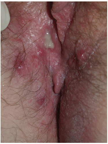 FIG. 8-10. Severe disease causes nearly confluent nodules and comedones. |
As occurs with other chronic inflammatory dermatoses, squamous cell carcinoma is a rare, late complication (5).
Diagnosis
The diagnosis is made on the basis of the history of chronic or recurrent draining nodules in the axillae and/or groin and the presence of comedones. A biopsy,
which is not indicated, reveals a distended follicle with surrounding inflammation consisting of neutrophils, lymphocytes, and histiocytes. Abscesses form and destroy pilosebaceous units, with resulting granulation tissue and epithelialized sinus tracts.
which is not indicated, reveals a distended follicle with surrounding inflammation consisting of neutrophils, lymphocytes, and histiocytes. Abscesses form and destroy pilosebaceous units, with resulting granulation tissue and epithelialized sinus tracts.
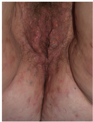 FIG. 8-11. Hidradenitis suppurativa is characterized by red nodules and purulent, draining sinuses, often with surrounding irritant dermatitis from the chronic exudative material. |
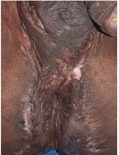 FIG. 8-12. Hidradenitis suppurativa is a disease well known to produce scarring, which can be severe. |
Staphylococcal furunculosis is the disease most easily confused with hidradenitis suppurativa. However, furunculosis is usually not limited to the axillae and groin. In addition, furuncles resolve quickly and completely with antistaphylococcal antibiotics although recurrence after discontinuation of antibiotics is common. In addition, comedones are not seen in association with furunculosis. A solitary inflamed epidermal cyst can mimic mild hidradenitis and may, in fact, represent mild hidradenitis.
HIDRADENITIS SUPPURATIVA: Diagnosis
Morphology of inflamed nodules, comedones, scarring of genitalia and/or axillae; scrotum, vulva, crural creases, perianal skin; sometimes medial thighs and buttocks
Location of the groin, buttocks, medial thighs, vulva, scrotum, lower abdomen, breast areas, and/or axillae
Chronicity
Cultures that show normal skin flora or mixed bacteria;
Incomplete response to antibiotics
Pathophysiology
Sometimes called inverse acne, or apocrine acne, hidradenitis suppurativa represents a foreign body inflammatory reaction to keratin within the dermis. The initial lesion is a comedone, in which the follicle is obstructed by keratin debris from desquamated stratum corneum of the follicular epithelium. This occurs primarily in those people who, as an inherited characteristic, exhibit follicles that have two or more rather than one outlet to the surface of the skin in areas containing apocrine glands. After the follicle is obstructed, it is then distended by keratin shed from deeper portions of the follicle to form an epidermal cyst. Eventually, the stretched, thin walls of the follicle rupture, allowing keratin to extrude into the surrounding dermis and to produce an inflammatory reaction.
Cultures often reveal multiple organisms, and although some physicians believe that bacteria play a pathogenic role, others believe that these organisms act as irritants. Clearly, lesions sometimes become secondarily infected.
Hormonal influence plays a role as well, with hidradenitis rarely occurring before puberty and often flaring premenstrually and postpartum.
Management
Weight loss can be helpful in many patients, and discontinuation of smoking is important.
Although incision and drainage of tense, fluctuant lesions make the patient more comfortable, this approach provides only temporary improvement of individual lesions. The intralesional injection of triamcinolone acetonide 3 mg/mL into acutely inflamed nodules can produce marked, although temporary, improvement.
Oral antibiotics with direct anti-inflammatory effects can inhibit the appearance of new nodules. These antibiotics include tetracycline 500 mg, erythromycin 500 mg, doxycycline or minocycline 100 mg, clindamycin 150 mg, or double-strength trimethoprim-sulfamethoxazole, all prescribed to be taken twice daily on a long-term basis. More recently, the combination of clindamycin and rifampin has been reported to be beneficial (6,7).
For those who do not improve adequately after several months with an antibiotic, surgical removal of the affected skin should be considered when practical and has the highest cure rate (8). The removal of apocrine gland-containing skin is curative. Excision is the most common mode of removal, but hidradenitis suppurativa occurring in the groin can be too extensive for excision. Alternatives include carbon dioxide laser ablation with healing by secondary intention, excision of only the most involved areas, and excision of individual fistulas (8).
Women with hidradenitis sometimes derive benefit from hormonal therapy. High-estrogen oral contraceptives
such as those with ethinyl estradiol 0.035 mg or higher and antiandrogens such a spironolactone can sometimes be useful.
such as those with ethinyl estradiol 0.035 mg or higher and antiandrogens such a spironolactone can sometimes be useful.
Many patients’ disease is not controlled with the foregoing measures. More recently, marked improvement of hidradenitis suppurativa has been reported with tumor necrosis factor alpha (TNF-α) antagonists such as infliximab (9), etanercept (10,11), and adalimumab (12). These extremely expensive medications are approved by the Food and Drug Administration for rheumatoid arthritis and psoriasis. Although infliximab must be given by intravenous infusion, etanercept and adalimumab are self-administered at home, with minimal monitoring. These medications may well become the treatment of choice in the future if early experience is confirmed.
Other therapies that have been used include oral retinoids, such as isotretinoin, which are primarily used for cystic acne of the face and trunk. Although a 4- to 5-month course of isotretinoin normally induces long-term remission of acne, incomplete clearing and prompt relapse of hidradenitis suppurativa is usual (13).
HIDRADENITIS SUPPURATIVA: Management
Mild disease (often, multiple concomitant medications)
oral anti-inflammatory antibiotics: doxycycline or minocycline 100 mg b.i.d., clindamycin 150 mg b.i.d., trimethoprim-sulfamethoxazole double strength b.i.d.
High-estrogen oral contraceptives (women)
Spironolactone 50 to 200 mg as anti-androgen (women)
Topical anti-inflammatory antibiotics: clindamycin or erythromycin solution twice a day.
Antiperspirants
Severe disease
Excision of affected skin: total excision or individual cysts
TNF-α antagonists – etanercept, adalimumab, infliximab
Mucocutaneous Candidiasis (see also Chapter 15)
Clinical Presentation
Cutaneous candidiasis occurs in women with severe vaginal candidiasis, but more often in individuals who are overweight, diabetic, and/or incontinent so that there is a warm, wet environment. Immunosuppression can be a major factor.
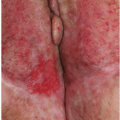
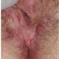
Although cutaneous candidiasis classically presents as a red plaque with surrounding satellite pustules, pustules actually are observed only rarely (Figs. 8-13 and 8-14). The extremely superficial nature of the pustules and the fragile character of the modified mucous membrane that covers part of the external genitalia result in early rupture of pustules. Therefore, the usual morphology is that of superficial collarettes or round erosions surrounding the typical intertriginous red plaque.
Candida vaginitis usually accompanies the skin disease in women. In men, intertriginous candidiasis presents with red, well-demarcated, polycyclic plaques in the crural crease, and multiple satellite l- to 3-mm papulopustules, erosions, or collarettes at the periphery. Eventually, the disease may spread to the scrotum and the proximal, medial thighs. Uncircumcised men often develop balanitis or balanoposthitis with flat, delicate, white-yellow, discrete 1-mm pustules, and surface erosions on the glans penis.
Candida vaginitis usually accompanies the skin disease in women. In men, intertriginous candidiasis presents with red, well-demarcated, polycyclic plaques in the crural crease, and multiple satellite l- to 3-mm papulopustules, erosions, or collarettes at the periphery. Eventually, the disease may spread to the scrotum and the proximal, medial thighs. Uncircumcised men often develop balanitis or balanoposthitis with flat, delicate, white-yellow, discrete 1-mm pustules, and surface erosions on the glans penis.
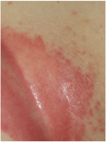 FIG. 8-13. Cutaneous candidiasis classically produces red plaques with peripheral pustules and erosions. |
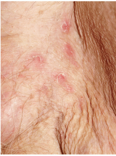 FIG. 8-14. Despite the initial lesion of a pustule, the pustule roof of Candida albicans is extremely fragile, so desquamation is more prominent than pustules. |
Diagnosis
The diagnosis of candidiasis is confirmed by direct microscopic examination of the skin scrapings or purulent exudate, using potassium hydroxide. A fungal culture may be performed from skin pustules as well as vaginal secretions to confirm candidal infection. Because the diagnosis of candidiasis can be confirmed by a microscopic examination of skin scrapings, a biopsy is not indicated. However, histology shows a subcorneal pustule although some pustules are spongiform, showing also basophilic hyphae with a periodic acid-Schiff stain within the upper layers of the epithelium.
Related posts:
Stay updated, free articles. Join our Telegram channel

Full access? Get Clinical Tree


