White Lesions


White Lesions


Sallie M. Neill


Skin pigmentation depends on melanin-containing melanosomes produced by melanocytes. Variation in pigmentation is due to differences in melanocyte numbers and to the numbers and size of melanosomes. White lesions may arise from damage to melanocytes, causing a temporary decrease in melanin production (hypopigmentation), or from total destruction of melanocytes, resulting in absent pigmentation (depigmentation). Loss of color of the skin is not always the result of melanocyte number or melanogenic activity and is seen in:

maceration of the skin due to hydration of top layer of the epidermis, produced when the stratum corneum is thickened by inflammation or friction.

scarring where the white appearance is due to a combination of both vascular and optical effects that are caused by changes of the dermis and epidermis.


WHITE PATCHES AND PLAQUES


Vitiligo

Vitiligo is a depigmenting condition of the skin and hair affecting approximately 1% to 2% of the population worldwide. Autoimmune, genetic, autocytotoxic, neural, environmental, and genetic factors have all been implicated (1). There is a strong genetic link as up to 30% of the affected individuals have a family history of the disorder and it seems that genetically predisposed individuals have melanocytes that are prone to damage which then undergo responses that lead to depigmentation. There are two forms of vitiligo, segmental which is unilateral occurring at a young age and nonsegmental which is generalized, bilateral running a chronic and unpredictable course. Patients with generalized vitiligo exhibit an increase in other autoimmune diseases, especially thyroid disease and have more circulating autoantibodies in general. Occupational vitiligo can occur in the genital area in men as a result of destruction of melanocytes by exposure to para-tertiary-butylphenol, a substance found in resins for adhesives in the car industry (2).


Clinical Presentation

Vitiligo is characterized by a total loss of pigmentation with no evidence of any textural change to the skin, that is, there is no scaling, crusting, or induration (Figs. 11-1, 11-2 and 11-3). There may be an accentuation of the pigment in the skin bordering the depigmented lesions. Wood’s light can be used to distinguish the depigmentation seen in vitiligo which appears brilliantly white rather than the hypopigmentation of other disorders where the lightening is more subtle. Patients usually present with extragenital lesions in the summer months when the areas burn or become more noticeable because of tanning of surrounding unaffected skin. There is a predilection for body sites that are normally hyperpigmented including the face, flexures, areola of the nipples, and the external genitalia. The pigment loss can be patchy or extensive and confluent. Vitiligo can exhibit the Köebner phenomenon, the occurrence of a skin disease in an area of injury or inflammation. The hairs in affected areas may lose their pigment and remain white even after spontaneous recovery of the skin. Spontaneous remission does occur, particularly in young children, but this is rarer in adults. Perifollicular hyperpigmentation is the first sign of recovery.


Diagnosis

The diagnosis of vitiligo is usually made clinically by the distribution of the depigmentation, absence of textural change, and the presence of a hyperpigmented border. Illumination of the skin with Wood’s light helps to delineate the extent of the vitiliginous areas, as it accentuates and highlights areas that may not be seen easily in visible light and furthermore helps in the differentiation of disorders characterized by hypopigmentation. A biopsy may be necessary in some cases.

Histopathologically, there is an absence of melanocytes and melanin in the epidermis. This can be difficult to discern on routine hematoxylin and eosin stains, but incubation with 0.01% 3,4-dihydroxyphenylalanine (also called dopa) stains enzymatically active melanocytes black in the basal layer, and electron microscopy can demonstrate the loss of the melanocytes. There is also a mild to moderate dermal lymphocytic infiltrate at the borders of some lesions, and melanocytes in this area often appear large with long dendrites containing melanin.

Confusion of vitiligo with postinflammatory hypopigmentation can arise, particularly in the early lesions of vitiligo. This can be an especially difficult
differentiation in black patients with hypopigmentation as a postinflammatory sequelae of the original inflammatory condition, for example, eczema or psoriasis. Pityriaisis versicolor enters the differential diagnosis at extragenital sites. The main diffential diagnosis in the anogenital area is lichen sclerosus. This has a similar marble white color, but the crinkled, rough textural changes in the skin help to differentiate this condition. A confusing picture arises with the relatively common coexistence of both vitiligo and lichen sclerosus. The anesthetic, hypopigmented patches of leprosy may also mimic vitiligo, but testing for sensation within the white skin is normal in vitiligo. Vitiligo-like change can be induced by the topical immunonmodulatory cream, imiquimod, used for treating genital warts (3).


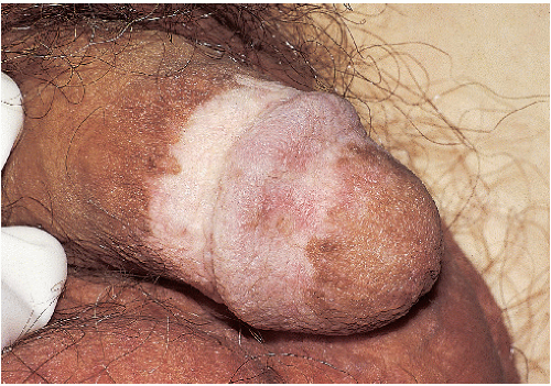
FIG. 11-1. This patient with vitiligo has depigmented patches without surface texture change and associated patchy peripheral hyperpigmentation.


VITILIGO:


Diagnosis


  • White, depigmented patches


  • No scale, surface change, or texture change of any kind


  • Biopsy generally not necessary, and routine histology is often normal; may require dopa stains to confirm absence of melanocytes


Management

Treatment to encourage repigmentation in vitiligo includes medical and surgical therapies (4). Medical treatment includes a trial of a potent topical corticosteroid (i.e., clobetasol propionate) for no longer than 8 weeks. Care must be taken, particularly in the genitocrural folds, inner upper thighs, and scrotum as these areas are susceptible to steroid atrophy. Topical calcipotriol, a vitamin D analog (5), or the calcineurin inhibitor pimecrolimus can be used if a topical steroid is ineffective. Depigme-ntation of normal skin should be reserved for those patients with more than 50% skin involvement. Phototherapy can be used for extragenital vitiligo. Surgical treatment involves split skin grafting or minigrafting. However, most of these treatments are unsuitable for genital skin, particularly phototherapy because of the increased risk of cutaneous malignancy. Reassurance and support should be offered.


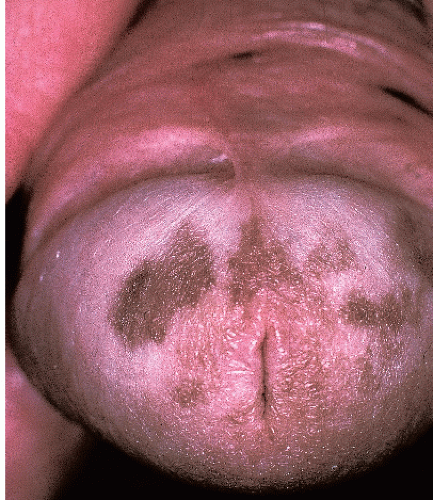
FIG. 11-2. Vitiligo tends to occur first on the glans, and patchy hyperpigmentation may or may not occur.


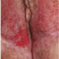
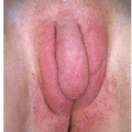
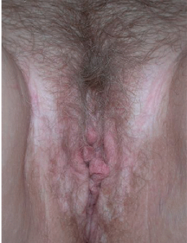
FIG. 11-3. Patchy vitiligo affecting the vulva and genito crural folds.


VITILIGO:


Management


  • No satisfactory therapy for repigmenting the genital area, but the following are occasionally useful, alone or in combination


    • Topical ultrapotent corticosteroid ointment applied b.i.d. in 6- to 8-week pulses


    • Tacrolimus or pimecrolimus applied b.i.d. ongoing


    • Calcipotriol (calcipotriene) cream applied b.i.d.


    • Ultraviolet light not practical, and confers risk of malignancy


  • When large areas of the body are affected, remaining normal skin can be depigmented permanently with 20% monobenzylether of hydroquinone applied twice a day to even the skin color


Postinflammatory Hypopigmentation and Depigmentation


Clinical Presentation

The loss of pigment is often very subtle, and usually there is no associated surrounding hyperpigmentation. Under Wood’s light examination, there is no enhancement of the hypopigmentation. However, patients with a dark complexion, and those who had past or present lesions of eczema or psoriasis, sometimes exhibit striking postinflammatory pigment loss that is accentuated by Wood’s light, almost indistinguishable from vitiligo. It is possible that these are examples of the Köebner phenomenon localized to the sites of the inflammatory disease.


Diagnosis

The diagnosis can usually be determined by the pattern of hypopigmentation in association with the past or present existence of skin disease or injury. A biopsy is only occasionally required and shows a decreased amount of melanin in the basal keratinocytes, but melanocytes are present. Pigment-ladened macrophages may be present in the underlying dermis.

The pallor of lichen sclerosus, lichen planus, and lichen simplex may be similar to postinflammatory hypopigmentation but the textural changes and scarring help to distinguish these disorders. Vitiligo due to depigmentation can mimic postinflammatory hypopigmentation and Wood’s lamp examination can usually differentiate between the two.


Pathophysiology

Any inflammatory dermatosis or injury can leave residual hypopigmentation or hyperpigmentation in the affected areas (Fig. 11-4). Postinflammatory hypopigmentation is more common in conditions that disrupt the integrity of the basement membrane zone, resulting in damage to the melanocytes and their function. In tinea versicolor, oxidation products are produced which are able to inhibit the tyrosinase activity of the melanocytes. Finally, permanent hypopigmentation may be a sequelae of destructive treatments in which the melanocytes may be more susceptible to damage, as seen after cryotherapy or radiotherapy.


POSTINFLAMMATORY HYPOPIGMENTATION:


Diagnosis


  • Usually, history of preceding inflammatory process; diaper dermatitis, lichen simplex chronicus, trauma of wart therapy, etc.


  • Pale, hypopigmented patches


  • No scale or surface change, except for that associated with any ongoing underlying etiologic inflammatory process


  • Distribution correlates with previous inflammatory process


  • Biopsy generally not necessary for diagnosis; stains show presence of melanocytes


Management

There is no treatment for postinflammatory hypopigmentation but the normal skin pigmentation returns spontaneously with time. If the underlying dermatosis
that caused the problem is still present, this should be treated to prevent further hypopigmentation.


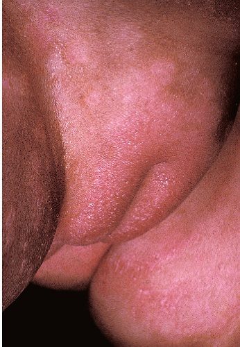
FIG. 11-4. This child still shows remaining erythema and roughness from her diaper rash; the area of pigment lightening is characteristic of postinflammatory hypopigmentation.


POSTINFLAMMATORY HYPOPIGMENTATION:


Management


  • Control of any ongoing underlying inflammatory process


  • Otherwise, self-resolving; no therapies hasten repigmentation


Lichen Sclerosus

Lichen sclerosus is a relatively common disease with a predilection for the anogenital skin. The terms leukoplakia, kraurosis vulvae, and balanitis xerotica used to describe genital atrophy and pallor in the past are now recognized as probable examples of lichen sclerosus. It is commoner in women with an estimated prevalence of between 1 in 300 and 1 in 1000 of the population.


Clinical Presentation

The peak times of presentation of lichen sclerosus are childhood and later life, particularly after menopause. In young boys, lichen sclerosus presents with phimosis and is probably the main reason why a medical circumcision is required around the age of 6 or 7 years. The lichen sclerosus often goes unrecognized, particularly if there is no histologic examination of the tissue removed at the time of surgery. Constipation is often the presenting symptom in young girls because lichen sclerosus around the rectal canal causes fissuring and painful defecation with consequent anal retention. Perianal involvement does not seem to occur in male patients. An intense itch is the primary symptom of anogenital lichen sclerosus with pain as a secondary symptom when there is ulceration and erosions.

The classic findings of early lichen sclerosus consist of pearly white papules and plaques. In the male these occur on the glans and prepuce of the penis and less commonly the shaft, whereas in the female the labia minora, clitoris, and interlabial sulci are the most frequent sites affected (Figs. 11-5, 11-6, 11-7, 11-8, 11-9, 11-10, 11-11 and 11-12). The lesions can become confluent and can spread out to involve the skin of the genitocrural folds and down to the perianal area in a figure-of-eight configuration. The epidermal atrophy when present gives the skin surface the texture of wrinkled cigarette paper and a shiny appearance. Ecchymosis when seen is pathognomonic for this disorder as the upper dermal vessels in the area of hyalinization are easily damaged. These ecchymoses can be mistaken for evidence of abuse in young girls. Additional signs include erosions, ulceration, and areas of hyperkeratosis.

The scarring that accompanies lichen sclerosus typically results in sealing of the clitoral hood concealing the glans clitoris underneath, shrinkage or loss of the labia minora, and more rarely some narrowing of the anterior and/or posterior aspects of the introitus (Fig. 11-13). Introital narrowing is unusual in women because lichen sclerosus appears to spare mucosal, non-cornified
stratified epithelium. There can be involvement of the modified mucous membrane at junctional zones such as the vestibule, but the vagina is never involved. Perianal involvement appears to occur exclusively in female patients.


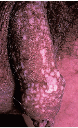
FIG. 11-5. Although at first glance, these very light papules mimic vitiligo, the crinkled texture change is pathognomonic for lichen sclerosus rather than vitiligo.


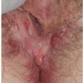
FIG. 11-6. This woman presented with severe itching, so scratching in the setting of the fragility typical of lichen sclerosus resulted in the classic finding of purpura.


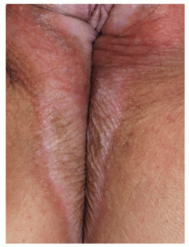
FIG. 11-7. Although hypopigmentation is the most striking abnormality in lichen sclerosus, the texture change is the key to diagnosis; the crinkled appearance seen here, and the distribution on the vulva and perianal skin are characteristic.


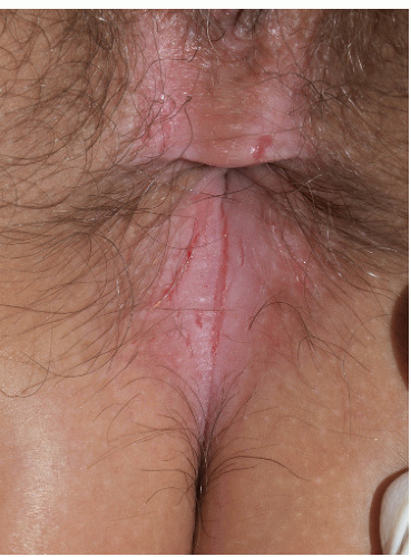
FIG. 11-8. Lichen sclerosus of perianal skin is very common in female patients, although it is not seen in male patients. Fissuring that produces pain with defecation contributes to constipation, a common presenting symptom in girls.


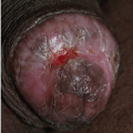
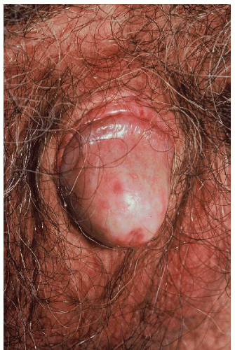
FIG. 11-9. The texture change of lichen sclerosus in this patient is manifested by shininess. The glans and inner prepuce are the most commonly affected areas.

Since lichen sclerosus is more likely to affect uncircumcised men, the scarring leads to a gradual tightening of the prepuce and eventually phimosis (Fig. 11-14). The coronal sulcus can also become obliterated with adhesions (Fig. 11-15). Lichen sclerosus sometimes involves the
cornified epithelium surrounding the urethral meatus and lower part of the urethra (Fig. 11-16).


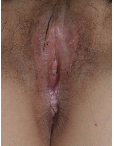
FIG. 11-10. Lichen sclerosus eventuates in resorption of architecture; the left labium minus has disappeared, and there is edema and midline scarring of the clitoral hood, an area of early and prominent involvement.


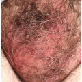
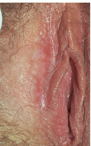
FIG. 11-11. Very early lichen sclerosus can be quite subtle, with mild hypopigmentation, poorly demarcated borders, and even a more waxy texture as pictured here in the anterior, interlabial fold.

Extragenital lichen sclerosus in men is rare but occurs in up to a third of women. Extragenital sites occur on the upper back, axillae, volar aspects of the wrists, and in areas exposed to friction such as the shoulders and inframammary areas. The distribution of lesions in the friction zones can be partly explained by the Köebner phenomenon. Very rare instances of face, scalp nails, and mouth have all been reported. Oral lesions of lichen
sclerosus are extremely uncommon, and most of the lesions seen involve the frictional sites which can become cornified, on the buccal mucosae, dorsal aspect of the tongue, and attached gingivae (6).


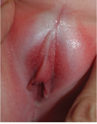
FIG. 11-12. Although lichen sclerosus is generally a disease of adults, its occurrence is well recognized in children, especially prepubertal girls, who are at risk of significant scarring of the vulva. Hypopigmentation, shiny texture, and purpura of the right labium minus are pathognomonic for lichen sclerosus. Edema of the clitoral hood is common as well.


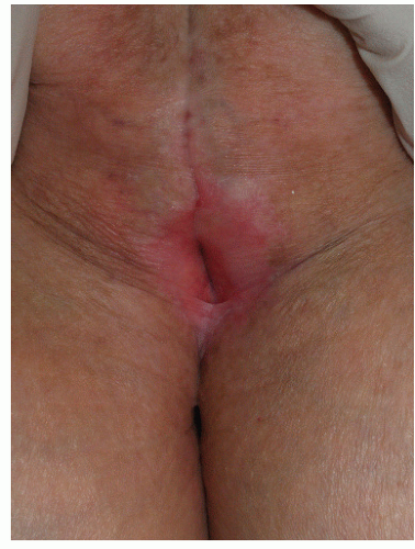
FIG. 11-13. Severe scarring occurs in some women as well as men; all vulvar architecture has been obliterated in this elderly woman with longstanding disease. Urinary obstruction occasionally occurs.


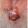
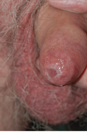
FIG. 11-14. Lichen sclerosus can produce phimosis, and it is the most common cause of phimosis in boys. (Courtesy of Errol Craig, MD.)


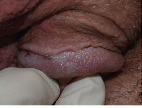
FIG. 11-15. Lichen sclerosus in male patients occurs most often on the glans, corona, and inner prepuce; scarring of the corona to the shaft has begun in this man.

There is an association with the other lymphocyte-mediated disorders such as lichen planus and morphoea and some patients can develop all three conditions. Vitiligo may also accompany lichen sclerosus.

There is a small risk, 4% or less, of neoplastic change and the development of a squamous cell carcinoma in long-standing untreated lichen sclerosus (Fig. 11-17). Verrucous carcinoma also has been associated with lichen sclerosus (Fig. 11-18).


Diagnosis

Although a diagnosis of lichen sclerosus can be made clinically in typical cases, histologic confirmation is ideal to differentiate lichen sclerosus from lichen planus and to detect if there is a thickened epidermis. However, the hyalinization of lichen sclerosus is lost if a potent topical steroid has been used. The skin that exhibits fine crinkling or ecchymosis is the ideal site for biopsy.


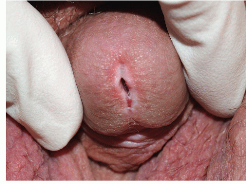
FIG. 11-16. Lichen sclerosus often affects urethral meatus and can cause strictures.


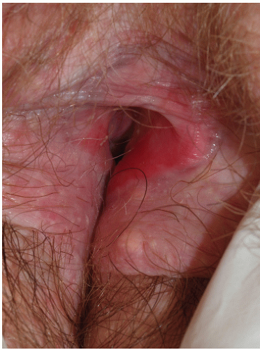
FIG. 11-17. Vulvar intraepithelial neoplasia (squamous cell carcinoma in situ) can present as red plaques within lichen sclerosus.

In uncomplicated lichen sclerosus, a biopsy shows an epidermis that is hyperkeratotic, thinned, and effaced. The epidermis
overlies a band of hyalinization in the upper dermis with a mixed lymphocytic infiltrate immediately below it. In some areas, small foci of lichenoid change are found where the infiltrate abuts the dermoepidermal junction. Rarely, the epidermis exhibits the histologic features of squamous cell hyperplasia, that is, it is thickened (acanthotic). This is important as there seems to be an association with this kind of change and development of squamous cell carcinoma.


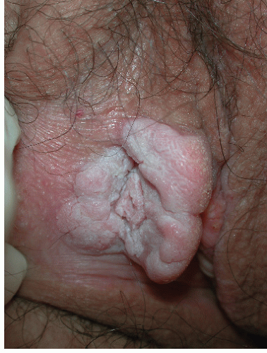
FIG. 11-18. This warty growth represents a verrucous carcinoma arising on a background of lichen sclerosus. Even clinically unimpressive thickened white areas of lichen sclerosus that do not promptly respond to therapy should be biopsied.

The pallor, atrophy, and loss of the normal anatomic features associated with well-established lichen sclerosus can be seen as a common end-stage picture of other inflammatory, scarring dermatoses, most notably lichen planus, cicatricial pemphigoid, and morphoea. Perianal itching with lichen simplex in children also raises the possibility of pinworm infestation.


LICHEN SCLEROSUS:


Diagnosis


  • White, crinkled, or shiny plaques


  • Cornified epithelium of the vulva, perineal body, and perianal skin of women; primarily of the glans and prepuce of men


  • Scarring often prominent


  • Confirmed by biopsy that shows upper dermal chronic inflammatory infiltrate, hyalinization of the upper dermis, and disruption of basement membrane


Jun 25, 2016 | Posted by in Dermatology | Comments Off on White Lesions

Full access? Get Clinical Tree

Get Clinical Tree app for offline access