Ulcers


Ulcers


Peter J. Lynch


Erosions and ulcers occur when tissue is lost from the surface of the skin. They are differentiated on the basis of depth. Tissue loss limited to the epithelium produces erosions, whereas tissue loss that extends into, or through, the dermis is termed an ulcer. The base of an erosion either may be red or may be covered with a loosely adherent yellow crust. The base of an ulcer, on the other hand, may be red or may be covered with a crust that has red, blue, or black heme pigment due to the destruction of blood vessels within the dermis. The crusts covering ulcers may also have considerable fibrin in them and for this reason are adherent and tenacious; this is sometimes termed eschar formation (see Chapter 9 for the coverage of erosions). Ulcers due to infection are discussed in the first portion of this chapter and those that are noninfectious in etiology are discussed in the latter half.


SYPHILIS

Syphilis is a sexually transmitted infection that is characterized by asymptomatic periods of varying duration, interrupted by three stages of clinical disease. In primary syphilis, a painless and nontender ulcer (“chancre”) develops, most often in or around the genitalia. The major discussion of syphilis will be found in this chapter with brief mention made of secondary syphilis in Chapter 12. Although syphilis is a global problem, the data in this chapter refer primarily to syphilis in Western countries.


Clinical Presentation

According to the 2007 Centers for Disease Control (CDC) report, the rate of primary and secondary syphilis in the United States declined 90% from 1990 to 2000 at which point it reached its nadir (1). Since then the rate has been steadily increasing. Most of this increase has been in men, primarily among men who have sex with men (MSM) (1). This population now accounts for about 65% of all of the cases of early syphilis. In 2007, the incidence of syphilis was 6.6 per 100,000 for men and 1.1 per 100,000 for women. The rate is appreciably higher in Hispanic and African American men than that in non-Hispanic white men. Syphilis occurs most frequently between the ages of 20 and 24 in women and 20 and 30 in men. The data for Australia and European countries is quite similar to that found in the United States (2,3). Coinfection with human immunodeficiency virus (HIV) is encountered frequently.

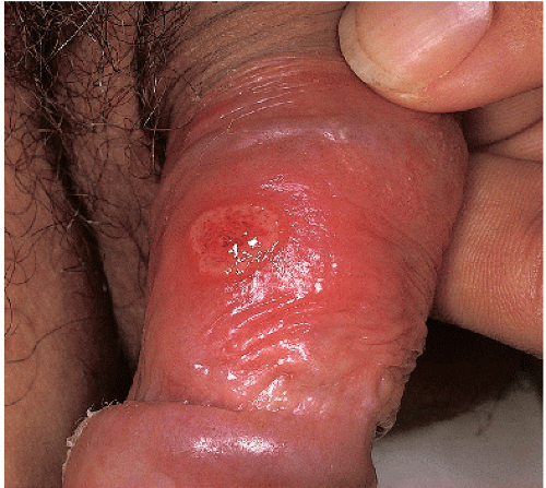
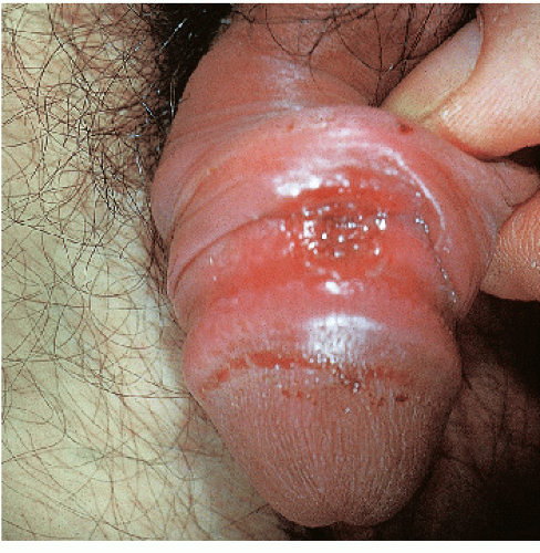
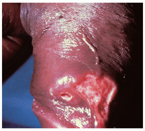
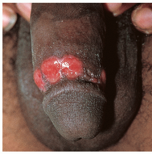
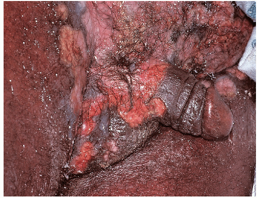
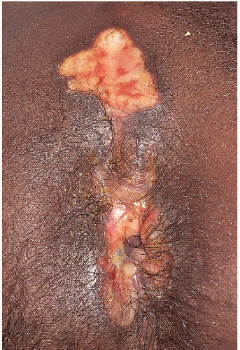
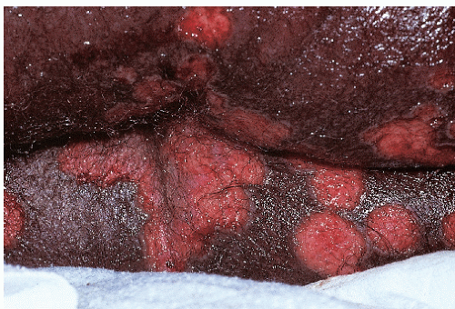
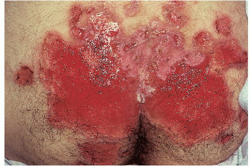
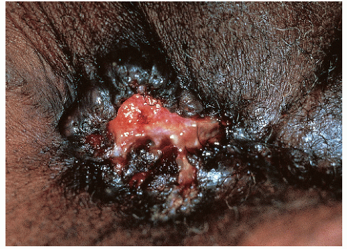
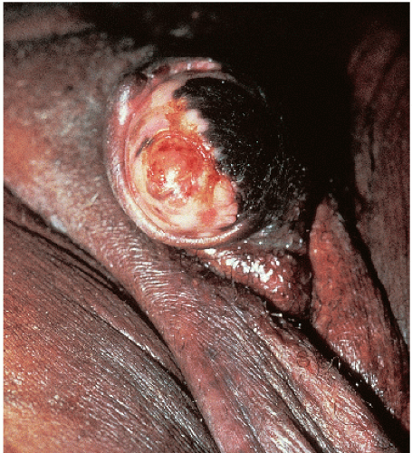
After an incubation period of 9 to 90 days (average of 3 weeks), the primary lesion occurs at the original site of inoculation. It begins as a small red papule that quickly enlarges and ulcerates to form a painless ulcer (“chancre”) that may be up to 2 cm in diameter. A chancre has a clean, red, glistening base (Figs. 10-1 and 10-2). A small amount of nonpurulent, watery exudate may be present. The ulcer borders are sharply demarcated and are characteristically raised and indurated. The chancre is painless and usually nontender. The base feels firm on palpation. Most patients present with a solitary ulcer, but two or even multiple lesions are possible. Chancres are located most often at the distal tip of the penis in men and on the labia majora or vestibule in women. However, it is likely that many chancres in women are never observed due to a location within the vagina or on the cervix. For this reason, the first clinical presentation of syphilis in women is more likely to be at the secondary stage than is true for men. Individuals who have receptive anal intercourse may develop chancres within or around the anus. Secondary bacterial infection of ulcerative lesions may distort the clinical presentation, causing tenderness and purulent discharge. Within about 1 week of chancre formation in the anogenital area, unilateral or bilateral inguinal regional lymphadenopathy develops as nontender, firm, rubbery, mobile lymph nodes.


Diagnosis

The presence of a solitary, nontender ulcer with a clean, firm base is highly likely to represent primary syphilis. Nevertheless, a clinical diagnosis must be confirmed by laboratory means. In years past, the quickest confirmation could be obtained by a dark-field examination carried out on site by the clinician. Unfortunately, this test has almost completely disappeared due to the lack of dark field microscopes in clinicians’ offices and the declining skills in carrying out this test. For this reason, a serologic test for syphilis needs to be obtained. The most commonly used nontreponemal serologic tests are the Venereal Disease Research Laboratory (VDRL) and the rapid plasma regain (RPR) tests, both of which detect antibodies to cardiolipin. These are readily available, cheap, and reliable. In addition, positive results can be titrated providing a valuable tool for determining response to
therapy. Unfortunately, there are several drawbacks to these tests: (1) the test may not become positive until 1 or 2 weeks after the chancre has appeared; (2) false-positive tests occur with some frequency; and (3) false-negative tests may occur due to the prozone phenomenon especially in those patients who are coinfected with HIV.


FIG. 10-1. This is a classical indurated, well-demarcated chancre with a glistening red base.

Because of these drawbacks, treponemal-specific tests such as the fluorescent treponemal antibody absorbed test (FTA-ABS), the Treponemal pallidum particle agglutination test (TP-PA), the T. pallidum hemagglutination assay (TP-HA), and Western blots are available. These tests offer nearly 99% sensitivity and 99% specificity. Interestingly, some large laboratories are reversing the algorithm and are using automated enzyme immunoassays (EIAs) for initial screening followed by a VDRL or RPR if positive results are obtained. This latter approach may offer some cost savings but can cause difficulties due to both occasional false-positive results with EIAs and the fact that unlike the nontreponemal tests, they may remain positive after previous effective treatment and thus cannot be used for determining a response to therapy (4).


FIG. 10-2. Clean, firm borders and a base with slight nonpurulent exudate are typical of a chancre.

Both the nonspecific and the specific serologic tests can remain negative for 7 to 14 days after a chancre appears. In this situation, biopsy should be considered for any lesion that is clinically suspicious for syphilis. The histologic presence of numerous plasma cells would then be followed by the use of a silver stain (e.g., Warthin-Starry stain) to identify the presence of spirochetes. Patients with syphilis are likely to have been exposed to other sexually transmitted diseases. For this reason, patients should be examined and tested for other infections when a diagnosis of syphilis has been established.

The two main considerations in the list of differential diagnoses are aphthous ulcers and genital herpes (HSV) infection, especially when the latter occurs in an immunosuppressed individual. Genital herpes occurring in those who are immunosuppressed may present with fairly deep, persistent, and relatively less painful ulcers rather than the more typical, transient, painful erosions seen in immunocompetent persons. Individual aphthous ulcers (as occur in complex aphthosis and Behçet disease) are similar in appearance to a chancre but, in contrast, several ulcers are usually present, they are extremely painful, and they tend to occur mostly in adolescent women. Chancroid is vanishingly rare in Western countries and is associated with a crusted base along with considerable pain and tenderness. Anogenital ulcers occur with Crohn disease, hidradenitis suppurativa, and granuloma inguinale, but the morphology is different as is the histopathology.


PRIMARY SYPHILIS:


Diagnosis


  • One or two relatively painless genital ulcers


  • Ulcer base is clean without crust or eschar


  • Base of the ulcer is firm and nontender on palpation


  • Positive RPR or VDRL; confirmed by positive FTA-ABS


  • If serology is negative, repeat tests in 1 week or biopsy


Pathophysiology

Syphilis is caused by the spirochete, Treponema pallidum. The level of contagion for syphilis is very high; it has been estimated that about one third of individuals having direct contact with a chancre will develop syphilis. The likelihood of infection is further enhanced in the presence of even minor breaks in the skin as well as in a setting of immunosuppression such as occurs with HIV infection (5). The reverse is also true. There is an increased risk for the acquisition of HIV in patients with chancres. Many (6), but not all (7), studies suggest that circumcision in males reduces the likelihood of acquiring
syphilis. The incubation period after exposure to T. pallidum is quite variable but averages about 3 weeks. Unfortunately, before the end of the incubation period and therefore before the development of a chancre, spirochetes have already gained access to the regional lymph nodes. For this reason, syphilis can be viewed as a systemic infection from its earliest inception.


Management

If untreated, a chancre undergoes spontaneous, nonscarring resolution within about 1 to 2 months. This leads the patient to the misconception that the process has healed and that it is not necessary to seek medical attention. However, as indicated above, syphilis becomes a systemic infection soon after the initial inoculation with the result that after a short latent period the generalized lesions of secondary syphilis are highly likely to appear. Rarely, secondary lesions may appear, without a latent period, while the chancre is still present. If the symptoms and signs of secondary syphilis are unrecognized and the patient remains untreated, the patient enters another latent period, leading some years later to the possibility of tertiary syphilis.

Most clinicians in the United States follow the treatment guidelines published by the CDC. The most recent update is from 2006 where benzathine penicillin G 2.4 million units, administered intramuscularly in a single dose, is the recommended therapy for primary and secondary syphilis (8). Note that Bicillin L-A (containing only benzathine penicillin) is to be used rather than Bicillin C-R that contains a combination of benzathine penicillin and the short-acting procaine penicillin. No penicillin resistance has yet been noted. Guidelines that are somewhat similar to those of the CDC have been developed for the United Kingdom and Europe (9,10). Because of their similarity, they will not be reviewed here.

For those who are allergic to penicillin, the CDC recommends desensitization rather than use of a second-line treatment with a nonpenicillin product. However, where desensitization is not feasible, it is appropriate to use alternative antibiotics such as doxycycline (100 mg orally twice daily for 14 days), ceftriaxone (1 g IM daily for 10 days), or azithromycin (2 g in a single oral dose) (8). The CDC recommendation for desensitization stems from a relative lack of published experience with all three of these agents together with some reports of azithromycin resistance. Patients who are pregnant should be treated with penicillin. Limited evidence suggests that in HIV infected persons, the response to therapy may be somewhat diminished. For this reason, some clinicians choose to repeat the 2.4 million units of benzathine penicillin weekly for 3 weeks.

The Jarisch-Herxheimer reaction is an acute febrile reaction that may occur within 24 hours of treatment for syphilis, regardless of the treatment regimen used. Symptoms include headache, malaise, myalgia, arthralgia, and fever. This transient reaction occurs most commonly after treatment for early syphilis and requires only reassurance, bed rest, and antipyretics for management.

Patients who are treated for primary or secondary syphilis require follow-up documentation of response. This is done by determining the titer of the RPR or VDRL antibodies at periodic intervals until at least a fourfold drop in titer has been obtained. Note that although either the RPR or the VDRL can be used, the same test must be used consistently as the titer levels differ between the two tests. Nearly all patients with early syphilis will eventually develop a negative RPR or VDRL test. However, this is not true for the treponemal specific tests (see above) where lifelong positivity usually continues.


PRIMARY SYPHILIS:


Management


  • Benzathine penicillin (Bicillin L-A) single dose 2.4 million units IM


  • Consider repeating the dose if the patient is HIV positive


  • Desensitize if the patient is allergic to penicillin


  • Or, doxycycline 100 mg b.i.d. × 14 days, azithromycin 2 g as single dose


  • Follow RPR or VDRL titer until negative


CHANCROID


Clinical Presentation

Chancroid is seldom encountered in Western societies. In the United States, fewer than 50 cases per year are reported to the CDC. However, within the last decade, the disease has occurred in clusters in several large cities of America. These mini-epidemics are usually associated with exposure to a single infected prostitute. Chancroid predisposes to the development of HIV infection and persons with chancroid are quite likely to have other sexually transmitted diseases as well.

After a short incubation period of 3 to 7 days, a small red papule or pustule appears at the site of inoculation. This inflammatory lesion may be either asymptomatic or tender. The area then quickly evolves into an extremely painful, deep, shaggy ulcer with ragged, soft, undermined margins (Figs. 10-3 and 10-4). In contrast to the chancre of syphilis, induration is not typically present. The base of the ulcer is friable and is often covered with a yellow-gray, foul-smelling, necrotic exudate. Most patients present with one or two ulcers, but multiple or giant ulcer formation is not uncommon. Fifty percent of patients develop painful, most often unilateral, inguinal lymphadenitis. Massively enlarged nodes (bubo formation) develop in 25% of cases and when untreated may become fluctuant with spontaneous rupture and chronic drainage.


FIG. 10-3. The ulcer of chancroid typically exhibits a shaggy border with a somewhat purulent red base. (Courtesy of Jack Mosley.)


Diagnosis

There are no easily available diagnostic tests for chancroid. The causative bacterium, Haemophilus ducreyi, is difficult to identify on smears taken from the ulcer even when special stains such as the Giemsa stain are used. H. ducreyi can only be cultured on special media and unfortunately this media is not available commercially. As a result, most laboratories do not offer the ability to culture this bacterium. Biopsy does reveal a fairly characteristic pattern consisting of three separate horizontal zones with distinctive vascular changes. The floor of the ulcer shows necrosis, red blood cells, neutrophils, and fibrin. Underlying this is a wide area of new vessel formation with proliferating endothelial cells that sometimes obstruct the lumina. The deepest portion of the histologic changes shows an inflammatory infiltrate of plasma cells and lymphoid cells.


FIG. 10-4. These well-demarcated round ulcerations of chancroid on the distal shaft are uniform and atypical, demonstrating that diagnosis often cannot be made on the basis of morphology.

The CDC indicates that a presumptive clinical diagnosis can be made if all of the following criteria are met: (1) the ulcers are painful and tender; (2) a serologic test for syphilis, taken at least 7 days after the ulcer has appeared, is negative; (3) the clinical presentation, including lymphadenopathy, is typical for chancroid; and (4) a test for HSV performed on the ulcer is negative (8). Primary syphilis, genital HSV infection, lymphogranuloma venereum, granuloma inguinale, ulcerated carcinoma, or secondarily infected traumatic lesions may all mimic chancroid.


CHANCROID:


Diagnosis


  • Single, sometimes two or three, painful ulcers


  • Base of ulcer is covered with purulent crust


  • Base is soft and very tender on palpation


  • Inguinal lymphadenopathy is usually present


  • Biopsy if confirmation of the diagnosis is needed


Pathophysiology

Chancroid is caused by the fastidious, gram-negative, aerobic or facultative anaerobic bacterium, H. ducreyi. It is highly contagious and experimental transfer from human to human is fairly easily carried out (11).


Management

Untreated, the ulcer of chancroid lasts about 2 months before spontaneous healing, generally with scarring, takes place. Ulceration and drainage from the enlarged nodes, if present, can last even longer. The CDC indicates that all of the following can be used for treatment: azithromycin (1 g orally in a single dose), ceftriaxone (250 mg IM in a single dose), ciprofloxacin (500 mg orally twice a day for 3 days), and erythromycin base (500 mg orally three times a day for 7 days) (8). With any of these treatments, healing of the ulcer occurs in about 1 week with lymph node response occurring much more slowly. Greatly enlarged, fluctuant nodes may require needle aspiration or incision and drainage.


CHANCROID:


Management


  • Azithromycin 1 g orally in a single dose or


  • Ceftriaxone 250 mg IM as a single dose


  • Incise and drain if lymph node is fluctuant


  • Follow to ensure complete healing


GENITAL HERPES IN IMMUNOSUPPRESSED INDIVIDUALS

HSV infection causes shallow, self-healing genital erosions in persons with normal immune status. This presentation of genital herpes is covered in Chapter 9. However, in those who are immunosuppressed, HSV infection becomes locally more severe as reflected by the development of chronic ulcers rather than transient erosions.


Clinical Presentation

In normal circumstances, control of HSV infection depends in large part on the presence of an intact cell-mediated immune system. For this reason, it is not surprising that evidence of genital HSV infection becomes prolonged and more severe in patients who are chronically immunosuppressed due to such disorders as HIV infection, malignancy (especially of hematopoietic origin), and those receiving long-term immunosuppressant medications for any reason. HSV infection, usually due to HSV type 2, then transforms from an erosive to an ulcerative disease.

In immunosuppressed patients, the lesions of HSV most often arise from reactivation of preexisting disease rather than from a primary infection. As is true for HSV infection occurring in immunocompetent patients, immunosuppressed patients describe accompanying pain (though frequently less severe) and careful questioning usually reveals prior, more typical recurrent episodes of genital herpes.

HSV outbreaks in immunosuppressed patients start as grouped vesicles on an erythematous base. The roofs of these vesicles disintegrate almost immediately leaving well-demarcated, discrete erosions. However, unlike the situation in immunocompetent patients, the erosions may not heal, but rather coalesce and deepen to form large, well-demarcated (“punched out”), somewhat painful, nonhealing ulcers (Figs. 10-5, 10-6, 10-7, 10-8 and 10-9). These ulcers may be superficial initially but often become deeper (Fig. 10-10). Secondary bacterial infection is possible but does not occur frequently. The configuration of the ulcers may be round but arcuate shapes are common due to coalescence of centrifugally expanding ulcers (Fig. 10-11). In men, chronic HSV ulcers are commonly found in a perianal distribution but also occur on the penis, scrotum, or in the groin. In women, ulcers may involve the mucosal portion of the vulva but also may extend to the labia majora and minora and even to the groin or inner thighs. As for men, HSV infection may also occur in a perianal location. In a small percentage of cases, HSV lesions in immunocompromised individuals may appear as eroded or ulcerated papules or nodules.


Diagnosis

A high index of suspicion should be present for HSV infection in any chronic, nonhealing ulcer in an immunosuppressed patient. A viral culture will usually confirm a clinically suspected diagnosis but viral growth may be slow and results may be delayed for a week or more (8). Examination of a stained smear taken from the base of the lesion (A Tzanck preparation) is less reliable for the diagnosis of a chronic ulcerative herpetic infection than is true for HSV infection in immunocompetent patients (8). Direct immunofluorescent antibody testing is available in most labs and offers a rapid diagnosis. This is accomplished by scraping the base of the ulcer with a number 15 blade and smearing the material collected on a microscope slide. The slide is then submitted to the laboratory and the results may be available within an hour or two.


FIG. 10-5. Herpes simplex virus infection in an immunosuppressed patient, such as this man with HIV, becomes chronic with relentless extension and ulceration.

The characteristic histologic features in HSV infection include ballooning degeneration of keratinocytes, reticular degeneration, and formation of multinucleated keratinocytic giant cells. Multinucleated keratinocytes are

pathognomonic for herpes virus infections, but they do not distinguish between HSV and varicella-zoster virus infection. Polymerase chain reaction (PCR) techniques, which are exquisitely sensitive, can be used in instances where the clinical picture highly suggests HSV infection but where other diagnostic tests are negative (8).


FIG. 10-6. Although this man has extension of his herpetic penile erosion, it has lost the classic arcuate borders from coalescing vesicles and now is only a nonspecific erosion.


FIG. 10-7. Perianal herpes simplex virus infection can also extend peripherally and deeply, producing a yellow fibrin base characteristic of ulceration of any cause.


FIG. 10-8. Deeper involvement of herpes simplex virus infection in an immunosuppressed patient such as this woman with chronic lymphocytic leukemia sometimes produces indurated lesions.


FIG. 10-9. This patient with chronic sacral herpes resistant to antiviral medication exhibits both extension and healing simultaneously.


FIG. 10-10. Herpes simplex virus infection resistant to antiviral agents in this man with AIDS has produced a peripherally enlarging perianal ulcer despite appropriate antiviral therapy.


FIG. 10-11. The most common cause of genital ulceration in an immunosuppressed patient is herpes simplex virus infection.

Although the presence of multiple well-demarcated, painful, chronic ulcers is highly suggestive of HSV infection, several other ulcerating infections must be considered in the list of differential diagnoses. Syphilis produces one or more relatively short-lived, firm, painless, noncrusted ulcers. Chancroid causes a painful, ragged crusted ulcer and may cause massive painful inguinal adenitis. Granuloma inguinale consists of one or more genital ulcers, often linear in configuration, containing exuberant granulation tissue. Rarely cytomegalovirus (CMV) has been reported to cause large, deep, necrotic ulcers in immunosuppressed patients, particularly those with acquired immunodeficiency syndrome (HIV/AIDS). Biopsy confirmation for CMV infection may be needed because HSV infection can occur concomitantly with CMV and may overgrow CMV in viral culture. Noninfectious causes such as aphthous ulcers and Crohn disease should also be considered.


HSV IN IMMUNOSUPPRESSED:


Diagnosis


  • Patient is significantly immunosuppressed


  • Punched out ulcers or ulcerated nodules


  • Arcuate configuration when confluence of ulcers occurs


  • Lesions are variable in terms of pain


  • HSV culture, immunofluorescent smear, or biopsy to confirm


Management

Left untreated, HSV ulcers in immunosuppressed patients may persist indefinitely. However, complete response to medical therapy is usually prompt. The CDC recommends acyclovir 400 mg orally three to five times daily until clinical resolution is attained (8). Famciclovir (500 mg orally twice daily) or valacyclovir (1.0 g orally twice daily) administered for 5 to 10 days is also effective (8). Rarely, intravenous acyclovir may be necessary if there is inadequate response to oral therapy (8). It is given as 5 to 10 mg/kg every 8 hours for 2 to 7 days or until clinical resolution is achieved.

Resistance to acyclovir has been reported to occur in up to 10% of immunocompromised individuals receiving long-term therapy to suppress outbreaks of HSV. All acyclovir-resistant strains are resistant to valacyclovir and most are resistant to famciclovir as well. The currently recommended treatment for acyclovir-resistant anogenital herpes is intravenous foscarnet in a dosage of 40 mg/kg every 8 hours until clinical resolution occurs. For HIV/AIDS patient not already receiving highly active retroviral therapy (HAART), starting such a regimen can lead to improvement though there is also some risk of exacerbation of latent HSV infection as part of the immune reconstitution inflammatory syndrome when this is done (12). Additional, new medications that are potentially usable are described in a recent review article on HSV therapy (13).


HSV IN IMMUNOSUPPRESSED:


Management


  • Start HAART therapy if not already underway


  • Acyclovir 400 mg orally 5 × per day until ulcers are healed or


  • Famciclovir 500 mg b.i.d. or


  • Valacyclovir 1.0 g b.i.d.


  • Foscarnet 40 mg/kg t.i.d. if the ulcer is acyclovir resistant


GRANULOMA INGUINALE (DONOVANOSIS)

Granuloma inguinale is a chronic, mildly contagious bacterial infection with a slowly progressive and destructive course. Four different clinical variants of the disease exist and this variability can result in a vast array of difficult-to-diagnose clinical presentations. These include ulcerovegetative (the most common), nodular, hypertrophic, and cicatricial forms of the disease.


Clinical Presentation

Granuloma inguinale is rarely encountered in North America and Europe, but it is endemic in some tropical and subtropical areas such as the Caribbean, Africa, Australia, southern India, South America, and Southeast Asia. It occurs most commonly in those of lower socioeconomic class. The majority of cases are found in adults where the transmission is sexually transmitted but nonvenereal transmission is possible and may account for most of the cases reported in babies and children.

The incubation period is typically less than 2 weeks but it can be up to 3 months in duration. The ulcerovegetative form of the disease is by far the most common clinical presentation. In this type of granuloma inguinale, the onset is insidious with formation of solitary or multiple, painless, firm papules or nodules at the site of inoculation. These lesions then erode to form soft, friable, painless ulcers that often involve skin folds (Figs. 10-12 and 10-13). In these sites, the ulcers characteristically have a linear, “knife cut,” morphology. The base of the ulcers is usually clean (noncrusted) with beefy-red granulation tissue. The margins are sharp and rolled borders are usually present. There is often progressive enlargement of some portion of
the ulcers while other portions are undergoing healing with fibrosis and scarring. In the less often encountered nodular variant of the disease, soft red papules and/or nodules develop and subsequently ulcerate. The ulcers on the surface of these lesions also contain appreciable amounts of granulation tissue. The hypertrophic form of granuloma inguinale consists of large vegetating masses, whereas the cicatricial variant is associated with expanding plaques of scar tissue.


FIG. 10-12. This perianal ulceration with heaped-up borders is typical of granuloma inguinale (Donovanosis). (Courtesy of Jack Mosley.)

Lesions in men most often occur in the anal and perianal area but also can be found in the coronal sulcus or on the inner aspect of the foreskin in uncircumcised individuals. In women, the vulva is most often involved but rarely lesions occur on the cervix. In both sexes, lesions may be found in the intertriginous folds of the perigenital skin. Typically, lymphadenopathy does not occur but spread of the infection to areas around the inguinal lymph nodes may cause swelling and ulceration. This process is termed pseudobubo. This lack of lymph node enlargement is characteristic enough to be a helpful in clinical diagnosis.


FIG. 10-13. Granuloma inguinale can produce a keratotic ulceration as seen on the glans penis. (Courtesy of Jack Mosley.)

Only gold members can continue reading. Log In or Register to continue

Stay updated, free articles. Join our Telegram channel

Jun 25, 2016 | Posted by in Dermatology | Comments Off on Ulcers

Full access? Get Clinical Tree

Get Clinical Tree app for offline access