The Skin and Internal Disease


The Skin and Internal Disease


Sarah Asch MS

Pascal Ferzli MD

Warren R. Heymann MD


A practicing dermatologist must be cognizant of the potential cutaneous manifestations of systemic diseases. Although certain skin findings are pathognomonic for particular maladies, more often than not cutaneous eruptions must be interpreted in the context of the complete clinical picture. Occasionally, a cutaneous finding may guide the clinician toward a previously undiagnosed systemic disease. It is the interplay between the skin and internal medicine that underscores the value of a cutaneous evaluation as a routine part of a complete physical examination.


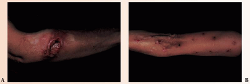
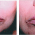
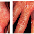
FIGURE 38-1 ▪ Nonspecific skin changes indicating an underlying psychologic disease state. (A) Delusional excoriation on the arm (“have to get the hairs out”). (B) Neurotic excoriations on the arm.


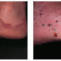
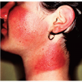
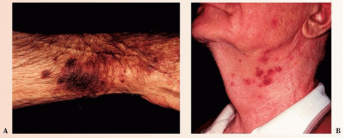
FIGURE 38-2 ▪ Nonspecific skin changes resulting from an internal disease. Purpura of the arm (A) and folliculitis of the neck (B) in a patient with myelogenous leukemia. (Courtesy of Syntext Laboratories, Inc.)

Cutaneous findings can be classified as specific and nonspecific. Specific changes demonstrate the same pathologic process as internal disease and can, therefore, be diagnostic of the disease. Nonspecific changes do not demonstrate the primary disease process (Figs. 38-1 and 38-2). These changes can be helpful in establishing the diagnosis only if interpreted within the context of the clinical data. The following cases are a few selected examples of the many systemic diseases with skin involvement. Table 38-1 lists some internal diseases and their dermatologic manifestations.


TABLE 38-1 ▪ Internal Malignancies with Cutaneous Manifestations


Disorder


Cutaneous Findings


Associated Malignancies


Acanthosis nigricans


Velvety hyperpigmentation of flexures and, less commonly, mucosal surfaces and palms (tripe palms)


Adenocarcinoma of genitourinary or gastrointestinal tract. Most commonly associated with adenocarcinoma of the stomach (55.5%)*


Acquired ichthyosis


Adult onset hyperkeratosis indistinguishable from ichthyosis vulgaris


Hodgkin’s lymphoma, mycosis fungoides, multiple myeloma, leiomyosarcoma


Acrokeratosis paraneoplastica (Bazex syndrome)


Acral psoriasiform plaques with nail dystrophy


Carcinomas of upper digestive and respiratory tracts. Also described in association with transitional cell bladder carcinoma


Carcinoid syndrome


Deeply erythematous or violaceous flushing of upper body associated with pruritus, diaphoresis, lacrimation, and facial edema


Foregut, midgut, and bronchial neuroendocrine tumors


Cushing’s syndrome


Generalized hyperpigmentation, including areolae, palmar creases, and scars; hirsutism; central obesity; moon facies, striae


Ectopic ACTH production by small cell lung cancer, bronchial carcinoid tumors and cancers of the thyroid, pancreas, and adrenals


Dermatomyositis


Heliotrope dermatitis, proximal nail fold telangiectasias, Gottron’s papules, cutaneous necrosis


Ovarian, gastrointestinal, and nasopharyngeal carcinomas; adenocarcinomas of the lung and prostate; hematologic malignancies


Erythema gyratum repens


Migratory figurate erythema with “wood grain” pattern


Malignancies of the lung, breast, female reproductive tract, gastrointestinal tract, and prostate


Hypertrichosis lanuginosa acquisita


Excessive growth of vellus hairs on neck and face, but can involve any body surface


Most commonly observed with colorectal, breast, and lung cancers


Necrolytic migratory erythema


Acral and intertriginous papulosquamous dermatitis with occasional vesiculation


Pancreatic α-cell tumor presents with glucagonoma syndrome


Paget’s disease


(1) Unilateral eczematous nipple plaque (2) Eczematous plaque of the anorectal, genital, and axillary regions (extramammary Paget’s disease)


(1) Associated with ductal adenocarcinoma (2) Regional associations are (a) anorectal—adenocarcinoma of the anus and colorectum, (b) vulvar—epithelial, eccrine, and apocrine neoplasms, and (c) male genitourinary reproductive tract malignancies


Paraneoplastic pemphigus


Diffuse mucocutaneous involvement with blisters, erosions, lichenoid, and erythema multiforme-like lesions. Head and neck skin is usually spared. Extensive oral erosions are notable.


Hematologic malignancies


Porphyria cutanea tarda


Vesicles and bullae with subsequent scarring, skin fragility on the dorsal hands, milia formation, and hypertrichosis on sun-exposed surfaces


Hepatocellular carcinoma, hematologic malignancies, myelodysplastic syndromes


Sweet’s syndrome—atypical bullous pyoderma gangrenosum overlap


Indurated erythematous to violaceous plaques with or without bulla formation and ulceration


Hematologic malignancies, myeloproliferative disorders


Sign of Leser-Trélat


Eruptive multiple seborrheic keratoses


Adenocarcinomas of the lung and gastrointestinal tract


* From Rigel DS, Jacobs MI. Malignant acanthosis nigricans: a review. J Dermatol Surg Oncol. 1980;6(11):923-927.

From Arregui MA, Raton JA, Landa N, et al. Bazex’s syndrome (acrokeratosis paraneoplastica)—first case report of association with a bladder carcinoma. Clin Exp Dermatol. 1993;18(5):445-448.


Abbreviations: ACTH, Adrenocorticotropic hormone.


Cardiology


Kawasaki’s Syndrome

Kawasaki’s syndrome, also known as mucocutaneous lymph node syndrome, is a self-limited acute vasculitis of childhood. It has a propensity for coronary artery involvement with aneurysms, angina pectoris, or myocardial infarction in up to 18% to 23% of untreated cases. With proper treatment, the percentage of coronary artery aneurysms decreases to 4% to 8%. Usually young children under the age of 5 are affected, with cases reported in infants and teenagers as well. There is a slight female preponderance. The diagnosis of Kawasaki’s syndrome is based on a constellation of clinical findings including fever lasting at least 5 days, nonsuppurative cervical adenopathy, bilateral nonpurulent conjunctival injection, reddening and fissuring of the lips, “strawberry tongue,” and several cutaneous findings. The skin changes begin with erythema of the palms and soles that may spread to the trunk. Then the syndrome progresses with the presence of an indurative edema and desquamation starting on the tips of the fingers and toes and around nails. A polymorphous rash that can vary from morbilliform to scarlatiniform may also be present.


LEOPARD Syndrome

Multiple lentigines syndrome is an autosomal dominant disorder with abnormalities of various clinical expressions. LEOPARD is an acronym for the following abnormalities that may be present in an individual patient with this syndrome:


  • Lentigines: multiple lentigines are present at birth and may cover the entire body, including the palms and soles but sparing the lips and oral mucosa. The pigment can be seen in the iris and retina as well


  • Electrocardiogram conduction defects


  • Ocular hypertelorism


  • Pulmonary stenosis


  • Abnormalities of genitalia


  • Retardation of growth


  • Deafness (sensorineural)


Pseudoxanthoma Elasticum

Pseudoxanthoma elasticum is a genetic connective tissue disease characterized by progressive mineralization of elastic fibers with cutaneous, cardiovascular, and ophthalmologic complications. The disease manifests as angioid streaks of the retina, retinal and gastrointestinal hemorrhages, hypertension, and occlusive vascular disease secondary to progressive calcification and fragmentation of the elastic fibers in the eye and blood vessels. Characteristic yellowish papules that appear like a “plucked chicken” are seen in the flexural areas of the neck as well as periumbilical areas. These lesions can also be seen in the oral, vaginal, and rectal mucosa.


Endocrinology


Diabetes Mellitus

Cutaneous manifestations associated with diabetes mellitus may correlate with metabolic derangements or may present as chronic degenerative changes with no apparent correlation to the degree of hyperglycemia. Metabolic changes in patients with poorly controlled diabetes tend to lead to a higher risk for the development of cutaneous infections by bacterial, fungal, and yeast pathogens. Diabetic dermopathy (atrophic, circumscribed, brownish plaques on the pretibial surfaces; Fig. 38-3) or bullous diabeticorum (spontaneous development of bullae on the extremities) can be seen as a result of chronic degenerative changes. Peripheral neuropathy, further compounded by
vascular compromise, may eventuate in neuropathic foot ulcers (Fig. 38-4).


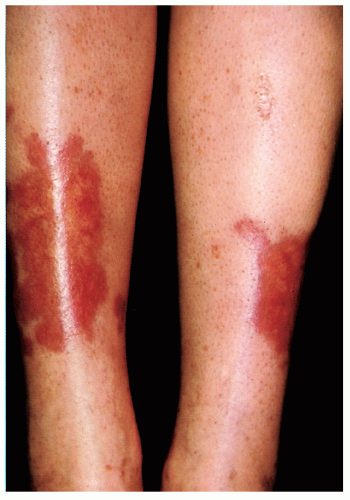
FIGURE 38-3 ▪ Necrobiosis lipoidica diabeticorum on the anterior tibial area of the legs. (Courtesy of Smith Kline & French Laboratories.)


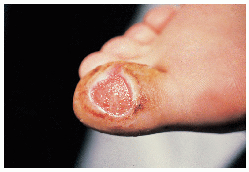
FIGURE 38-4 ▪ Mal perforans ulcer on the great toe of a diabetic man.


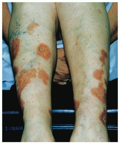
FIGURE 38-5 ▪ Necrobiosis lipoidica.

Necrobiosis lipoidica (NL) is seen in less than 1% of diabetics, but the majority of patients with NL have diabetes mellitus (Fig. 38-5). NL begins as sharply circumscribed, dusky-red nodules or papules located most commonly on the anterior and lateral surfaces of the lower extremities. The lesions expand to form atrophic, waxy, yellowish, telangiectatic plaques with active brawny borders. The plaques occasionally ulcerate. New NL lesions may appear at the site of surgery or trauma (Koebner phenomenon).


Disorders of the Hypothalamic-Pituitary-Adrenal Axis

Cushing’s syndrome of glucocorticoid excess is caused by either endogenous overproduction of corticosteroids by the adrenal glands or by iatrogenically administered steroids, whereas Cushing’s disease is caused by an adrenocorticotropic hormone-secreting anterior pituitary or nonpituitary neoplasm. The most profound cutaneous manifestations of Cushing’s syndrome (and disease) include epidermal atrophy, striae, plethoric moon facies, buffalo hump, supraclavicular fat pads, and central obesity. There is a marked susceptibility to cutaneous fungal infections. The following findings may be observed in Cushing’s syndrome:


  • Addisonian hyperpigmentation


  • Precocious puberty


  • Virilization


  • Pattern alopecia in females

Patients with Addison’s disease, or primary adrenocortical insufficiency, suffer from a deficiency of glucocorticoids as well as mineralocorticoids. There is a hyperpigmentation of sun-exposed surfaces, flexural areas, pressure points, scars, and palmar creases (Fig. 38-6). Women may experience loss of axillary and pubic hair.


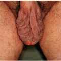
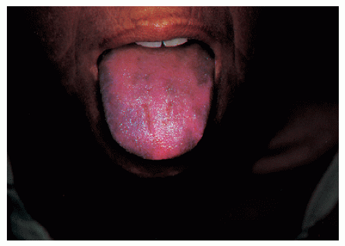
FIGURE 38-6 ▪ Hyperpigmentation of skin and tongue in a white woman with Addison’s disease.


Multiple Mucosal Neuroma Syndrome

Multiple mucosal neuroma syndrome is also known as multiple endocrine neoplasia IIB. In this autosomal dominant disorder, medullary thyroid carcinoma (MTC) and pheochromocytoma are associated with oral, nasal, upper gastrointestinal tract, and conjunctival neuromas. The skin lesions typically range from soft to firm intradermal nodules that tend to precede the MTC. However, MTC may occur in early childhood prior to the development of neuromas. Additional skin findings include “blubbery” lips, lentigines, café-au-lait macules, and localized intense unilateral pruritus on the back (notalgia paresthetica).


Thyroid Disease

The activity of the thyroid gland is intimately reflected by changes in the skin and its appendages. In hyperthyroidism, the skin is thin, warm, moist, and flushed secondary to vasodilation of the dermal vasculature. Erythema and hyperhidrosis of the palms and soles may be present. Adnexal changes include rapidly growing, fine, soft hair with
attendant nonscarring alopecia and soft nails with distal onycholysis. Graves’ disease is associated with ophthalmopathy (proptosis, exophthalmos, and lid lag), thyroid dermopathy (pretibial myxedema), and acropachy.


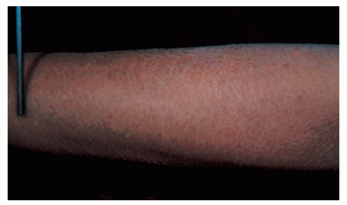
FIGURE 38-7 ▪ Year-round dry skin associated with hypothyroidism. (Courtesy of Reed and Carnick.)

In hypothyroidism, the skin is cold, dry, and pale secondary to vasoconstriction of the cutaneous vessels (Fig. 38-7). There is a generalized thinning and hyperkeratosis of the epidermis. Fine wrinkling and a yellow discoloration of the skin are also sometimes present. The hair is coarse, dry, brittle, and slow growing. Patchy or diffuse alopecia may be seen. Loss of the outer third of the eyebrow (madarosis) is a characteristic finding. Myxedema may be generalized in its distribution. Hypothyroid facies typically is expressionless, with thickening of the lips, broadening of the nose, drooping of the upper eyelids, and overall puffiness.


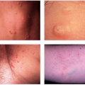
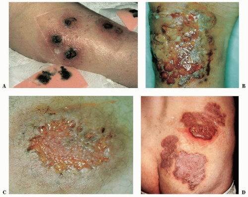
FIGURE 38-8 ▪ Pyoderma gangrenosum associated with ulcerative colitis. (Part D courtesy of Schering Corp.)


Gastroenterology


Dermatitis Herpetiformis

Also known as Duhring’s disease, dermatitis herpetiformis manifests as vesicles on the buttocks, elbows, and knees. Dermatitis herpetiformis is associated with celiac sprue (gluten-sensitive enteropathy). Celiac sprue is often subclinical but can be confirmed by jejunal biopsy. Dermatitis herpetiformis should be considered in the differential diagnosis of children presenting with recalcitrant eczema or individuals with recalcitrant pruritus and ill-defined dermatoses.


Inflammatory Bowel Disease

Ulcerative colitis is most commonly associated with pyoderma gangrenosum, a destructive neutrophilic dermatosis (Fig. 38-8). In pyoderma gangrenosum, a painful violaceous
nodule or pustule breaks down to form an enlarging ulcer with a raised, undetermined border and a boggy, necrotic base. Pyoderma gangrenosum has also been observed with Crohn’s disease as well as hematologic malignancies, monoclonal gammopathies, and various arthritides.


Peutz-Jeghers Syndrome

Peutz-Jeghers syndrome is an autosomal dominant disorder characterized by hamartomatous gastrointestinal polyposis and mucocutaneous pigmentation. Patients may present with abdominal pain, rectal bleeding, rectal prolapse, or intussusception. There is an increased risk of gastrointestinal tumors, ovarian and breast malignancies in females, and Sertoli cell tumors in males. Lentigines may be present at birth or develop in early childhood. Discrete brown, blue, or blue-brown macules are almost always located on the lips and oral mucosa, most commonly on buccal surfaces. Lentigines may also appear on the nail beds, hands, and feet, especially on palmar and plantar areas. With age, the cutaneous lesions may fade and even disappear, but the buccal mucosal lesions tend to persist into adulthood.

May 28, 2016 | Posted by in Dermatology | Comments Off on The Skin and Internal Disease

Full access? Get Clinical Tree

Get Clinical Tree app for offline access