Dermatologic Reactions to Ultraviolet Radiation and Visible Light
Laurie L. Kohen MD
Henry W. Lim MD
The Ultraviolet Spectrum
By convention, ultraviolet (UV) radiation is divided into UVA, UVB, and UVC. UVA ranges from 320 to 400 nm. UVB spans from 290 to 320 nm, and UVC includes wavelengths measuring from 200 to 290 nm. UVC radiation emitted by the sun is absorbed by the atmosphere. Therefore, it does not reach the earth’s surface and has no medical relevance. Sixty-five percent of UV radiation reaches the earth’s surface between 10:00 AM and 2:00 PM when the sun is most directly overhead. UV radiation in noonday sun consists of 95% UVA and 5% UVB. This is the reason that, for optimal photoprotection, broad-spectrum sunscreens that absorb in both the UVA and UVB ranges are recommended. The type of UV light and chromophores in the skin, such as nucleic acids, melanin, and aromatic amino acids, determine the depth of penetration of UV radiation. UVA, being of a longer wavelength, penetrates deeper than UVB. Twenty to thirty percent of UVA radiation reaches the deep dermis, whereas only 10% of UVB reaches the superficial dermis. UVA, but not UVB radiation, can penetrate window glass.
Sunburn and Tanning: Acute Effects of UV Radiation
Skin type plays an important role in the clinical outcome of sun exposure. Fitzpatrick’s classification of skin types is widely used (Table 39-1). Sensitivity to UV is best assessed by the determination of minimal erythema dose (MED), which is defined as the smallest dose of radiation causing perceptible erythema covering the entire irradiated area. For individuals with skin type I, the MED for broadband UVB (MED-B) is between 20 and 40 mJ/cm2 and for broadband UVA (MED-A), it is 20 to 40 J/cm2, illustrating the 1,000-fold less efficiency of UVA radiation in causing erythema.
Presentation and Characteristics
UVA-induced erythema is typically apparent by the end of the irradiation period and fades gradually in the next 24 to 72 hours. UVA is much more effective at inducing pigmentary alteration than causing erythema. Exposure to UVA radiation results in three types of pigmentation changes: immediate pigment darkening (IPD), persistent pigment darkening (PPD), and delayed tanning. IPD occurs immediately after exposure and fades within 10 to 20 minutes; it presents as a bluish-gray discoloration of the skin. The mechanism of IPD is oxidation of preexisting melanin in the epidermis; there is no neomelanogenesis. PPD occurs by the same mechanism and follows IPD if the UVA dose is sufficiently high. It lasts from 2 to 24 hours. Delayed tanning is due to the formation of new melanin in the epidermis; it may blend with IPD and PPD and lasts for days.
UVB-induced erythema consists of an immediate and a delayed phase; the former starts in 2 to 6 hours and peaks in 24 to 36 hours. There is no apparent IPD/PPD seen with UVB, just a delayed tanning reaction that is always preceded by erythema. UVB-induced delayed tanning peaks at 72 hours and fades rapidly. Similar to UVA-induced delayed tanning, neomelanogenesis also takes place.
TABLE 39-1 ▪ Fitzpatrick Skin Types | ||||||||||||||
|---|---|---|---|---|---|---|---|---|---|---|---|---|---|---|
|
SAUER’S NOTES
1. UVB is most efficient at inducing sunburn, while UVA is most efficient at inducing delayed tanning.
2. Photoprotection includes seeking shade between 10 AM and 4 PM, the use of photoprotective clothing, wide-brimmed hats, and sun protective glasses, and the application of sunscreen.
3. Broad-spectrum sunscreen with SPF greater than 30 should be applied generously and reapplied every 2 hours when outdoors.
Sunburn reactions present as erythema, edema, vesiculation, and pain, followed by scaling, desquamation, and hyperpigmentation. Acute reactions, when severe, may be accompanied by weakness, fatigue, and pruritus.
Treatment
Photoprotection: Photoprotection consists of minimizing sun exposure between 10 AM and 4 PM, the use of photoprotective clothing, wide-brimmed hats, and sunglasses, and the application of broad-spectrum sunscreens. Sunscreens with a sun protection factor (SPF) of 15 or greater should be applied 20 minutes before sun exposure and reapplied every 2 hours, especially after sweating or swimming. Sunscreens should be applied generously: 1 oz (30 mL) is needed to cover the entire body surface. Broad-spectrum sunscreens, which protect against both UVB and UVA radiation, are recommended. Commonly used sunscreen ingredients in the United States are listed in Table 39-2.
Nonsteroidal anti-inflammatory agents and corticosteroids: These should be taken within 4 to 6 hours after sun exposure. Topical corticosteroids and cool compresses are helpful in reducing the inflammation. Oral prednisone (1 mg/kg) may be used for 5 to 7 days in severe cases.
TABLE 39-2 ▪ Commonly Used Sunscreen Ingredients in the United States | ||||||||||||||||
|---|---|---|---|---|---|---|---|---|---|---|---|---|---|---|---|---|
| ||||||||||||||||
Photoaging: Chronic Effects of UV Radiation
Presentation and Characteristics
Photoaging accounts for 90% of age-associated cosmetic problems. The effects of photoaging can be broken down into the following categories:
Pigmentation changes
Texture changes
Vascular changes
Papillary changes
Pigmentation changes result from UV damage to the epidermis; the other changes result from dermal pathology. Both UVA and UVB radiation contribute to the process of photoaging.
The prototypical pigmentary change seen in older adults is a solar lentigo. Solar lentigines, or “age spots,” appear in chronically sun-exposed areas, usually starting at around 40 years of age. They are macules with well-demarcated borders and vary in color from yellowish brown to dark brown. The mechanism of occurrence is thought to be an increase in melanin content within the keratinocytes and possibly reactive hyperplasia of melanocytes. Areas near the lentigines may be hypopigmented, giving the skin an overall mottled appearance.
The leathery texture and deep wrinkling of the skin from photoaging is called solar elastosis and is very characteristic of severe chronic sun damage. This typically occurs on the face and neck and gives the skin a yellowish hue. The pathologic hallmark is deposition in the papillary dermis of amorphous elastotic material that does not form functional elastic fibers. This altered connective tissue does not demonstrate the resilient properties of normal elastic tissue. There is also epidermal acanthosis seen on histology. Furthermore, collagen destruction, induced by downstream effects of oxidative and direct DNA damage from UV radiation, plays a role in the loss of the skin’s tensile strength.
Blood vessel damage occurs with photoaging as well. Thinning of vessel walls and a decrease in vessel number are observed. Connective tissue support of the vasculature is diminished. Thus, fragility of vessels is demonstrated by the development of ecchymoses after minimal trauma. Telangiectasias are also seen in chronically sun-exposed regions.
A common example of a papillary change seen in photoaging is a seborrheic keratosis. This “wisdom spot” results from disrupted keratinocyte maturation imposed by accumulated UV radiation. Seborrheic keratoses appear “stuck on” to the skin and are more frequent on sun-exposed skin of the face, trunk, and extremities. They are completely benign growths and pose no risk of malignant transformation.
Treatment
Topical retinoids: These can cause slight reversal of photoaged skin. They increase collagen levels, which effaces wrinkles. Retinoids also stimulate epidermal hyperplasia, which manifests clinically as smoother
skin with fewer fine lines. Deeper wrinkles caused by chronologic aging persist. Retinoids also lighten pigmentary changes associated with photodamage. Side effects include peeling, erythema, and dryness. Topical retinoids are not recommended during pregnancy.
Photorejuvenation: This entails stimulation of dermal collagen synthesis by exposure to laser, intense pulse (visible) light, radiofrequency, or photodynamic therapy. This is a rapidly evolving area with numerous methods and equipment on the market. More studies are needed for many of the methods used.
Resurfacing: This can occur at a superficial, medium, or deep level. Techniques employed include microdermabrasion, chemical peels, and, less commonly, laser resurfacing. Efficacy depends on the depth of wound infliction. The mechanism of wrinkle reduction is stimulation of wound healing with new collagen formation. Re-epithelization occurs from stem cells located in adnexal appendages. Side effects of resurfacing include permanent pigmentary changes and scarring.
SAUER’S NOTES
1. Photoaging changes include solar lentigines, solar elastosis, loss of elasticity of the skin, and telangiectasias.
2. Treatment includes topical retinoids, photorejuvenation, and resurfacing.
Photocarcinogenesis
Actinic Keratoses
Presentation and Characteristics
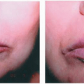
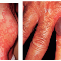
Actinic keratoses (AKs) are premalignant lesions that predominately form on the chronically sun-exposed areas of skin type I and II (Table 39-1) patients. They occasionally appear in type III and IV individuals as well. Clinically, they present as discrete, rough, hyperkeratotic areas with a scale. They may be brown, yellowish-brown, flesh-colored, or red. When the lower lip is involved, the term actinic cheilitis is used (Fig. 39-1). Texture is the key to diagnosis. Histologically, an AK is an abnormal proliferation of cells confined to the epidermis with some evidence of cellular atypia. It has been estimated that over 10 years, 10.2% of AKs would evolve into invasive squamous cell carcinoma, thus necessitating the treatment of lesions that do not spontaneously remit. AKs are considered to be precursors of squamous cell carcinoma; hence, they should be treated appropriately.
Treatment
Cryotherapy: This is the treatment of choice for most superficial lesions. For AK lesions that appear indurated, painful, or with a thick crust, surgical removal may be required and a specimen should be sent for pathologic examination to rule out squamous cell carcinoma.
Topical agents: 5-Fluorouracil, imiquimod cream, and diclofenac sodium gel are useful for patients with multiple or recurrent lesions.
Photodynamic therapy: This modality utilizes 5-aminolevulinic acid (which gets converted into protoporphyrin) and blue light, or methyl aminolevulinate and red light. It has been shown to successfully eliminate AKs.
Related posts:
Stay updated, free articles. Join our Telegram channel

Full access? Get Clinical Tree


