Skin Diseases in Ethnic Skin


Skin Diseases in Ethnic Skin


Cheryl M. Burgess MD

Beverly A. Johnson MD


Learning Objectives/Goals


  • Recognize diversity among skin types.


  • Describe the changing demographics among ethnic populations in the United States.


  • Recognize the market growth of ethnic consumers of dermatologic services.


  • Recognize differences between white skin and ethnic skin.


  • Identify skin types using established classification systems.


  • Assess therapeutic considerations and identify potential for adverse effects in ethnic skin.


  • Select treatment plans with respect to the potential for tissue response in ethnic skin.


  • Identify the most common cosmetic concerns in ethnic skin.


Chapter Overview

Dermatologists must take adequate steps for maintaining the necessary knowledge and instruction to provide all patients with thorough and comprehensive services. Therefore, additional knowledge and training must be considered to deliver the greatest efficacy with the lowest risk of adverse events when using state-of-the-art products and procedures in ethnic skin. This is especially true in light of daily advances in dermatologic services and growing diversity of skin types in the ethnic patient population. Clinicians must be fully prepared to recognize common problems in ethnic skin, identify appropriate treatments, and take steps to limit risk for any adverse events within this patient population.


Terminology

Throughout the burgeoning research on ethnic skin, it has been difficult to provide consistent terminology in the medical literature that conveys appropriate categorization and accurate description of skin types. To address this challenge, the following conventions are used throughout this chapter: The term “white skin” refers to skin having a Fitzpatrick classification of I, II, or III. The term “Caucasian” is avoided because this term refers to a larger group of skin types, beyond people with white skin. The term “ethnic skin” refers to skin that has a Fitzpatrick classification of IV, V, and VI, which applies to skin types in people of black, Latino, and Asian ethnic groups. The term “blacks” is used in place of “African Americans” because the latter term refers to only a subgroup of a much larger category of people with pigmented skin. These conventions have been selected to provide consistency, accuracy, and clarity in the following discussion. These conventions should not be substituted for careful clinical descriptions.


Introduction and Background


Demographic Trends and the Growth of Dermatologic Services in Ethnic Consumers


Diversity of Skin Types

The buzz phrase, “Skin of Color,” has often been thought to refer primarily to black skin. However, in the United States, individuals with pigmented skin come from a variety of racial and ethnic groups. These groups are summarized in Table 35-1. The diversity of ethnic and racial groups yields a broad spectrum of pigmented skin types that is continuously changing. For example, the 1990 US Census Bureau listed 6 races with 23 racial subtypes. Only 10 years later, the same 6 race categories included at least 67 subtypes. The largest ethnic populations—black, Hispanic, and Asian—are expected to make up almost half of the total US population by the year 2050; the predictions are 25% Hispanic, 14% black, 8% Asian, and 1% other. Trends in the
growth of diversity must drive the practice of dermatology to new levels of knowledge and awareness, so that clinicians are prepared to meet the challenges of today’s patient populations and provide the highest level of care to the largest possible number of patients.


TABLE 35-1 ▪ Racial and Ethnic Groups in the United States


African-American black persons (including Caribbean-American black persons)


Asian and Pacific Islanders (including those of Filipino, Chinese, Japanese, Korean, Vietnamese, Thai, Malaysian, Laotian, Hmong)


Latino or Hispanic (including those of Mexican, Cuban, Puerto Rican, Central American, Spanish descent)


People traditionally categorized as Caucasoid (including a majority of Indians, Pakistanis, and those of Middle Eastern origin)


Native Americans


Alaskans


Aleuts


Categorizing Pigmented Skin Types


Fitzpatrick Skin Phototype Classification System

In the United States, individuals with pigmented skin come from a large collection of racial and ethnic groups. The diversity of racial and ethnic groups yields a broad spectrum of pigmented skin types that defy easy categorization. Throughout the years, the fields of dermatology and cosmetics have struggled to characterize pigmented skin types adequately. After its development in the 1970s, the Fitzpatrick Skin Phototype classification became a surrogate classification system for this purpose.

The Fitzpatrick Skin Phototype classification system was originally developed to categorize the skin’s response to UV radiation. Over time, dermatologists became accustomed to using the system to classify both UV sensitivity and skin color. However, the system has limited utility for accurately communicating patient information for either research or clinical purposes and is of almost no value for helping clinicians treat ethnic skin effectively and safely. Patients with ethnic skin would benefit more from a classification system based on the propensity of the skin to scar and/or become hyperpigmented—a unique characteristic of pigmented skin. Several classification systems have been developed or proposed to meet these obvious needs. However, none have risen to an industry standard.


TABLE 35-2 ▪ The Roberts Classification System


Name (units)


Fitzpatrick Scale (FZ)


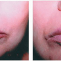
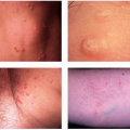
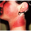
Roberts Hyperpigmentation Scale (H)
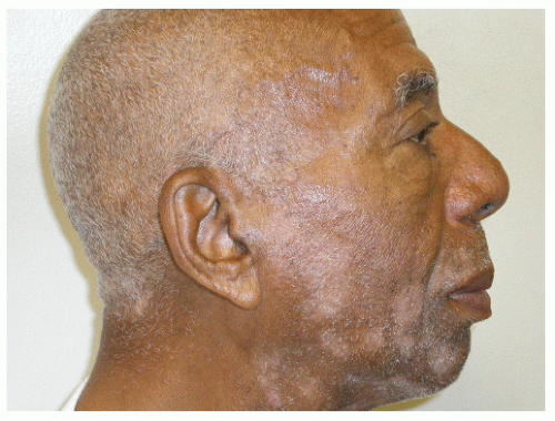

Glogau Scale (G)


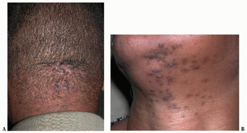
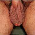
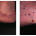
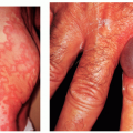
Roberts Scarring Scale (S)


Scale


Measures skin phototypes


Propensity for pigmentation


Describes photoaging


Describes scar morphology


Categories


FZ1 White skin. Always burns, never tans


H0 Hypopigmentation


G1 No wrinkles, early photoaging


S0 Atrophy


FZ2 White skin. Always burns, minimal tan


H1 Minimal and transient (<1 year) hyperpigmentation


G2 Wrinkles with motion, early to moderate photoaging


S1 None


FZ3 White skin. Burns minimally, tans moderately and gradually


H2 Minimal and permanent (>1 year) hyperpigmentation


G3 Wrinkles at rest, advanced photoaging


S2 Macule


FZ4 Light brown skin. Burns minimally, tans well


H3 Moderate and transient (<1 year) hyperpigmentation


G4 Only wrinkles, severe photoaging


S3 Plaque within scarred boundaries


FZ5 Brown skin. Rarely burns, tans deeply


H4 Severe and transient (>1 year) hyperpigmentation


S4 Keloid


FZ6 Dark brown/black skin


H5 Severe and transient (<1 year) hyperpigmentation


S5 Keloid nodule


H6 Severe and permanent (>1 year) hyperpigmentation


Note: Reprinted with permission from Roberts WE. The Roberts skin type classification system. J Drugs Dermatol. 2008;7(5):452-456.


Roberts Skin Type Classification System

The recently introduced Roberts Skin Type classification system (Table 35-2) may provide the most comprehensive information to meet the needs of clinicians. This system uses a four-part serial profile to characterize the skin’s likely response to insult, injury, and inflammation through a quantitative and qualitative assessment that includes a review of ancestral and clinical history, visual examination, test site reactions, and physical examination of the patient’s skin. Skin is categorized using a numeric descriptor that provides information on the phototype, hyperpigmentation, photoaging, and scarring characteristics. The Roberts classification system can provide a means to help facilitate study designs and communicate data in the medical literature.


TABLE 35-3 ▪ Characteristics of Darker Complexions


Black skin has larger, more dispersed melanosomes


The minimal erythema dose of black skin is 30-fold greater than that of white skin


The skin becomes darker in response to injury


Thicker, more compact stratum corneum


Thicker collagen bundles in the dermis


Blacks have increased apocrine and sebaceous glands associated with increased follicular responses


Increased transepidermal water loss


Note: Stephens TJ, Oresajo C. Ethnic sensitive skin: a review. Cosmet Toiletries. 1994;109(February):75-80.


Draelos ZD. Is all skin alike? Cosmet Dermatol. 2002;15(9):81-83.


Distinguishing Characteristics of Black Skin


Structure and Function

There are several distinguishing characteristics of black skin (Table 35-3). For example, in the skin of blacks, the stratum corneum layer counts are significantly higher and more compact, with thicker collagen bundles present in the dermis. The most evident difference between ethnic skin and white skin is epidermal melanin content. While no differences exist in the number of melanocytes, variations do exist in the number, size, packaging, and distribution of melanosomes. In the skin of blacks, melanosomes are larger and more dispersed. Moreover, the epidermal melanin unit in ethnic skin contains more melanin overall and may undergo slower degradation. These differences in melanin and melanosomes provide superior UV protection in ethnic skin. In fact, the minimal erythema dose in black skin is 30-fold greater than that of white skin. In addition, black skin has increased apocrine and sebaceous glands that are associated with increased follicular responses. Transdermal water loss is also increased in the skin of blacks.


FIGURE 35-1(A) Acne keloidalis nuchae. (B) Pseudofolliculitis barbae.

Because of these differences in skin structure and function, ethnic people suffer less photodamage than whites do. In fact, one study of adults living in Tucson, Arizona, found that the epidermis of black participants was largely spared the gross photodamage observed in white participants. Most of the white women, aged 45 to 50 years, had wrinkles in the area of the lateral epicanthus (this is also known as crow’s feet) and on the corners of the mouth, while none of the black women of comparable age had obvious crow’s feet wrinkles or perioral rhytides. The skin of blacks was also felt firmer, and the histology of the dermal elastic fibers in black skin was similar to the appearance of these fibers in sun-protected white skin.


Common Dermatologic Concerns in Ethnic Skin


Pseudofolliculitis Barbae and Acne Keloidalis Nuchae

These chronic inflammatory disorders are among the most common dermatologic concerns in ethnic skin (Table 35-4). It is observed primarily in individuals with tightly curled hair, and found most commonly in black men, followed by Hispanic men. It is also found in women of all races due to shaving in the bikini region where natural folds of the crural area in friction from underwear promote epidermal reentry of even straight hair (Fig. 35-1).


TABLE 35-4 ▪ Most Common Dermatologic Concerns in Ethnic Skin


Pseudofolliculitis barbae and acne keloidalis nuchae


Acne vulgaris and acne rosacea


Seborrheic dermatitis


Scarring alopecia


Scalp folliculitis


All treatments should target elimination or reduction of the foreign body reaction surrounding an ingrown hair. Cessation of shaving is the first choice of treatment but is typically insufficient to break the cycle of ingrown hairs because many men must have a clean-shaven face, and discontinuation of shaving can continue to cause problems in approximately 10% to 20% of affected individuals. Shaving with single-blade razors can result in the extrafollicular reentry of the hair follicle. New or multiple blade systems can lead to transfollicular penetration. Plucking hairs can also lead to transfollicular penetration. An inflammatory response associated with these processes can lead to papules and pustules, which has the propensity to leave hyperpigmented macules and keloids.


Treatment

To minimize pseudofolliculitis barbae, it is necessary to discontinue close shaves. This can be achieved by the use of a correct shaving tool with proper training of the patient. Single-bladed razors or The Bump Fighter Razor is an effective shaving tool for this purpose. Treatment can also be accomplished with fairly good success by the AM/PM alternation of a topical steroid and a topical retinoid. Treatment may also consist of depilatories, adjunctive topicals, electrolysis, chemical peels, and laser hair removal therapy. Ingrown hair follicles may be prevented by the use of retinoids, glycolic acid or salicylic acid preparations, topical anti-inflammatory agents, or the use of a soft toothbrush to lift the hair follicles above the skin.


Seborrheic Dermatitis

Seborrheic dermatitis occurs in all types of skin and is commonly found in areas of the scalp and along the hairline; in and behind the ears; along the side of the nostrils, eyebrows, or T-zone of the face; midchest; and in the beard and mustache of men. Contrary to what the references have said regarding black skin, applying pomades and oils and lotions to these areas only exacerbates the condition. Seborrheic dermatitis may cause hypo- or hyperpigmented changes in skin color in affected areas of black skin (Fig. 35-2).


Treatment

Treatment for seborrheic dermatitis includes topical medications and dandruff shampoos containing selenium sulfide, coal tar, sulfur, salicylic acid pyrithione zinc, ketoconazole, and mild steroids. When dandruff shampoos are recommended, patients must routinely shampoo their scalp for a minimum of 5 minutes once or twice a week.


FIGURE 35-2 ▪ Seborrheic dermatitis.

Frequent shampooing can lead to dry brittle hair and subsequent breakage; therefore, dandruff shampoos are not commonly used or recommended. Topical preparations with active ingredients containing ketoconazole or corticosteroids can be used as an alternative to the daily use of dandruff shampoos. Topical preparations are applied directly to the scalp several times a week until the condition is brought under control. The use of topical preparations limits the need to shampoo the scalp on a daily basis. With proper treatment, skin discoloration typically returns to normal.


Scarring Alopecia


General Information

Scarring alopecia is a condition that commonly occurs in black women and some Hispanic women and is typically the result of some unique biologic attribute. For men and women of all ages, hair loss can be devastating and can cause serious psychologic and financial consequences. The spectrum of hair loss is broad. Causes of hair loss vary from

Only gold members can continue reading. Log In or Register to continue

Stay updated, free articles. Join our Telegram channel

May 28, 2016 | Posted by in Dermatology | Comments Off on Skin Diseases in Ethnic Skin

Full access? Get Clinical Tree

Get Clinical Tree app for offline access