Key Points
- ▪
Chest radiography in all subsets of patients with myocardial infarction increases the sensitivity and specificity of determination of left-sided heart failure.
- ▪
A false aneurysm of the anterior, apical, or lateral wall may be apparent by a bulging contour of the left ventricular silhouette.
- ▪
Postinfarction ruptures of papillary muscle result in severe left-sided heart failure.
- ▪
Ventricular septal ruptures result in pulmonary vascular plethora.
Left Ventricular Aneurysm
Signs include the following:
- □
Bulge of the left ventricular contour
- □
Calcification of the aneurysm ( Graphic 17-1 ; Figs. 17-1 and 17-2 )
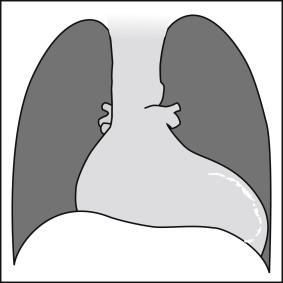
Graphic 17-1
Posteroanterior projection: calcified left ventricular aneurysm. Note a thin and well-defined line of calcification toward the left ventricular apex/free wall, as well as cardiomegaly.
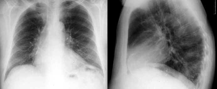
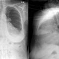
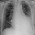
Figure 17-1
There is borderline cardiomegaly and definite calcification of a left ventricular aneurysm. There is increased pulmonary venous vascular prominence due to heart failure associated with the left ventricular aneurysm and systolic dysfunction.