Radiographic Findings by Diagnosis: Valvular Heart Disease


Key Points


  • The single most important role of chest radiography in valvular disease is to establish or document the presence of heart failure.


  • The different valvular lesions reveal themselves through their respective influence on chamber dilation that is specific to the disorder.


Acquired Valvulopathies
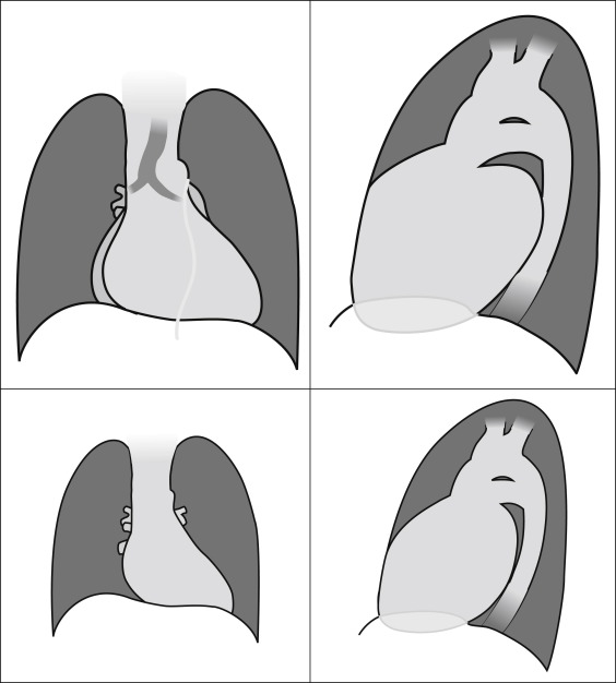
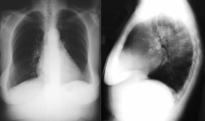
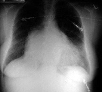
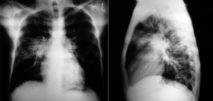
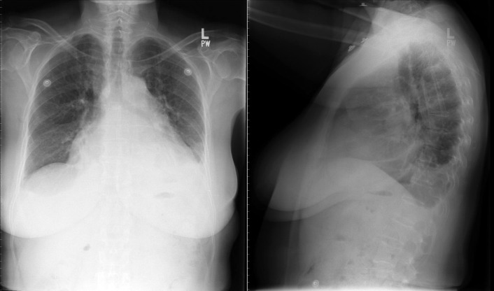
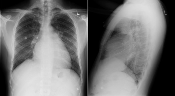
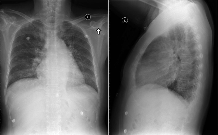
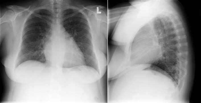
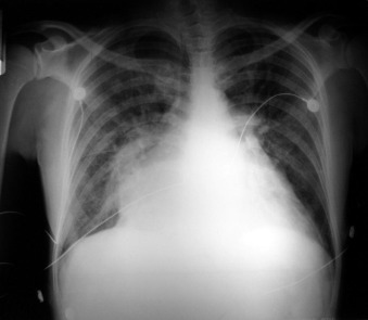
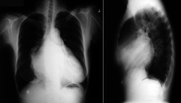
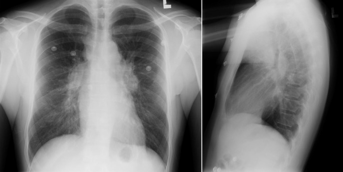
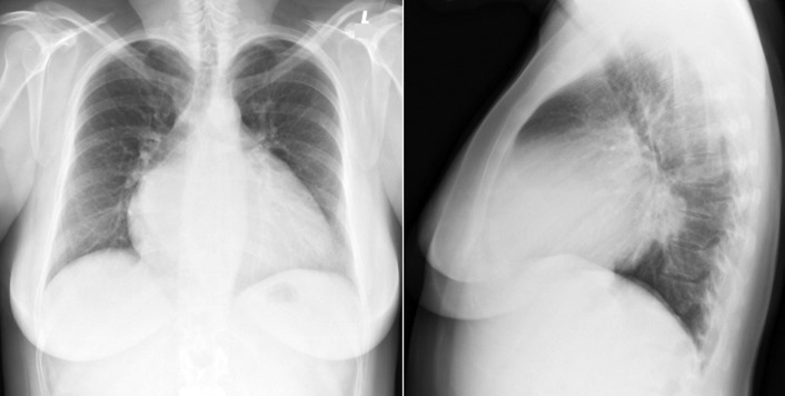
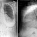
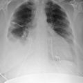
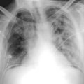
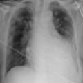

Mitral Stenosis


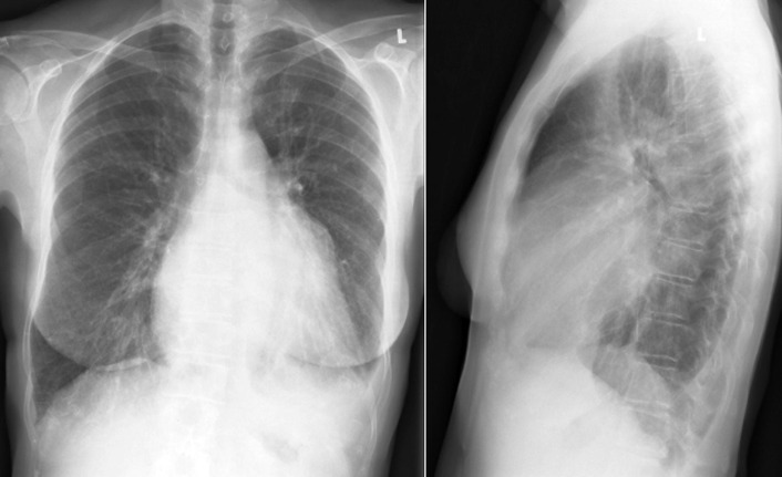
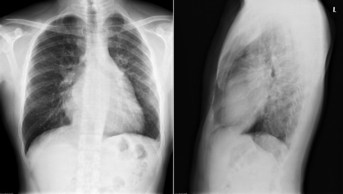
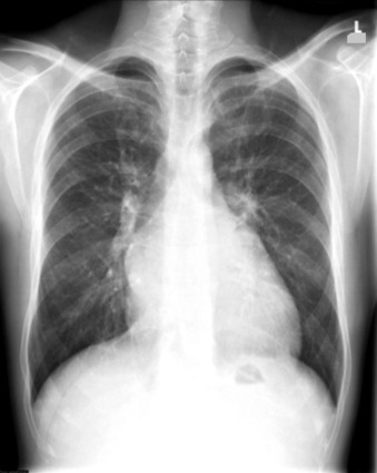
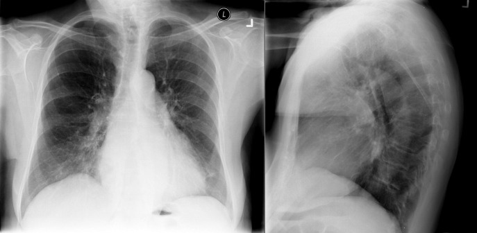
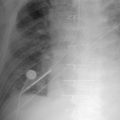
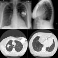
The radiographic findings of mitral stenosis ( Graphic 15-1 ; Figs. 15-1 to 15-16 ) reflect the pressure overload of the left atrium and pulmonary veins, and later of the right heart. As well, the commonly associated chronic atrial fibrillation contributes to (bi)atrial dilation. Associated rheumatic valvular lesions such as mitral regurgitation, tricuspid regurgitation, aortic insufficiency, and aortic stenosis/aortic insufficiency are common, and they alter the appearance of the heart.


Graphic 15-1


Posteroanterior and lateral projections of findings of mitral stenosis ( upper graphics ), and mitral insufficiency ( lower graphics ). In mitral stenosis, the left atrium is dilated but the left ventricle is not. Often there is significant associated pulmonary hypertension. Associated right-sided heart enlargement is common, due to both pulmonary hypertension and associated tricuspid insufficiency. In mitral insufficiency, the volume overload dilates both the left atrium and the left ventricle.


Figure 15-1


Severe mitral stenosis. In the posteroanterior radiograph, the upper left heart border is straightened due to left atrial appendage enlargement. In the lateral radiograph, prominent posterior left atrial dilation is apparent.


Figure 15-2


Several signs of left atrial enlargement persist in this case of post–mitral valve replacement for mitral stenosis: (1) the left upper heart border is full due to left atrial appendage enlargement; (2) the “carinal angle” is greater than 90°; and (3) the right (upper) heart border is displaced rightward, showing dilation of the left atrium.


Figure 15-3


Severe mitral stenosis. The upper left heart border is straightened due to left atrial appendage enlargement, and the overall cardiothoracic ratio is normal. The left atrium is posteriorly enlarged on the lateral radiograph. The pulmonary arteries are enlarged. There is air-space infiltration within the mid-lung zones due to pulmonary (bronchial artery) hemorrhage.


Figure 15-4


Severe mitral stenosis and severe tricuspid insufficiency. On the posteroanterior radiograph, there is straightening of the left heart border and fullness in the area of the left atrial appendage. As well, the lower left heart border is elongated, suggesting left ventricular enlargement, and there is right lower prominence consistent with right atrial enlargement.


Figure 15-5


Severe mitral stenosis. On the posteroanterior radiograph, there is straightening of the upper left heart border and particular fullness in the area of the left atrial appendage. On the lateral radiograph, there is left atrial posterior displacement.


Figure 15-6


Severe mitral stenosis. On the posteroanterior radiograph, there is straightening of the upper left heart border and particular fullness in the area of the left atrial appendage. On the lateral radiograph, there is left atrial posterior displacement.


Figure 15-7


Severe mitral stenosis. On the posteroanterior radiograph, the overall cardiothoracic ratio is not much increased because the left atrium is not well represented on this projection. The left upper heart border is full/straightened due to left atrial appendage enlargement. There is pulmonary venous prominence. On the lateral radiograph, which depicts the left atrial size more directly, the heart size is obviously increased, mainly from posterior displacement of the left atrium.


Figure 15-8


Severe mitral stenosis. The cardiothoracic ratio is increased, the left upper heart border is full/straightened, the carina is splayed, and there is a “double contour”/silhouette of the right heart border, all from left atrial enlargement. As well, there is enlargement of the pulmonary arteries and pulmonary edema.


Figure 15-9


Severe mitral stenosis and moderately severe tricuspid insufficiency. The cardiothoracic ratio is increased. The lung fields are clear. There is straightening of the left heart border, near splaying of the carina, and a “double contour” over the right heart border, all due to left atrial enlargement. The double border on the right heart is superior, consistent with left atrial enlargement. The lower right heart border is bulging to the right, consistent with right atrial dilation. On the lateral film, left atrial posterior displacement is obvious.


Figure 15-10


On the posteroanterior radiograph, there is pulmonary artery enlargement and fullness of the upper left heart border due to left atrial appendage enlargement. On the lateral radiograph, there is posterior left atrial enlargement, without left ventricular enlargement, consistent with, and due to, severe mitral stenosis.


Figure 15-11


Severe mitral stenosis, with moderate pulmonary hypertension and 3+ tricuspid regurgitation. The carina is splayed by left atrial dilation, the left upper heart border is bulging due to left atrial appendage dilation, and there is a “double contour” on the right side of the heart from left atrial dilation (upper bulge) and right atrial dilation (lower bulge). On the lateral radiograph, the left atrium is severely enlarged and dilated posteriorly, and the heart is pressed against the sternum from both left atrial dilation and right ventricular dilation due to mitral stenosis and tricuspid regurgitation, respectively.


Figure 15-12


Severe mitral stenosis, with moderate pulmonary hypertension and 3+ tricuspid regurgitation (TR). The carina is splayed by left atrial dilation, the left upper heart border is markedly bulging due to left atrial appendage dilation, and there are soft double contours on the right side of the heart from left atrial dilation (upper bulge) and right atrial dilation (lower bulge). On the lateral radiograph, the left atrium is severely enlarged and dilated posteriorly, and the heart is pressed against the sternum from both left atrial dilation and right ventricular dilation from TR and pulmonary hypertension.


Figure 15-13


Severe mitral stenosis, with moderate pulmonary hypertension and 3+ tricuspid regurgitation (TR). The carina is splayed by left atrial dilation, the left upper heart border is moderately bulging due to left atrial appendage dilation, and there are definite double contours on the right side of the heart from left atrial dilation (upper bulge) and right atrial dilation (lower bulge). On the lateral radiograph, the left atrium is mildly enlarged and dilated posteriorly, and the heart is pressed against the sternum from both left atrial dilation and right ventricular dilation from TR and pulmonary hypertension.


Figure 15-14


Severe mitral stenosis, with moderate pulmonary hypertension and 3+ tricuspid regurgitation. The carina is splayed by left atrial dilation, the left upper heart border is moderately bulging due to left atrial appendage dilation, and there are definite “double contours” on the right side of the heart from left atrial dilation (upper bulge) and right atrial dilation (lower bulge).


Figure 15-15


Severe mitral stenosis with moderately severe pulmonary hypertension. On the frontal radiograph, there is straightening of the left upper heart border due to dilation of the main pulmonary artery and of the left atrial appendage. The pulmonary veins are distended. On the lateral radiograph, the left atrium is dilated posteriorly, and the right ventricle is either enlarged or displaced anteriorly.

Apr 10, 2019 | Posted by in General Surgery | Comments Off on Radiographic Findings by Diagnosis: Valvular Heart Disease

Full access? Get Clinical Tree

Get Clinical Tree app for offline access