Pacemakers and Implantable Cardioverter Defibrillators


Key Points


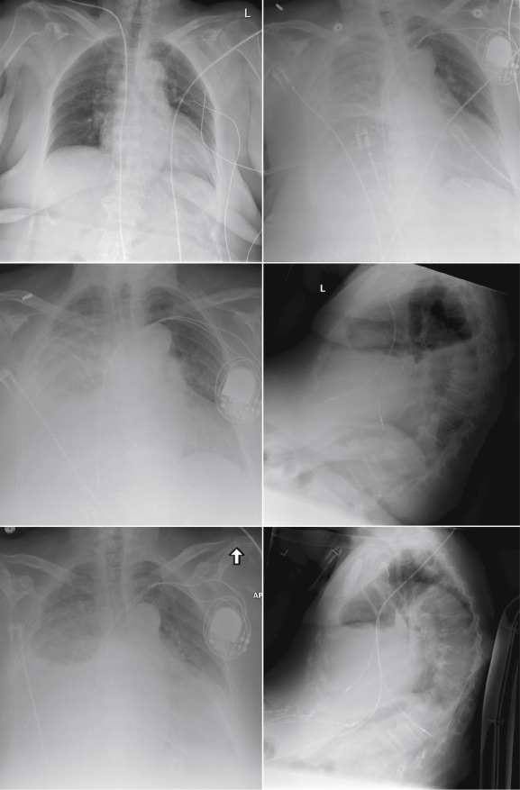
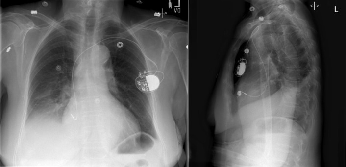
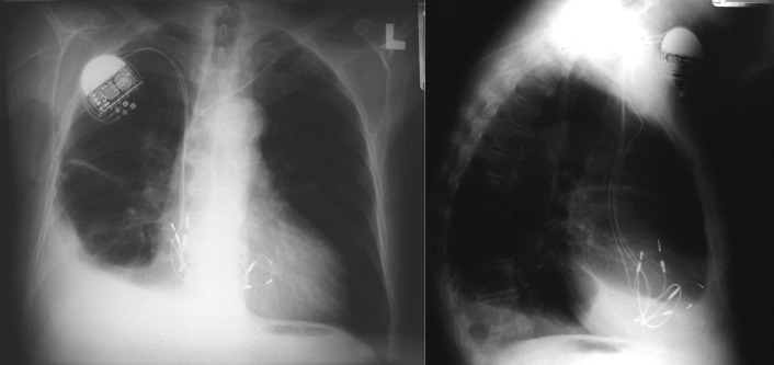
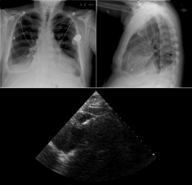
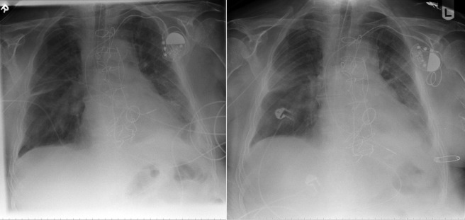
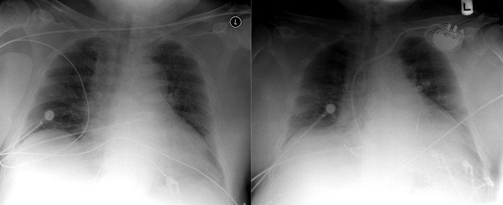
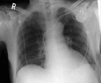
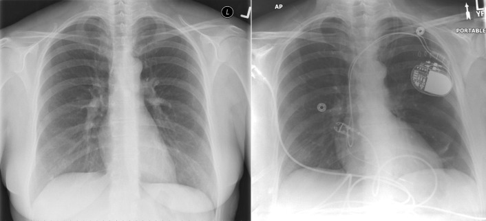
  • Chest radiography has a standard role in the identification of several potential complications such as pneumothorax, hemothorax, and several forms of malposition after insertion of pacemakers, implantable cardioverter defibrillators (ICDs), and cardiac resynchronization therapy devices.


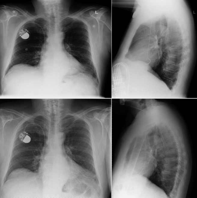
  • Pacemaker leads are apparent, and ICDs are more obvious because of their larger coils. Coronary sinus leads for cardiac resynchronization therapy are finer leads.


Endocardial Pacer Leads


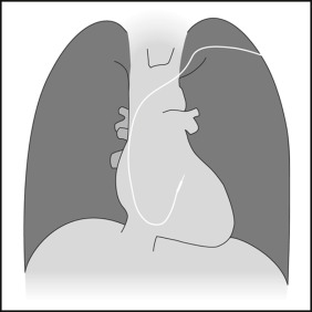
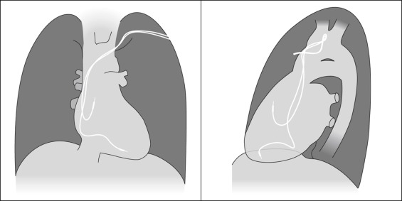
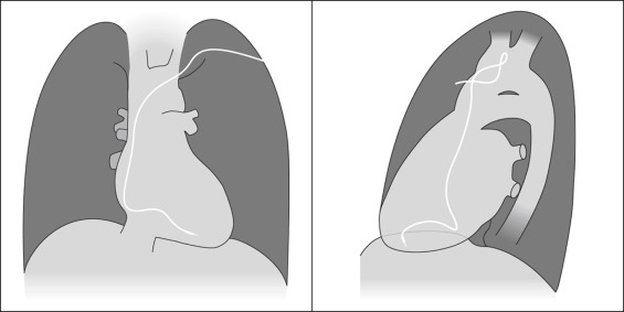
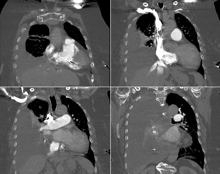
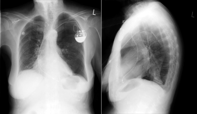
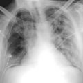
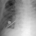
The position and integrity of endocardial pacer leads should be verified, especially when pacemaker dysfunction is clinically suspected ( Graphics 23-1 to 23-4 ; Figs. 23-1 to 23-15 ).The most common cause of pacemaker dysfunction that is apparent on the chest radiography is distal lead displacement/misplacement.


  • With a right atrial lead , the tip should be in the right atrial appendage.


  • With a right ventricular lead , the tip should lie anteriorly at the apex of the heart.


Graphic 23-1


Posteroanterior projection: coronary sinus misplacement of a ventricular pacemaker lead.


Graphic 23-2


Posteroanterior and lateral projection graphics of dual chamber (atrial and ventricular)/atrioventricular sequential endocardial pacer leads. Note the respective positions of the atrial lead in the atrial appendage and the ventricular lead.


Graphic 23-3


Posteroanterior and lateral projection graphics of a single ventricular endocardial pacer lead.


Graphic 23-4


Posteroanterior and lateral projection graphics of a single atrial endocardial pacer lead.


Figure 23-1


The left upper image is an anteroposterior chest radiograph of a patient presenting with symptomatic sinus node pauses. Transcutaneous pacer leads are present over the left chest. The right upper image is a chest radiograph several weeks after insertion of a dual chamber pacemaker. A very large right-sided pleural effusion has evolved. The ventricular endocardial pacemaker lead may be in the correct position, but the right atrial endocardial lead appears to be extracardiac. The middle images are posteroanterior (PA) and lateral chest radiographs at the time of re-presentation, again showing the right ventricular endocardial lead likely in the right position but the right atrial lead appearing too far inferior (PA radiograph) and too far posterior (lateral radiograph). The lower images (a few days later) show that the left pleural effusion has further enlarged and the right atrial lead has migrated further inferiorly and posteriorly. The site of perforation of the right atrial lead was the inferior aspect of the superior vena cava.


Figure 23-2


Same patient as in Figure 23-1. Contrast-enhanced computed tomography scans along coronal planes at different levels. The study is not electrocardiography-gated, and therefore there are motion artifacts. In the left upper image, the right ventricular endocardial lead is within the right ventricular cavity. In the right upper image, one of the pacemaker leads is external to the superior vena cava on the right side. In the lower images, the right atrial pacemaker lead is external to the heart, inferiorly and posteriorly located within the left pleural effusion.


Figure 23-3


Atrial-only pacing (for sinoatrial nodal disease). There is a single endocardial lead inserted into the right atrial cavity and located in the right atrial appendage.


Figure 23-4


A dual chamber pacing system had previously been inserted via the left subclavian vein. The system was changed to the right subclavian side and the old leads cut, enabling the old ventricular lead to migrate into the right ventricle, resulting in prominent tricuspid insufficiency as the lead held the tricuspid valve open. The resulting right-sided heart failure was responsible for the right pleural effusion.


Figure 23-5


Following mitral and tricuspid valve repairs (mitral and tricuspid annuloplasty rings), the patient developed complete atrioventricular block and underwent insertion of a dual chamber pacemaker. The position of the ventricular lead tip is unremarkable on the chest radiographs, but by transthoracic echocardiography the ventricular lead is seen to have migrated through the right ventricular apex into the epicardial fat over the left ventricular apex.


Figure 23-6


Ventricular lead insertion through a patent foramen ovale (PFO) and also across a mechanical mitral prosthesis. The left image shows postventricular pacemaker lead insertion, inadvertently through the PFO and mitral prosthesis. The right image is after revision and reinsertion. The pacer had functioned well when pacing via the left ventricle, with a right bundle branch block pattern at that time, as expected. The lower position of the right heart chambers compared with the left heart chambers is denoted by the differing position of the lead at the two times.


Figure 23-7


Ventricular lead insertion into the right ventricular outflow tract. Preinsertion and postinsertion images.


Figure 23-8


Pacer lead into the coronary sinus ( upper images ), and after revision and ( lower images ). The curvature of the pacer lead when in the coronary sinus is not strikingly abnormal on the posteroanterior (PA) radiograph; however, the lateral radiograph reveals the posterior location of the lead and in where the atrioventricular groove would plausibly be. When repositioned, the curvature is only slightly different on the PA radiograph, but far more anterior on the lateral radiograph.


Figure 23-9


Right ventricular perforation. The atrial lead is in the correct position. The distal position of the ventricular lead, which is somewhat more inferior than usual but not clearly abnormal, has perforated the right ventricular apex, resulting in tamponade.


Figure 23-10


Left: Pre–pacemaker insertion. Right: Post–pacemaker insertion. Note the left-sided pneumothorax.

Apr 10, 2019 | Posted by in General Surgery | Comments Off on Pacemakers and Implantable Cardioverter Defibrillators

Full access? Get Clinical Tree

Get Clinical Tree app for offline access