Keywords
Facial rejuvenation, Fillers, Neuromodulators, Periorbital hollows, Facial aging, Eyebrow, Eyelid
Introduction
The shift towards less nonsurgical techniques to rejuvenate the periorbital area continues, and patient requests for procedures involving minimal downtime with less “surgical” results increase each year. The American Society of Aesthetic Plastic Surgeons reported that patients received a total of 8.9 million cosmetic nonsurgical procedures, including 5.5 million injectable treatments and 2.1 million skin rejuvenation treatments . Understanding the underlying anatomy and aging changes represents a critical component to successful nonsurgical treatment of the periorbital region.
The aging face changes in all layers including the skin, muscle, fat, and bone; addressing these changes will allow for optimal facial rejuvenation . The three-dimensional contours of the face should also be kept in mind when addressing facial aging. Deflation of the brow and unveiling of periorbital hollows is a major concern for patients seeking periorbital rejuvenation. Because of this, soft tissue filling with hyaluronic acid gel (HAG) fillers has reached an all-time high of 1.7 million procedures. In line with this, botulinum toxin (BT) treatments have increased to 3.6 million procedures and skin rejuvenation to 2.1 million procedures in the United States alone .
Surgical and nonsurgical procedures can be complementary and can often be used in conjunction for periorbital rejuvenation. Surgical treatments will be covered elsewhere, and this chapter will focus on nonsurgical rejuvenation of the brow and periorbital complex.
Anatomy
When performing facial rejuvenation, it is important to understand the anatomy of the area. A detailed description of the anatomy is beyond the scope of this chapter but a general overview of the layers will be described.
The skin in the periorbital area has different thicknesses and elasticity, with the brow and forehead having thicker skin and the eyelid having thinner skin. The elasticity of the skin decreases with age. This, with subcutaneous volume loss, can also affect the apparent skin quality and wrinkling.
The muscles of the periorbital area include the frontalis, corrugator, procerus, and orbicularis oculi. Manipulation of the action of these various muscles can be used to change the position of both the eyebrows and eyelids. The frontalis is the main elevator of the eyebrow, and interdigitates with the main depressors of the eyebrow: the procerus, corrugator supercilii, depressor supercilii, and orbicularis oculi . Furthermore, laterally, the frontalis may change slightly during aging, accounting for some of the lateral brow descent, in addition to brow volume deflation, found in older patients .
Volume deflation also unveils the underlying structures including the periorbital ligaments. The orbicularis retaining ligament (ORL) encircles the entire bony orbit and inserts into the dermis of the skin . The ORL contributes to hollows in both the upper and lower eyelid depending on the adjacent volume loss or fat herniation. In the upper eyelid superior volume loss can unveil the ORL especially in Asian patients. In the lower eyelid, other periorbital hollows include the septal confluence and the zygomaticomalar ligament . Deflation in the adjacent areas, such as the midface and temple, can also affect the appearance of the periorbital area. Therefore the periorbital area should not be treated in isolation but rather in conjunction with these areas of the face.
Skin Rejuvenation
Skin ages via both intrinsic and extrinsic factors, which result in changes including skin wrinkling, loss of elasticity, the appearance of excess skin, and pigmentation changes . To address these changes, various procedures can be performed including surgical or nonsurgical procedures. Surgery alone, however, cannot change the quality, texture, or elastic properties of the skin, which is where chemical peeling and laser resurfacing can help .
Nonsurgical skin rejuvenation procedures essentially remove different layers of the skin and thus induce the genesis of new collagen and elastin. Treatments can be aimed at either superficial or deep treatments. The most superficial layer of the skin can be addressed with various exfoliants, retinoids, mild α-hydroxy acids, or abrasives. Treating the deeper skin layers is usually accomplished with chemical peeling or laser resurfacing.
In the periorbital area, 20% trichloroacetic acid peel is commonly used. When used carefully with a frost and feathering technique, light acid peels have minimal complications. Other deeper chemical peels such as phenol present a higher risk for complications in the periorbital area and are therefore not used as frequently .
Laser treatments can also be used in the periorbital area but with care. Laser treatments have been classified into superficial and deep. Superficial lasers produce injury of the epidermis and dermis less than 750 µm, and deep lasers produce injury greater than 750 µm . The eyelid skin is very thin and more superficial treatments are generally favored to prevent full-thickness dermal injury, which could lead to eyelid malposition. Near-infrared, intense pulsed light devices, and nonablative lasers have a very low complication rate, but unfortunately they have minimal effects on wrinkle reduction and collagen formation. Fully ablative lasers such as carbon dioxide (CO 2 ) and erbium:yttrium-aluminum-garnet have been successfully used in periorbital rejuvenation, but they carry a higher complication rate (prolonged healing, erythema, and pigmentary changes). Fractional CO 2 lasers are intermediate in risk and reward, and may have more of a role in the periorbital area. They produce small columns of thermal injury with sparing of adjacent untreated skin, allowing for a more rapid healing process .
Neurotoxins
Neurotoxins, or BTs, have become the mainstay in the treatment of dynamic lines and wrinkles. BT is produced by the bacteria, Clostridium botulinum . Serotypes A and B are used for clinical procedures, with serotype A (BTA) being used for most cosmetic purposes . The most prevalent BTA products include Botox Cosmetic (Allergan, Irvine, CA, USA), Dysport (Medicis, Scottsdale, AZ, USA), and Xeomin (Merz Aesthetics, San Mateo, CA, USA). These neurotoxins inhibit the release of acetylcholine at the presynaptic neuromuscular junction, producing a localized and temporary reduction of function of the muscle. The onset of these effects usually occurs over the first 7 days and last 3 to 6 months. Botox, Dysport, and Xeomin differ slightly in their storage, protein, and unit measurement, and their use depends largely on injector preference. Reconstitution of the product can be performed with 0.9% sterile saline. The authors use 0.9% sterile bacteriostatic saline to reconstitute the various products, as it has been suggested that this may decrease patient discomfort . The traditional locations for periocular BT injections include the following:
- 1.
Glabella: the most common cosmetic use for BT and the only one currently indicated by the Food and Drug Administration. A typical treatment of the glabellar lines involves intramuscular injection into four sites: one injection in the central procerus, two injections to the depressor supercilii on both sides, and another two in the corrugators bilaterally ( Figs. 9.1 and 9.2A ). Approximately 20 U of onabotulinum toxin A or incobotulinum toxin A is typically injected into the glabellar region, with a range of 10 to 30 U .
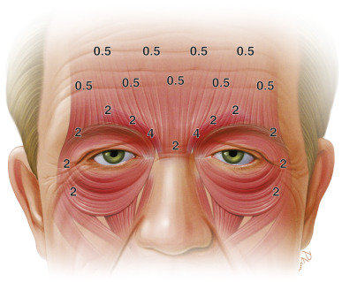
Figure 9.1
Pattern of Botox treatment to the periorbital and forehead areas.
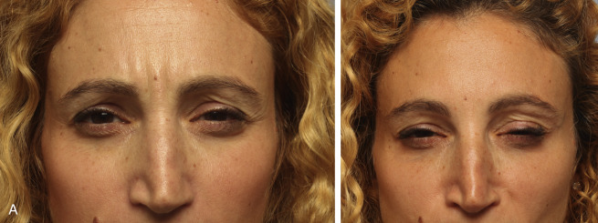
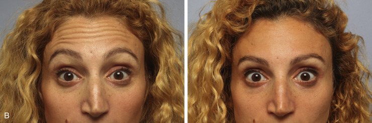
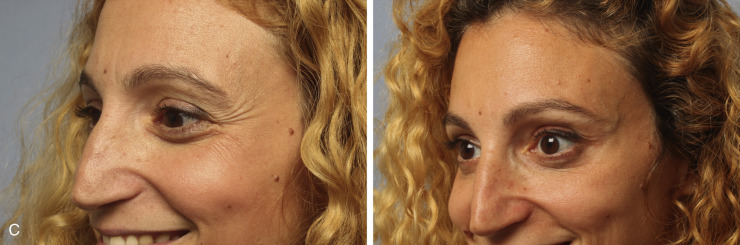
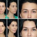
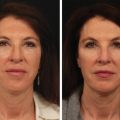
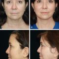
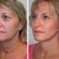
Figure 9.2
Two weeks after Botox injection to the forehead and periorbital area. (A) Preinjection (left) and postinjection (right) to the glabella; (B) preinjection (left) and postinjection (right) to the frontalis muscle; (C) preinjection (left) and postinjection (right) to the lateral canthal rhytids.
- 2.
Forehead area: injection into the frontalis muscle that elevates the brows. Neurotoxins will lessen lines and wrinkles. Injection consists of several subcutaneous injections along the frontalis, but one should take into consideration that weakening the frontalis muscle will also lower the brows and may exacerbate brow or eyelid ptosis. To avoid this, dilute BTA can be used in a grid pattern across the upper two-thirds of the forehead or in various patterns in order to achieve brow reshaping ( Figs. 9.1 and 9.2B ). Also, one should pay attention to the temporal portion of the frontalis muscle, to avoid the quizzical look of lateral eyebrow elevation (“Spock” brow appearance).
- 3.
The crow’s feet or lateral canthal rhytids: these are caused by contraction of the lateral orbital and septal rings of the orbicularis oculi muscle. Treating them with neurotoxins is usually done with two to four subcutaneous injections at a dose of 10 to 15 U, 1 cm lateral to the lateral orbital rim ( Fig. 9.2C ).
- 4.
“Bunny lines”: lines on the lateral aspect of the nasal dorsum caused by the action of the nasalis muscle. Typical dose in this area is usually 2 to 4 U on each side of the nose.
The most common adverse effects of neurotoxin injections are injection site pain and localized bruising. Asymmetry can occur, often related to underlying facial asymmetry, and usually responds to “touch up” injections followed by adjustment of the subsequent dosing. True complications are rare, and they include eyelid ptosis, brow ptosis, “Spock look,” diplopia, ectropion, and psychological depression .
Soft Tissue Fillers
Soft tissue fillers have been used increasingly in the periorbital area to address volume deflation often seen with aging. Patients seek soft tissue fillers for a more natural result and less downtime. In addition, fillers have been proposed to have positive effects on skin quality.
Fillers can be categorized into temporary or permanent, and reversible or nonreversible ( Table 9.1 ). The main temporary and reversible filler is HAG fillers. The HAG fillers are reversed by the use of hyaluronidase (HYAL), which is an enzyme that breaks down hyaluronic acid. There are two commercially available HYALs, Hylenex (Halozyme, San Diego, CA, USA) and Vitrase (Bausch & Lomb, Bridgewater, NJ, USA), which can be used to help reverse the HA filler products and should be available whenever HA fillers are used. By varying the amount of HYAL injected, some or all of the filler can be dissolved .
Related posts:
Stay updated, free articles. Join our Telegram channel

Full access? Get Clinical Tree


