Abstract
- 1.
Lateral canthal suspension is an integral adjunct to lower blepharoplasty surgery.
- 2.
These procedures include a canthoplasty where the lateral canthal tendon and/or temporal tarsus is modified and secured to the lateral orbital rim; or a canthopexy where the canthal tendon, and/or orbicularis muscle, is plicated to the lateral orbital rim.
- 3.
Indications for canthal suspension include preexistent lower eyelid laxity, and prevention of lower eyelid malposition.
- 4.
In aesthetic surgery less disruption of canthal architecture/integrity is best.
- 5.
A thorough understanding of canthal and lower eyelid anatomy is essential to avoid surgical complications.
- 6.
A careful preoperative evaluation of the eyelid position, tone, laxity, and globe/midface configuration (orbitofacial vector) are critical to attain appropriate outcomes.
- 7.
Familiarization with both open (involves canthal incision) and closed (no canthal incision) canthal suspension techniques provides options for the aesthetic lower eyelid surgeon.
- 8.
The risk of postblepharoplasty lower eyelid retraction, ectropion, and rounding of the canthal angle can be reduced with lateral canthal suspension.
- 9.
The expectations and goals of lateral canthal suspension should be discussed with each patient before surgery. Patients unhappy with this form of surgery are often difficult to manage.
Keywords
Lateral canthal suspension, Open canthal suspension, Closed canthal suspension, Lateral canthal surgery, Canthoplasty, Canthopexy, Commissure, Lower blepharoplasty, Lateral tarsal strip
Introduction
Traditional lower blepharoplasty consists of an open approach, transcutaneous procedure in which variable amounts of skin, muscle, and fat are excised. Inherent to this technique is a predilection for rounding of the canthal angle and lower eyelid retraction , noted to occur in 6% to 20% of such cases . In the authors’ experience these are very difficult problems to address and cause tremendous dissatisfaction in the aesthetic blepharoplasty population. Traditionally, postblepharoplasty lower eyelid retraction has been thought to be primarily related to unaddressed lower eyelid laxity, anterior lamellar shortage, and the development of a middle lamellar scar . More recently, it has been shown that weakness of the orbicularis oculi muscle and negative orbitofacial vector eyelid configuration also play an important role in postblepharoplasty eyelid malposition . Irrespective of the primary cause, canthal suspension, as a means of addressing preexistent lower eyelid laxity, has become an integral part of lower blepharoplasty and reduces the incidence of postoperative eyelid malposition and aesthetic deficit . In addition to maintaining aesthetics, when canthal suspension is added to surgery, eyelid function is preserved. Unaddressed eyelid laxity can biomechanically weaken orbicularis oculi function and lead to an inadequate blink, ocular surface exposure, altered tear clearance, foreign body sensation, and tearing .
In 1969 Tenzel was the first to suggest lateral canthal suspension as a method to correct lower lid malposition. He described a lateral canthotomy (open procedure) and inferior cantholysis to gain access to the lower tarsus for suspension to the lateral orbital rim . Webster then described canthal suspension as an adjunct to blepharoplasty to prevent lid malposition . As it became clear that the incidence of lower lid malposition could be reduced with adjunctive canthal suspension during blepharoplasty, this addition to surgery became commonplace . In 1979, Anderson et al. described the lateral tarsal strip (LTS), which gained favor as a standard canthal suspension technique. In this procedure the lateral canthal tendon (LCT) is accessed through a canthotomy, and horizontal eyelid shortening is a common step. While the LTS and its variations are a landmark development in the management of reconstructive eyelid procedures, its invasiveness and disruption of canthal architecture and anatomy have limited its integration into contemporary aesthetic blepharoplasty. Some of the limits of this procedure include the potential development of horizontal eyelid length disparity between the upper and lower lid, alteration of the canthal angle and commissure, lateral canthal dystopia, and poor overall cosmesis . In the setting of ectropion or entropion, patients are often accepting of these issues as their premorbid condition has been improved significantly. The aesthetic patient is much less forgiving. As such, open canthopexy procedures , in which the canthal tendon is plicated to the orbital rim, gained favor as they better preserve lateral canthal anatomy and are less disruptive. In an effort to further preserve canthal integrity and maintain canthal aesthetics, closed canthal suspension techniques have evolved . In this setting the canthus is accessed from a distant site without a canthal incision (hence a closed procedure). This better preserves native canthal anatomy and avoids the aforementioned complications of the LTS procedure, as well as the development of canthal webs and scars, which can occur when canthal incisions are made.
In this chapter the authors attempt to simplify canthal suspension surgery by detailing a spectrum of procedures, including: (1) traditional open canthoplasty and open canthopexy ; and (2) more contemporary closed canthoplasty and closed canthopexy . The authors will also review canthal nomenclature, relevant canthal anatomy, discuss canthal aging changes, highlight key preoperative examination pearls, and review postoperative care and complications.
Canthal Nomenclature
The description of canthal surgery in the literature can be confusing for both the novice and/or experienced surgeon as numerous procedures are reported, most being simple modifications of one another. What makes this even more complex is the naming of these surgical variations and the anatomic structures modified during surgery. For simplification and clarity, the authors prefer the general term “canthal suspension,” as all canthal procedures have a common goal of “supporting or tightening” the lower lid at the canthus. In addition, to develop a proper foundation and framework for understanding and planning these procedures, a mental flowchart of the options available is often helpful. To this vein, the authors subdivide canthal suspension techniques into two broad categories: (1) open, and (2) closed, with each category further subdivided into either a canthoplasty or canthopexy variant.
An open canthal procedure involves a canthal incision, or canthotomy, while a closed procedure does not. In open surgery, as anatomy is obvious, precise placement of the lower eyelid is enhanced. However, because the canthus is disarticulated and reformed, canthal deformity can occur. A closed canthal suspension, on the other hand, accesses and suspends the canthal tendon or terminal tarsus through a distant site (typically the upper lid crease). In this technique, exposure is limited, anatomy is better preserved, but placement of the canthus can be less precise. Its main advantages are that it is less traumatic and avoids lateral canthal deformity. A canthoplasty is a procedure in which the LCT or temporal lower lid is incised, plus/minus shortened, and secured to the lateral orbital rim. Conversely, a canthopexy is a procedure in which the lower eyelid is suspended to the lateral orbital rim with a plication suture without modification of the LCT or terminal tarsus.
Attaining clarity of anatomic terminology of the canthus is also important to understanding canthal suspension surgery. The connective tissue structure that secures the terminal eyelid to the orbit is called the LCT. It is derived/originates from distal fibers of the palpebral orbicularis muscle and inserts onto the lateral orbital rim. A synonymous name for the LCT is the lateral palpebral ligament. This is actually a better description of the structure, as a tendon connects a muscle to bone to move the bone, while a ligament attaches bones to bone to secure their alignment, which is more descriptive of what is occurring here. The lateral retinaculum is another name used to describe the LCT. A retinaculum, by definition, is a band around tendons to stabilize them and hold them in place. As the LCT has contributions or connections with many other structures (lateral horn of levator aponeurosis, check ligament of lateral rectus, orbital septum, etc.), the confluence of these structures can be considered a retinaculum. Finally, the lateral canthus is the general area where the upper and lower lids meet, whereas the commissure is the point of this union. The lateral raphe is an area of fine fibrous bands where the terminal orbicularis muscle of the upper and lower lids meet. Additional nomenclature specific to lateral canthal surgery includes the terms ab-interno (originating from the inside) and ab-externo (originating from the outside). Refer to Table 14.1 for a simplified naming and description of relevant canthal surgical concepts and anatomic structures.
| Nomenclature | Definition |
|---|---|
| Open canthal suspension | Requires a canthal incision (canthotomy) to access the LCT or terminal tarsus |
| Closed canthal suspension | Requires no canthotomy to access the LCT or terminal tarsus. These structures are accessed from a distant site (i.e., upper lid crease) |
| Canthoplasty | The terminal tarsus or LCT is modified and secured to the lateral orbital rim |
| Canthopexy | The terminal tarsus, orbicularis muscle, or LCT are not modified, but are suspended to the lateral orbital rim with a plication suture |
| Canthotomy | Lateral canthal incision |
| Cantholysis | Dynamic release of the LCT from the lateral orbital rim |
| Lateral canthus | The general area where the upper and lower lids meet laterally |
| Lateral commissure | The point of union of the upper and lower lids at the lateral canthus |
| LCT | Connective tissue structure that secures the upper and lower terminal eyelid to the lateral orbital rim |
| Lateral retinaculum | Another name used to describe the LCT. The confluence of several of the soft-tissue structures of the lateral upper and lower eyelid that have connections with the LCT |
| LPL | Another synonymous name for the LCT |
| Lateral raphe | The area of fine fibrous bands where the terminal orbicularis muscle of the upper and lower lids meet |
| Orbitofacial vector | The relationship of globe projection to the lower lid and midface. When the globe and midface are aligned in a horizontal plane the vector is neutral. When the globe projects more anterior than the midface, the vector is negative; when the globe sits posterior to the midface the vector is positive |
| Ab-externo | Suture passage starts outside the wound (outside in) to secure the LCT to the lateral orbital rim |
| Ab-interno | Suture passage starts inside the wound (inside out) to secure the LCT to the lateral orbital rim |
Canthal Anatomy
The lower eyelid is suspended medially and laterally at the respective canthi by the medial canthal tendons and LCTs. As stated, the LCT is a connective tissue structure derived from the terminal pretarsal and preseptal orbicularis fibers that inset onto the lateral orbital rim. It is a three-dimensional structure anchored horizontally, but with a vertical component, as the lateral canthus is typically 2 mm higher than its medial counterpart (normal canthal tilt) . In addition, it has an anterior and posterior component. The posterior portion attaches 3 mm posterior to the orbital rim at Whitnall’s tubercle 10 mm below the frontozygomatic suture . It is important to recreate this attachment during canthal suspension surgery, as it assures the lower lid’s appropriate apposition to the globe ( Fig. 14.1A ). The anterior portion of the tendon inserts at the anterior edge of the orbital rim. This lends support to canthal integrity, but plays a secondary role to the more critical posterior attachment.
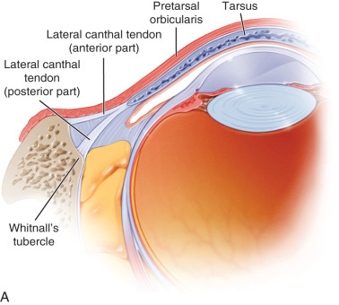
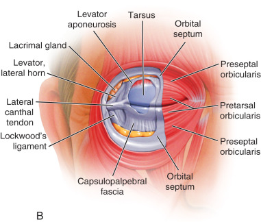
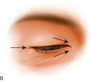
Traditionally, the lateral canthus has been considered a support structure only; however, it has other functions and many moving parts. It is associated with the orbital septum, the lateral horn of the levator aponeurosis, and plays a role in native upper lid tension ( Fig. 14.1B ). For this reason, on occasion, surgery has led to temporary or permanent ptosis (when the upper limb of the tendon is imbricated). The canthus is also intimately associated with the check ligament of the lateral rectus muscle, and in the normal state moves laterally a few millimeters with abduction. Finally, the orbicularis muscle (eyelid protractor) is oriented circumferentially around the eye. This normally would allow a circular contraction pattern of the muscle. As the canthal tendons secure the orbicularis laterally and medially, this biomechanically imparts a vertical contraction pattern of the palpebral aperture. When canthal integrity is lost with senescence or from eyelid surgery, this contraction pattern becomes deficient, leading to an altered blink, poor eyelid closure, and “fishmouthing” ( Fig. 14.2 ) , or a medialization of the lateral canthus with a shortened horizontal fissure and impaired eyelid dynamics.
Canthal Aging Changes
With aging, tissue descent, deflation (bone and fat), and cutaneous degeneration contribute to an alteration of the dimensions of the palpebral aperture and the associated position, tone, and function of the lower eyelid. In general, the eyelid aperture changes from one that is a horizontal oval (longer in this dimension) to one that is more vertical as the lateral canthus migrates medially . Canthal alterations resulting from tissue involution is a multifactorial process that occurs in three dimensions. It is very difficult to recreate a youthful appearance and function when the sole intervention is tissue excision and suspension in one plane. This is especially true of the canthus, whose purpose is so much more than maintaining appropriate lid position (i.e., blink, check ligament to the lateral rectus, eyelid excursion, lacrimal pump function, etc.). For this reason, the “belt and suspenders” procedures we often employ in this setting can cause havoc among the aesthetic patient population, even in the presence of what appears to be a nice result . This is also why, in this particular patient population, less tissue disruption is better and consideration should be given to the more contemporary closed suspension techniques elaborated on later.
Preoperative Assessment
A careful analysis of the lower lid position and function in patients undergoing blepharoplasty is essential to determine the need for canthal suspension. In addition, identification of patients at high risk of postoperative eyelid malposition is critical. For example, patients with a history of thyroid disorder, facial nerve paresis, previous blepharoplasty, or trauma require special attention on examination to assure surgical feasibility. The physical examination should include both a static and dynamic examination of the lower eyelid and canthus. The clinical features evaluated include: eyelid position, tone, and an assessment of laxity. Also, eyelid fissure dimensions, lateral canthal angle configuration, direction of movement of the lateral commissure with blink, adequacy of eyelid closure, and the morphologic relationship of the globe to the cheek (orbitofacial vector) should be assessed. In the following paragraphs the authors will focus on the essential elements of the physical assessment.
The lower eyelid margin normally rests just above the inferior limbus in neutral gaze. The margin reflex distance-2 (MRD-2), which is the distance between the corneal light reflex and the lower eyelid margin with the patient staring at a light in primary gaze, is used to measure the lower lid position . It is normally 6 mm and can increase in patients with lower lid laxity. When lower lid retraction is present the lid rests below the limbus, scleral show is manifest, and the MRD-2 increases. The horizontal palpebral fissure normally measures 30 mm and the canthus lies 2 mm higher temporally than nasally. Documenting these measurements is important for assessment, surgical decision-making, and photographic reference after surgery if needed.
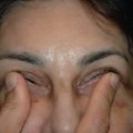
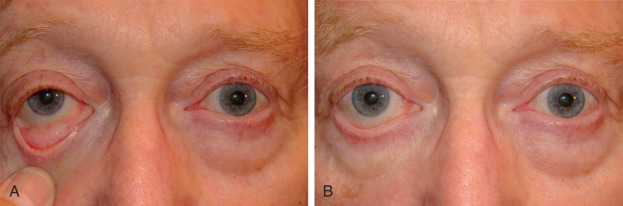
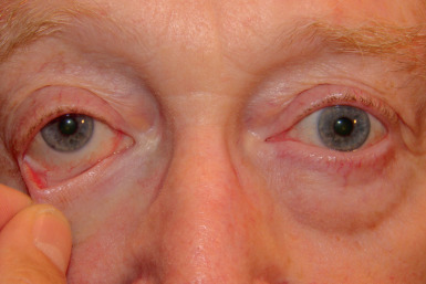
The eyelid snap-back and distraction tests are performed to evaluate the integrity of the lower eyelid canthal complex. The snap-back test is performed by pulling the lower eyelid down and away from the globe. The eyelid should return to its normal resting position in less than 1 second without blink. If this is delayed the test is positive and generally signifies orbicularis weakness ( Fig. 14.3 ). In the eyelid distraction test, the distance that the lower eyelid can be pulled away from the globe is measured ( Fig. 14.4 ). A distance of 8 mm or more is defined as a positive test and indicates clinically significant eyelid laxity. In the setting of aesthetic blepharoplasty, the examination modalities described may miss subtle degrees of eyelid laxity that can still predispose to postoperative complications. Therefore, for the purposes of preserving eyelid appearance and function after surgery, canthal suspension should be considered even when small deficiencies of canthal tendon integrity are present.
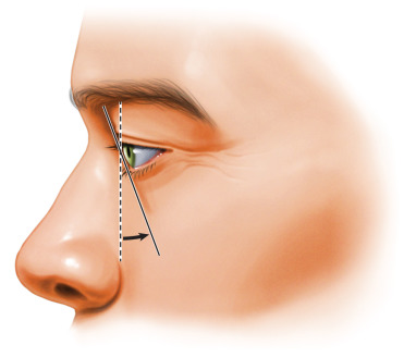
Another critical factor on examination is the relationship of globe projection to the lower lid and midface. This relationship is referred to as the orbitofacial vector. When the globe and midface are aligned in a vertical plane the vector is neutral. When the globe projects more anterior than the midface, the vector is negative; when the globe sits posterior to the midface the vector is positive ( Fig. 14.5 ). A negative vector configuration places the eyelid at a mechanical disadvantage, as the lower lid must work against a gradient to maintain normal resting position. This is a delicate equilibrium that can be negatively impacted by even a small manipulation of the lower lid/canthal complex. In fact, it has recently been shown that negative vector globe/lid topography can be a significant contributor to postblepharoplasty eyelid retraction . Special care and attention must be given to patients presenting with negative vector topography, which is typically seen in the setting of shallow orbits, long eyes (myopia), proptosis, or a hypoplastic midface.