Introduction
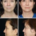
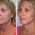
The transcutaneous skin-muscle flap blepharoplasty has a long history in the field of cosmetic eyelid surgery. As with any surgical approach, it has distinct advantages and disadvantages. The chief advantage of the transcutaneous approach is the capacity to treat excess skin and orbicularis muscle that requires redraping for adequate lid recontouring and superior lower eyelid rejuvenation, the major limitation of the transconjunctival approach. The transcutaneous approach also allows for broad exposure for wide release of the orbitomalar retaining ligaments, fat transposition (or septal reset), as well as a myriad of midface lift procedures. The disadvantages to this approach include a higher risk of lower eyelid malposition reported in the 10% to 15% ranges, orbicularis denervation atrophy, and a frank ectropion rate of 1%.
It is our goal in this chapter to elucidate how the aesthetic surgeon can still utilize the transcutaneous technique and minimize risk. In the modern era of plastic surgery, studies have revealed key anatomic risk factors, intraoperative technical details, and postoperative care regimens that allow for safe employment of the transcutaneous technique. What follows is a description of the relevant anatomy, indications and contraindications, our modified approach to transcutaneous skin-muscle flap blepharoplasty, and how to avoid complications with meticulous postoperative care.
Aging Eyelid Anatomy
The lower eyelid is formed by three lamellae: the anterior, middle, and posterior lamella. The anterior lamella is the skin and orbicularis oculi muscle, the middle lamella is the tarsal plate and orbital septum, and the posterior lamella is the conjunctiva and the lower lid retractors. The orbicularis oculi muscle is comprised of two parts, the outer orbital and inner palpebral portion. The palpebral portion contains a pretarsal component and a preseptal component. The upper and lower pretarsal components of the orbicularis oculi muscle join medially and insert onto the lacrimal crest as the medial canthal tendon. The two lateral components of the pretarsal orbicularis join together to form the lateral canthal tendon, which inserts onto Whitnall’s tubercle. Immediately deep to the orbicularis oculi muscle is the orbital septum, a continuation of the orbital periosteum. The lower eyelid retractors insert onto the orbital septum approximately 5 mm inferior to the inferior-most aspect of the tarsal plate.
Orbital fat is contained within the orbital septum. The lower eyelid has three fat pads: medial, central, and lateral. The inferior oblique muscle runs between the medial and central fat compartments, and is an important anatomical marker to identify and preserve. The medial fat pad is distinct in that it contains denser fat that appears whiter in coloration. Both genetics and aging play a role in the size of these fat pads, and these fat pads do not fluctuate in size with changes in body habitus.
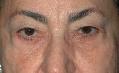
Age-related changes in the periorbital area are multifactorial, and are best understood by the contributing counterparts. The aged periorbital appearance is the result of cumulative changes in skin texture, volume depletion, loss of elasticity, formation of rhytids, drooping of the skin, and ptosis. This milieu of changes result in the characteristic findings of the aged lower eyelid, one that is marred by dermatochalasia, pseudoherniation of the orbital fat, malar festooning, and atonia of the lower eyelid that affects lateral canthal position.
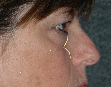
The tear trough is an area of particular concern in the aging eyes. The tear trough is the concave area caudal to the inferior orbital fat. First described as the nasojugal fold by Duke-Elder and Wybar in 1961 , the modern-day name “tear trough deformity” was coined in 1969 by Flowers , when it was observed that tears would track down this dependent area. There have been several explanations regarding the etiology of this tear trough deformity, which was considered to be multifactorial . More recently, evidence has suggested that the tear trough occurs as a result of skin tethering by an osteocutaneous ligament, which is situated between the origins of the palpebral and orbital parts of the orbicularis muscle. This ligament extends from the level of the insertion of the medial canthal tendon to the line of the medial pupil, where it continues laterally as the orbitomalar retaining ligament . This tethering effect is exacerbated with aging due to an increasing bulge of orbital fat from above, and atrophy with descent of the malar fat below ( Fig. 13.1 ) . This ligament also tethers the redundant orbicularis muscle that develops with age, creating malar festoons and mounding.
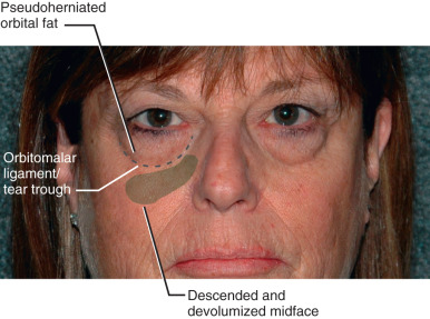
The midface is an important yet often overlooked contributing factor to periorbital aged appearance. Gravitational descent and volume depletion of the midface indirectly affects the periorbita and plays a significant role in the aged appearance of the lower lids. The midface descends with synchronous deflation of the suborbicularis oculi fat, creating a deepening of the tear trough. The ptotic midface creates another bulge inferior to the tear trough. The cumulative effect of these changes is psuedoherniation and apparent orbital fat excess that accentuates the tear trough, volume loss, and hollowing in the infraorbital and lateral orbital areas, and a midface bulge creating a double convexity of profile view ( Fig. 13.2 ).
Addressing the tear trough is important for successful rejuvenation of the lower periorbital area, which is why we recommend release of the orbitomalar retaining ligaments to allow for both redraping excess orbicularis muscle as well as volumizing the depth of the tear trough with transposed orbital fat.
Preoperative Evaluation, Indications, and Contraindications
Preoperative planning has been lauded by some as the most important aspect of blepharoplasty, and is vital to achieving successful outcomes . The eyes are a focal point of the face and serve as the natural transition between the upper and middle face. Prior to surgical manipulation of the eyes, it is prudent to evaluate the aging processes of the upper face and midface, specifically with regards to their contribution to the stigmata of periorbital aging. Oftentimes there is contemporaneous aging of the midface and brow that influence how the periorbital area is perceived, and it is important to diagnose the contribution of the brow and midface in order to address them concurrently. Thorough preoperative evaluation will identify the best surgical candidates for blepharoplasty, as well as steer the surgeon and patient away from unwanted potential postoperative pitfalls. This evaluation is inclusive of the medical comorbidities of patients, as well as their psychological well-being and emotional disposition. It is important to have a frank discussion with patients and record consultations with photographic documentation. This process is aimed towards setting realistic expectations, as well as pointing out any preexisting asymmetries for both patients and physicians to acknowledge. Smoking should be discontinued for 2 weeks prior to surgery. All anticoagulants or herbal medications that can interfere with blood clotting should be discontinued 2 weeks prior to surgery. Thyroid disorders should be investigated and noted, as hypothyroid and hyperthyroid states can cause disparate influences on the periorbital area that cannot be addressed with routine blepharoplasty. Any history of dry eyes should be investigated, with comprehensive ophthalmological assessment including Schirmer’s test, visual acuity, extraocular movements, intraocular pressure, cornea and ocular adnexa evaluation. Complaint of dry eyes despite normal Schirmer’s test should prompt further ophthalmologic and systemic evaluation to seek out any potential underlying collagen vascular disorders or autoimmune diseases.
Physical examination warrants special observation of all dermatological and structural changes in the periorbital area. Volume changes in the orbit should be evaluated. A relative volume excess is present in the pseudoherniation of the orbital fat. This accentuates the relative volume deficiency in the nasojugal groove and infraorbital hollows immediately inferior to the orbital fat pseudoherniation. In cases where the lower eyelids have less significant dermatochalasia and orbicularis muscle redundancy, the transconjunctival approach is a more appropriate choice, leaving the orbicularis muscle untouched. Mild skin redundancy can then be treated with resurfacing with a peel or laser.
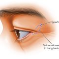
Excessive lower eyelid laxity should be assessed, defined as a snap test greater than 1 second and distraction test (being able to distract the lower eyelid from the globe) greater than 10 mm. On close inspection of those patients with excessive skin laxity, preoperative scleral show is often seen ( Fig. 13.3 ). Excessive laxity is a relative contraindication to transcutaneous lower blepharoplasty. When utilizing the transcutaneous approach, lower eyelids with increased laxity could lead to postoperative eyelid malposition on a spectrum from lateral canthal rounding to frank ectropion. In these cases the surgical plan should include a concomitant canthopexy or horizontal lower eyelid-shortening procedure (such as a lateral tarsal strip procedure), depending upon the degree of laxity. It is our preference to utilize a closed canthopexy (described later), suturing the lateral tarsus to the orbital rim, to tighten the lower eyelid margin because it does not change the horizontal aperture of the eye. In cases of more extreme laxity, a lower eyelid-shortening procedure will be required.
Indications for transcutaneous lower blepharoplasty include excess skin and orbicularis muscle that requires redraping for adequate lid recontouring and superior lower eyelid rejuvenation. This approach also provides broad exposure for wide release of the orbitomalar retaining ligaments and orbital fat transposition.
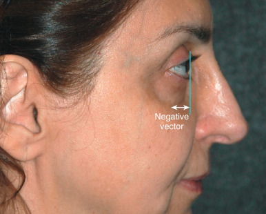
In our practice absolute contraindications include patients with a negative vector and preoperative orbicularis weakness. These two preoperative physical findings have been noted in studies on postblepharoplasty lower eyelid retraction to have a high risk of this complication . A negative vector eyelid is noted when the patient’s profile is examined (sagittal view) and the cornea projects more anteriorly than the midface ( Fig. 13.4 ). This is often present in patients with thyroid eye disease. When tightening the eyelid skin and orbicularis muscle, the tendency is for the redraped tissue to fall under the globe in a negative vector eyelid, similar to the way pants fall underneath a “beer gut”. Patients with preoperative orbicularis weakness are also predisposed to lower eyelid retraction as skin-muscle flap surgery further weakens the muscle, causing denervation, loss of strength, and lower eyelid margin eversion. Orbicularis strength can be assessed by trying to pry the patient’s eyelids open during forceful closure by the patient. In a normal situation the examiner cannot open the patient’s eyelids. With more degenerative changes and age, the eyes are easily pried apart. In both of these situations we elect to approach the orbitomalar ligament and orbital fat transconjunctivally, and address the skin externally by pinch technique and suspending the orbicularis muscle redundancy with suture fiction to the orbital rim and/or plicating the lateral orbicularis muscle.
Surgical Procedure
Our lower blepharoplasty technique is an extended transcutaneous submuscular blepharoplasty with orbitomalar ligament release and orbital fat repositioning. Depending upon the patient’s preoperative degree of infraorbital volume loss, this approach can be utilized including infraorbital ligament release with fat removal, partial removal and transposition, or complete transposition. Utilizing this method of periorbital volume replenishment is predicated on the presence of sufficient orbital fat volume for transposition. In cases where there is a lack of orbital fat, autologous fat injections can be utilized. We prefer orbital fat transposition when orbital fat is available because it is a vascularized, pedicled fat flap that has an essentially 100% take rate, whereas injected autologous fat is a free graft and fails to incorporate in as high as 35% of patients, in our experience. To minimize the risk of lower eyelid malposition, our approach preserves a robust orbicularis oculi sling, and incorporates periosteal fixation of the lateral canthus and skin-muscle flap. What follows is our description of the techniques in six steps.
Step 1
Skin Flap Elevation and Preservation of Orbicularis Muscle
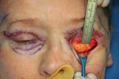
The patient is marked in the upright position, making note of the pseudoherniated orbital fat and the hollowed tear trough. The lower lid is injected with 1% lidocaine with epinephrine 1 : 100,000. The skin is incised with a #15 blade 1 mm below the lash line from 2 mm lateral to the punctum past the lateral canthus for approximately 10 mm in a crow’s foot rhytid.
A retraction suture is placed through the midline lower eyelid and retracted superiorly. A skin flap is elevated inferiorly with sharp iris scissors for approximately 1.5 cm, preserving an intact sling of functional orbicularis muscle ( Figs. 13.5 and 13.6 ). In traditional skin-muscle flap blepharoplasty, only 2 to 3 mm of pretarsal orbicularis is preserved. We believe that maintaining this wider functional sling of orbicularis oculi and minimizing trauma to the muscle minimizes risk of lower lid retraction.