History and Introduction
As noted by Pessa, “there are many arbitrary definitions of what constitutes a youthful face but the appearance of youth is not arbitrary; it is simply difficult to define” . In an effort to solve any problem, one must first define the problem, come up with a solution, and then successfully execute the solution. The progression of facial shape with aging is the subject of many theories and hypotheses, but much remains to be understood. Current understanding of the facial aging process remains largely empirical, given that it has traditionally been based on the effectiveness of various treatments aimed at rejuvenation, some resulting in an odd or “done” appearance. Defining the problem has proved challenging, as facial aging is a complex process that is the cumulative effect of simultaneous changes of the many components of the face, as well as the interaction of these components with each other.
A growing understanding of this complex process has been ongoing in the world of surgery and surgical techniques since its inception, and has informed and driven the change from an empiric approach to an anatomic one, enabling improved and more natural-appearing results. Fig. 3.1 , adapted from an article on facial aging from Cotofana et al., provides an impressive illustration of how innovation and advances in technology, which have given us newer and faster ways to both gather and share information, are accelerating our understanding of facial anatomy . As a result our understanding of the anatomic changes observed in the aging face has progressed considerably over the last couple of decades, leading to a paradigm shift in the way we both perceive and approach these changes. The answer to the question of whether we sink or we sag has become a “yes” to both, as we begin to see aging as a complex and interdependent interplay between all structural layers culminating in the collapse of a three-dimensional (3D) construct. Newer understanding of volume loss as a critical component of facial aging and the integration of volume replacement into the surgical and nonsurgical therapeutic algorithm is arguably the most significant recent development in the field of facial rejuvenation. The ability to accurately recognize where volume has been lost (or sometimes lacking in the first place) in each individual at a given point in time will greatly enhance our ability to address the loss with site-specific corrections in order to achieve optimal and natural-looking results. However, most of us would agree with Glasgold that the recent rapid and widespread adoption of “off the shelf” volume replacement has outpaced a sophisticated understanding of its goals, resulting in a new and different, but equally undesirable category of “looking done” .
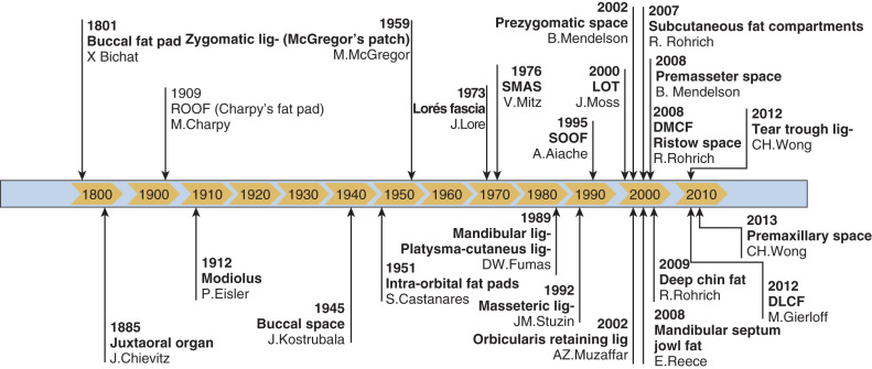
So the problem can be defined as the attainment of natural-looking results in the rejuvenation of the aging face, and the solution to that problem, as well as its execution, lies in understanding its pathogenesis, which is anatomic. Recent insights and gains in our anatomic understanding enhance our current ability to come closer to this goal. For this reason, this chapter is not written on the various types of fillers or techniques of filler injection, about which much has been written, but rather on how to decide where to use it and why in different faces, using anatomy as a guide. This approach is rational, practical, teachable, and reproducible, as it is simply the result of the recognition, and targeted correction, of currently recognized specific anatomic deficiencies. Using this approach has improved my results and resulted in much higher patient satisfaction. That said, it is not the only way. Many other approaches have been employed successfully (using specific landmarks or masks or phi ratios, for example), in order to obtain pleasing results; however, some aspects of these approaches also use anatomy. The purpose of this chapter is to provide an introduction and brief summary of some of the recent literature concerning facial anatomy and the anatomy of facial aging, which serve as the basis and foundation for predictable, specific, reproducible, and natural-appearing results with the use of injectables. This summary of current concepts will be presented along with clinical examples exhibiting primarily congenital absence or aging changes in the tissue layer discussed in order to better illustrate the discussion. The practical use of these concepts in injectable treatment of the face will then be illustrated using a number of case reports of patients of different ages, gender, and ethnic backgrounds along with a short description of where each face was treated and why, using both a layered anatomic (tissue structures) and regional approach (upper, mid, and lower face, shape, proportions). In order to look at a number of cases, as well as to compare and contrast different faces, these cases are presented in a “composite” format. This smaller format additionally makes it easier to recognize facial shape and proportions, and to determine what is present or missing that may be moving the face away from the ideal shape and proportions, which will be discussed below.
Finally, as aging is a complex multimodal process, multimodal therapy must be used to address it. Despite the widespread popularity of injectable treatments as an “immediate gratification no downtime option,” they have their limitations and risks like everything else, and are not a panacea (or stand alone) treatment of the aging face.
Personal Philosophy
Although a new patient may present pointing to a wrinkle, line, or fold they have noticed seemingly overnight, as stated above, we are now increasingly aware that these first obvious signs of aging noted by the patient are in fact downstream markers of a slow progressive change taking place in all structures of the face. This represents a paradigm shift in our current approach to facial rejuvenation. This concept will be addressed, discussed, and illustrated in this chapter.
Facial beauty and attractiveness are important cross-cultural social concepts as they tend to dictate how individuals are judged and treated . Research has shown that facial beauty is perceived and processed rapidly by the brain, and this perception biases subsequent cognitive processes . A recent extensive review of research on facial beauty determined that four characteristics emerge as the most statistically significant determinants of attractiveness: averageness (prototypicality), sexual dimorphism, youthfulness, and symmetry .
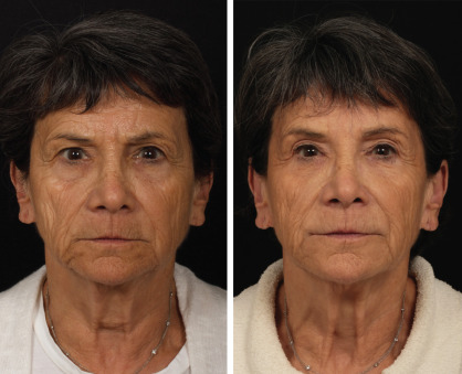
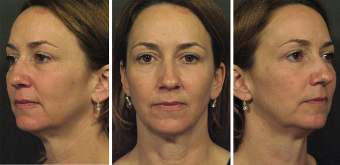
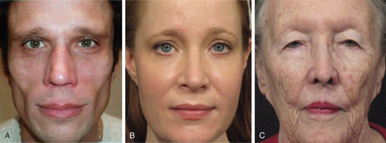
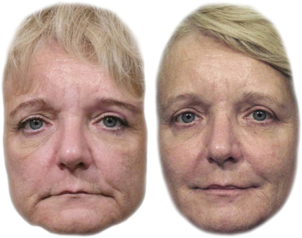
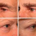
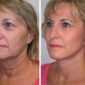
Not surprisingly, all of these have something to do with optimizing mate selection. Youth and sexual dimorphism are obvious. Prototypicality likely signifies a good mix of genes (avoiding autosomal recessive disease), while symmetry may indicate a history of maternal stability and health during development. Additionally, changes seen with aging may lead to an unintended and undesirable misinterpretation of mood by others that is unwelcome to most all of us as we age and is one of the most common presenting complaints, i.e., “I don’t mind getting older I just don’t want to look mad, sad, and tired.” This can often be remedied with glabellar neuromodulators as well as fillers infraorbitally as well as around the mouth, resulting in a surprisingly different first impression of a face, as seen in Fig. 3.2 . Looks matter because they can have a great impact on quality of life.
Facial Anatomy: Introduction and Overview
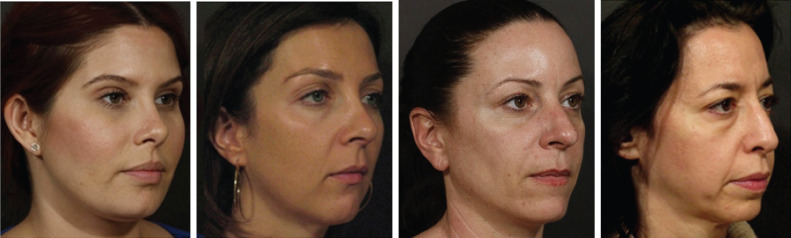
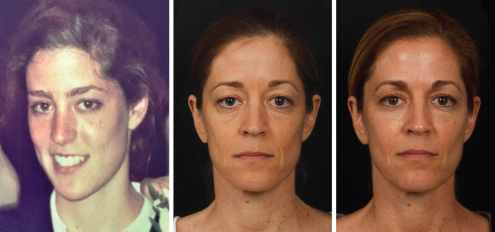
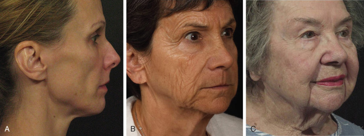
The traditional approach to assessing the face is to consider the upper, middle, and lower thirds, regionally. Other newer useful approaches using structural layers or functional differences reflect our recognition that the pathogenesis of facial aging is a multifactorial process that can be explained on an anatomic basis, and likely accounts for the variations in the onset and outcome of aging seen in different individuals. We will look at the face with these different approaches in the next section. Regardless of approach, cumulative changes in all structural tissue layers of the face with time lead to a change in the morphology of the entire face in terms of its shape, proportions, and topography. Morphologic changes seen in women from different decades of life (30s to 60s) are illustrated in Fig. 3.3 . Although these types of figures were initially used to visualize the differences in the depth of a tear trough (TT), nasolabial fold (NLF), or marionette line with advancing age, they can now be appreciated as evidence that these folds and lines are downstream markers of a global change rather than isolated entities. This figure also illustrates how changes in facial topography seen with aging sharpen the once smooth transition between anatomic units, by greatly magnifying light reflection and/or shadow. This concept is critical to our understanding, as seemingly subtle changes in light and shadow over time can have an enormous impact on our perception of a face in an almost indiscernible way. The rationale behind restoring 3D contours to the face as it ages, whether by lifting, tightening, or volume restoration, is easy to appreciate when looking at photographs illustrating how aging takes us from 3D to 2D, as shown in the woman in Fig. 3.4 shown at college graduation and 30 years later, both before and after injectable treatment. The youthful oval face dramatically flattens with age, and restoration of previous arcs and convexities restore youthful light and shadow patterns.
Although the sequence of events observed in aging is somewhat predictable, its pace among individuals is variable and progresses in each person from a unique starting point. Additionally, changes in different tissue layers within a single individual occur interdependently. The lack of, or loss, of structural integrity in one area may worsen the appearance of a neighboring area. Conversely, the presence, or restoration, of structural integrity in one area may improve the appearance of a neighboring area .
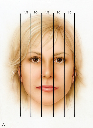
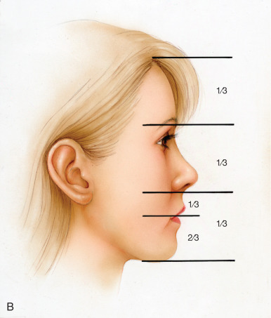
We know that almost all faces develop with slight asymmetry following development of the neural tube embryologically, and the aforementioned concept can be readily appreciated when looking at the facial asymmetry in the woman in Fig. 3.5 who appears more aged on her right, “sunken,” side compared with her fuller left side. Note that the less volumized side (right) of her face shows a clear delineation of her temple, lid, and cheek as separate entities, while the more volumized side (left) does not—one area seems to blend seamlessly with another, reflecting light uninterrupted by the shadows seen on the right. Note that the amount of volume loss with aging on the initially smaller right side has now resulted in an outer skin envelope slightly too large for its now “smaller” face, and contributes to a more pronounced ptosis and loss of jawline contour on this side earlier than on the left side (which commonly has more solar elastosis because its the driver’s side window). Less bony support and soft tissue leads to a lower brow position, leading to lid lag laterally and an early hollowing “A-frame” deformity somewhat camouflaged by a slackened upper lid. A TT and lid-cheek junction are seen only on the less volumized side. There is also less anterior and lateral cheek projection, a slightly deeper nasal sulcus, a longer upper lip, and an increased mental hollowing on the less volumized side, whereas no such demarcation is visible on the more volumized side. This combines to make the perioral proportions in the lower third of her face look slightly less youthful on the less volumized side. Finally, the peripheral contours on the less volumized side of her face are more abrupt than those on the fuller side. The convexity of the temple and the preauricular volume on the full side lends an overall oval shape to that side of her face that is lost due to the atrophy on the right. Compare this with the ideal proportions of a youthful face, as shown in Fig. 3.6 , depicting a width of five eyes across in vertical fifths and an equal volume in the upper, mid, and lower face when measured in horizontal thirds. Additionally, this schematic depicts the golden phi ratio of 1:1.6 in the perioral region of the lower third of the face. Note that the fuller side of the face in Fig. 3.5 is closer to these ideal proportions, providing a “roadmap” of where to revolumize the other side.
Over a decade ago, in an effort to visualize the aging face in linear examples, Lambros used computer animation to compare current photographs of patients matched for light and position with photographs taken 10 to 50 years previously to gain insight into midfacial aging, showing that deflation can mimic descent . Recent work by Lambros and Amos now provides an invaluable tool to visualize the facial aging process using 3D facial averaging . They have published animations made from 3D facial images amassed over the past 10 years, using a 3D camera system (Vectra; Canfield Scientific, NJ, USA). These are shown as static images in Fig. 3.7 . The image on the left shows the average of the 3D facial surfaces of 116 female subjects aged 20 to 30 years, and the image on the right shows the average of the 3D facial surfaces of 100 female subjects aged 68 to 91 years. The static images illustrate the differences seen in the morphology of the younger and older averages well; however, this image may be viewed in animation online at http://links.lww.com.easyaccess1.lib.cuhk.edu.hk/PRS/B922 . It is interesting to compare the similarities between the youngest and oldest women in Fig. 3.3 to the 3D-averaged image of similar age. Look at the shape of the orbits, the bony support under the brow and the nose, the flattening of the midface and lateral cheeks, and the change in proportions in the lower third of the face. Note how the TT and NLF deepen as the craniofacial support changes and the cheek flattens. Look at the eversion versus inversion of the lips. Look at light and shadow, and how it plays off areas of depression and prominence (convexities and concavities) in both the younger and older face. Do not focus on just “lines and folds,” but consider all the structural changes in the face. Consider the interdependency between them by treating the whole face as a 3D interlocking puzzle where losing or correcting one component may have a negative or positive impact on another.
Facial Anatomy and Aging: Regional Thirds of the Face
The traditional regional approach to assessing the face is to consider the upper, middle, and lower thirds (as shown in Fig. 3.6B ). Glasgold, Glasgold, and Lam have greatly increased our appreciation that a detailed examination of the shadows and shadow patterns that develop in all areas of the face with volumetric facial aging will lead to a better understanding of how to apply volumetric techniques to create a natural-appearing result. Although they have worked mostly with fat augmentation, the same concepts apply to filler (although filler may not be a cost-effective option for those needing a lot of volume) . Although every face is unique, the shadows that develop as we age are consistent. Not everyone develops every shadow, but the typical shadows of aging are universal. Glasgold notes the ease with which an artist can depict an aging face with a few shadow strokes makes this concept easy to grasp. Studies documenting the consistent patterns of volume loss in the aging face are reviewed in the following sections. The skin of the face has consistent attachment points to the underlying structures through the facial retaining ligaments, and as the volume of the face deflates, these attachment points will define most of the shadows that develop with age . Advancing age accounts for specific areas of volume loss in all thirds of the face. These changes in each third of the face are summarized here, paraphrasing the (“can’t be improved upon”) descriptions published by Glasgold, Glasgold, and Lam. Compare characteristics of younger and older faces as you read through these by looking again at the women pictured in Figs. 3.3, 3.4, and 3.5 and the averages of the 3D facial surfaces in Fig. 3.7 .
Upper Face
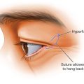
In the upper face the young eye appears full, the bony orbit is not visible, the skin is elastic and thick, and most of the upper lid is concealed by the full brow, with only a few millimeters of upper lid show. The youthful upper lid sulcus lacks a shadow, and the eye has an overall “almond” configuration, with the lid margin, lid crease, and eyebrow all parallel. As the upper lid deflates, a fold of skin develops where there was once fullness, and the shadow of the upper lid sulcus emerges. With increasing age, this fold of upper lid skin often droops and may encroach on the lash line, completely effacing any visibility of the upper lid (“hooding”). The eyelid skin may also slip into the lid crease, revealing the upper lid veiled in youth by the full brow. Often this is initially most pronounced medially, resulting in the so-called “A-frame” deformity . With aging, shadows also develop in the temple and upper orbit. A deep shadow of the temple sets off the lateral orbital rim and zygomatic arch. Filling the temple and lateral brow will affect the appearance of the tail of the brow as well as the upper lid. The temple additionally relates to the entire lateral face contour including the zygoma, buccal regions, and lateral mandible.
Midface
The manifestations of midfacial aging are largely due to changes in facial volume that transition the midface from a youthful convex platform dominated by highlights to an aged flattened platform segmented by shadows (concavities) . Younger midfaces have an unbroken convexity running from the lower eyelid to the NLF, creating a dominant cheek highlight. Soft tissue covers the bony skeletal components of the midface, providing a softer appearance; the inferior orbital rim is masked, minimizing any delineation between the lower eyelid and cheek . The zygomatic arch, providing the foundation of lateral cheek volume, is adequately covered by soft tissue to hide the shadows that delineate its superior and inferior margins . Advancing age is associated with a generalized deflation of the midface, particularly in the upper aspects. The combination of volume loss and the effect of underlying facial retaining ligaments contribute to the hallmarks of midface aging. As we will see in the next section, the most relevant ligaments in the midface are the orbicularis retaining ligament (ORL), malar septum (zygomaticocutaneous ligament), and the McGregor patch (zygomatic ligament) . Volume loss at the inferior orbital rim creates a concavity and overlying shadow, separating the lower eyelid from the cheek. Volume loss in the anterior cheek converts the youthful convexity into a concavity with its base tethered by the malar septum (zygomaticocutaneous ligament). Lateral cheek volume loss diminishes the dominance of midface volume and skeletonizes the zygomatic arch, creating a harsh submalar shadow . In the midface, augmentation of the cheek alone will worsen the separation from the eye, upper lip, buccal area, and temple, often contributing to an unnatural appearance. Addressing the shadow group of the midface as a whole will allow the creation of a unified cheek highlight with no separation between the cheek, the eye, and the upper perioral unit. Adding volume in the inferior orbital rim will reunify the lower eyelid and cheek segments. Filling the cheek, with a focus on the malar septal (zygomaticocutaneous ligament) depression, will recreate a convex cheek with a strong highlight. Volume may need to be added to the lateral cheek when there is deficient lateral projection, but most important is filling around the zygomatic arch to restore youthful soft contours. The buccal region transitions the lateral facial contour of the zygoma into the lateral mandible .
Lower Face
The lower face has two distinct components, the jawline and the perioral region. The hallmarks of a youthful lower face include a smooth transition from the cheek to chin, devoid of shadowing at the labiomandibular fold. The jawline is well defined by a curvilinear shadow coursing from the mandibular angle to the anterior chin; on oblique view the shadow framing the jawline has a “hockey stick” shape. This youthful jawline shape is dependent on an adequate bony foundation providing sufficient volume at the prejowl sulcus and angle of mandible. In the perioral area the labiomental hollow creates an upside down U-shaped shadow that separates the lower lip from the chin and the labiomental fold creates a distinct shadow that typifies the frown. The prejowl sulcus appears as volume loss progresses at the inferior portion of the mandible, anterior to the jowl, and corresponds to the attachment of the mandibular ligament causing a shadow in front of the jowl. The typical jowl can be thought of as a highlight that exists between the shadows of the prejowl sulcus and (if present) shadowing of the lateral mandible. Congenital lower face volume deficiencies are most common in the chin and mandibular angle. Deficiencies in the anterior chin and prejowl sulcus create a relative middle jawline dominance, which manifests in an appearance of early jowl formation. These patients tend to present at an earlier age for lower face rejuvenation as even early volume changes more easily highlight their skeletal deficiencies .
Finally, Glasgold et al. have introduced an additional regional concept they term the “three dominant frames in the face” . The first is the global facial frame, which extends along the jawline to offset the face from the neck and then flows up the lateral contour of the face from the angle of the mandible along the contour of the buccal, zygomatic, and temple line. The global facial frame strongly affects our perception of a face on many levels: aging, gender, and attractiveness. A sharp, uninterrupted shadow separating the face from the neck is desirable. A soft inverted egg-shaped lateral contour suggests youthful femininity, while a more angular/rectangular line is more masculine. The other two frames highlight the eyes and the mouth. As noted earlier, the traditional surgical approach to the eyes has been of volume and tissue removal, typically leaving the eye offset in deep shadows looking aged and unhealthy. Volume replacement to the upper eye, particularly the medial orbit A-frame shadow, is, in the opinion of Glasgold, one of the most effective uses of volume in the face, as it eliminates these aging shadows. In the perioral region the natural aging process invariably creates a ring of shadow around the lips and mouth, which contribute to an aged appearance. The effect of creating a frame of light in as many of the small subunits of the perioral region as possible will enhance the beauty of the mouth. Conversely, adding volume to the lips without addressing the surrounding area serves to deepen the shadows and further disconnect the lip and mouth from the perioral region, resulting in one of the odd appearances that patients fear . Many of these changes have been described earlier on the smaller, more aged-appearing side of the patient pictured in Fig. 3.5 .
Facial Anatomy and Aging: Functional and Structural
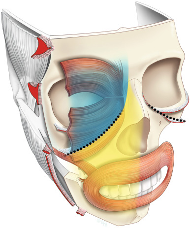
Despite the recent advances revealing the aging changes seen in facial soft tissue anatomy, and it’s underlying skeletal support, our understanding of this complex process is still in its infancy. Mendelson and Wong suggest that in addition to the traditional assessment of facial thirds, a more global understanding is facilitated by distinguishing between the different functional regions of the face, as well as by considering the anatomy in terms of a layered construct. They consider the face can be divided into the highly mobile anterior face, which is functionally adapted for facial expressions and the fixed lateral face, which overlies masticatory structures . A vertical line of retaining ligaments separates the anterior and lateral face ( Fig. 3.8 ). These ligaments are, from above: temporal, lateral orbital, zygomatic, masseteric, and mandibular ligaments. The orbicularis retaining ligament is seen running along the inferior orbital rim, and the zygomaticocutaneous ligament along the inferior zygoma. In the anterior face, the midcheek is split obliquely into two separate functional parts by the midcheek groove (which runs along the inferior zygoma correlating to the zygomaticocutaneous ligament) related to two cavities: the periorbital part above and the perioral part below. The transitions between these areas, while not seen in youth, become increasingly evident with aging. This is illustrated in Fig. 3.9 , where the ligaments themselves can be seen in the parotidomasseteric area in an emaciated face in Fig. 3.9A , and the pull on the skin from the various ligaments can be appreciated in both a thin and a full face in Fig. 3.9B and C . These transitions (or their lack) can also be appreciated in the faces in Figs. 3.3, 3.4, 3.5, and 3.7 .
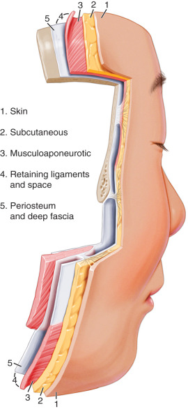
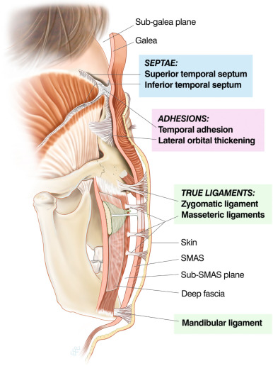
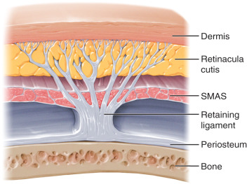
These authors conceptualize the soft tissues of the face as arranged concentrically into five basic layers that are bound together by a system of facial retaining ligaments, as first hypothesized by Stuzin et al. . The layers are pictured in Fig. 3.10 and consist of (1) skin; (2) subcutaneous fat; (3) the musculoaponeurotic layer; (4) areolar tissue, including facial retaining ligaments and facial spaces; and (5) periosteum and deep fascia . As noted earlier, to secure the superficial fascia (defined as the composite flap of layers 1–3) to the facial skeleton, a system of retaining ligaments binds the skeleton to the dermis, and the components of this system pass through all layers, as illustrated in Fig. 3.11 . Like a tree, these ligaments start as a thicker trunk, and subsequently fan out in a series of thinner branches as they insert into the subcutaneous fat and the dermis. At different levels of dissection, the ligament system is given different names, such as the retinacula cutis in the subcutaneous layer and ligaments under the superficial musculoaponeurotic system (SMAS) level, as seen in Fig. 3.12 . The density of these branches determine the difficulty encountered when dissecting the tissue and accounts for the fact that it is easier to dissect in the deep subcutaneous layer than the dermis. The nerves and vessels are always located in close proximity to the retaining ligaments, as these vital structures are protected by traveling through other layers in concert with these structures. The retinacula cutis fibers are not uniform across the face, but vary in orientation and density according to the anatomy of the underlying deeper structures. As will be apparent when the anatomy of the underlying layer 4 is described, at the location of the retaining ligaments, the vertically orientated retinacula cutis fibers are the most dense and are the most effective in supporting the overlying soft tissues, and in so doing, form the anatomic boundaries that compartmentalize the subcutaneous fat . Additionally, Mendelson and Wong’s extensive work support that a large part of the sub-SMAS layer 4 consists of soft tissue “spaces” that have defined boundaries that are strategically reinforced by retaining ligaments. As the roof of each space is the least supported part, it is more prone to developing laxity with aging, compared with the ligament-reinforced boundaries. This differential laxity accounts for much of the characteristic changes that occur with aging of the face.
Each of the aforementioned layers will be discussed in more detail (with an emphasis on fat and bone as it relates to injectable treatments) in the sections here.
Preprocedural Assessment
As all structural tissues play a role in the aging face, restoring youthful characteristics (or establishing them where they are congenitally absent) starts from the skeletal framework and builds progressively to the canvas of the face. Therefore, current literature pertaining to the morphological changes of the facial skeletal framework, retaining ligaments, facial muscles, fat compartments, and skin envelope will be presented in the next section. All contribute to facial aging in variable degrees, some of it primary, some secondary, and what is known about which is which, as well as the relative contributions of each, is in a constant state of evolution and refinement. Even with those limitations in mind, with careful evaluation some specific age-related changes or congenital deficiencies can now be addressed in a site-specific manner to achieve natural-looking results.
This section on assessment will discuss the importance of patient selection and introduce an approach to evaluating the face. As noted by Pessa, although anatomy is remarkably consistent between individuals, the variable sizes and shapes of the different structures in each individual gives everyone their own unique appearance and has tremendous influence on the variations in the onset and outcome of aging seen in different individuals . This is made more complex by the recognition that although the sequence of events as we age is somewhat predictable, its pace is variable between individuals and even between tissue layers in one individual. Subsequently, there is no one algorithm to address facial aging. As mentioned earlier four characteristics emerge as the most significant determinants of attractiveness: prototypicality (averageness), sexual dimorphism, symmetry, and youthfulness . Averageness requires harmony of all regions of the face. Sexual dimorphism requires recognition of gender differences. A detailed discussion of gender differences is out of the scope of this chapter, but a few well-accepted norms are worth mentioning here. Males often have a stronger forehead and straighter brow, a cheek apex that is lower and more medial, and a stronger chin and jawline than females. Conversely, female faces have a higher, more lateral cheek apex and a more tapered lower face than males. Obviously, you do not want to feminize a male face with high lateral cheekbones, or fail to treat a masculinizing masseter hypertrophy in a female. Regarding symmetry, after the neural tube develops early in our embryologic development, the two sides of the body develop like siblings, not twins, and the vast majority of us have a shorter fuller side and a thinner longer side. The discrepancy between these sides becomes more apparent with aging. The contribution of symmetry to beauty is often maligned by showing a photograph of a face next to photographs made using the two right and two left sides of that face, often showing a bizarre looking thin face and fat face. However, this results from comparing the two sides of an asymmetric face. If the original face photographed is truly symmetric, then all three photographs would be identical. It is interesting to note that the fuller shorter side is usually the more attractive side in youth, and the younger appearing side with age. Look back at Fig. 3.5 . In my experience, augmenting volume around the temple, brow, orbit, and zygomatic arch just enough to restore more symmetrical light reflection from both sides can make a surprising difference in our perception of that face.
In relatively young faces with early aging changes (including the face pictured in Fig. 3.5 ), addressing a TT, NLF, or marionette line as an isolated entity will often yield good results. However, as these folds represent downstream markers of global changes, in those individuals with congenital deficiencies, and those further along in the aging process, this approach may yield suboptimal results by taking one area of the face out of harmony with another. Figuring out what you want to treat is a process of observation and palpation/provocation that allows us to determine the nature and extent of the structural tissue changes affecting the face in front of us at that particular point in time, as well as how those structural changes have affected the shape, proportions, topography, and frames of the entire face. It is, of course, more of a “read” than a “recipe,” as there is no one algorithm that fits all faces. It is useful to think of what is deviating that face from the ideal proportions shown in Fig. 3.6 , which shows an ovalized “upside down egg” shape, an anterior convexity, an oval frame, youthful proportions of “five eyes across” and three relatively equal thirds of the face. What you choose to address depends on the extent of the changes seen in each structural layer or region, and the parity of these changes between layers or regions. Try to figure out “which tissue is the biggest issue” or, if regionally, “one of these things is not like the other.” If there is just a little change in all layers, almost any interventional approach will work. If there is regional disparity, try to blend them all back to a more similar place. The most common disparities are creating young lips or cheeks that “stick out” rather than blend in to an otherwise aging face, or placing too much filler too high under the eyes, which looks odd.
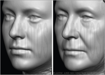
Figs. 3.13 and 3.14 may help clarify this approach. The faces pictured in Fig. 3.13 all have a clear-cut “one tissue issue” (see figure legend), while the face in Fig. 3.14 has a discrepancy in the size of her upper, mid and lower face. The lack of volume in the lower two-thirds of her face make her forehead seem too large in the before picture, while it looks quite normal following treatment of the cheeks and chin in the after photo.
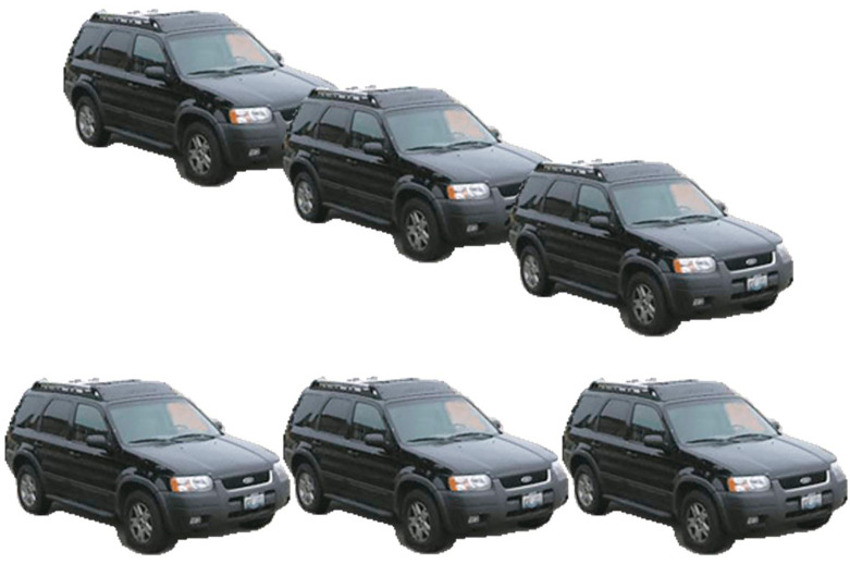
This kind of optical illusion is not uncommon in the face. It is termed a “perspective illusion” and is illustrated well in the classic three cars example seen in Fig. 3.15 . If there is a lot of loss of integrity in multiple layers, then multiple interventions and a great deal of product may be needed to obtain optimal results, as seen in Fig. 3.16 . It is interesting to note on the cropped close-up view of this face shown in Fig. 3.17 how treatment of the areas of skeletal remodeling and fat loss in this patient affected the overall shape, topography, and proportions of this face towards the ideal proportions pictured in Fig. 3.6 .

Recognize that a patient’s final outcome, and the amount of product and work it will take to get there, is a reflection of the quality of tissues with which they start. Wasted faces (associated with HIV or endurance exercise) are harder to fill, and it is harder to sustain the fill. Older faces with advanced craniofacial remodeling, fat loss, and very poor skin quality can be treated successfully; however, fillers of any kind may not be the most cost-effective choice in a patient who would best benefit from fat augmentation and a face lift. Discussing this and setting realistic expectations before treatment will decrease frustration for both the patient and the practitioner. Conversely, fuller and younger faces “bring in their own volume” and are therefore easily reshaped with a conservative amount of filler of any kind. This is, of course, an issue of patient selection and not product selection. For these reasons, for the novice, I strongly recommend starting with younger patients with mild-to-moderate volume changes.
Finally, as with any procedure, there is no guarantee of a specific or perfect result, and serious complications such as delayed nodules, ischemia and necrosis, or even blindness, although extremely rare, can occur. As with any procedure, the patient must demonstrate sufficient “psychosocial maturity” to weather any potential complications. The patient should be educated and a true informed consent obtained. Manage expectations. Anxious, demanding, or unhappy patients are poor candidates. Although there are no absolute contraindications, patients with active autoimmune disease, poor dental hygiene, or a history of previous permanent filler (especially if done outside the USA) may be at increased risk for complications.
Current Concepts in the Anatomy of Aging
Skin: Layer 1
Skin appearance (including elasticity, the absence of wrinkles, a smooth texture, and clarity and evenness of color) is a primary indicator of age. The epidermis is a cell-rich layer composed mainly of differentiating keratinocytes and a smaller number of pigment-producing melanocytes and antigen-presenting Langerhans cells. The dermis comprises predominantly the extracellular matrix secreted by fibroblasts. Type I collagen is the most abundant protein. Other collagen types (III, V, VII), elastin, proteoglycans, and fibronectins are present in smaller quantities. A rich vascular plexus is an important component of the dermis and it provides support and nutrients to the epidermis. The thickness of the dermis relates to its function and tends to be inversely proportionate to its mobility. The dermis is thinnest in the eyelids and thickest over the forehead and the nasal tip. The thinner the dermis, the more susceptible it is to qualitative deterioration aging changes . The vast majority of skin aging is photoaging from exposure to ultraviolet (UV) light . The shorter UVB rays cause a pathognomonic lesion (thymine dimers) in the epidermal keratinocytes, which may lead to the development of skin cancers. The longer UVA ray exposure causes oxidative damage in the dermis, leading to the elaboration of collagenase (matrix metalloproteinases [MMPs]), which fragments existing collagen . Research now reveals that collagen deficit in both photoaged and chronologically aged human skin derives primarily from altered mechanical properties of the fragmented collagenous extracellular matrix of the dermis rather than from time and/or UV irradiation-derived genetic damage to fibroblasts .
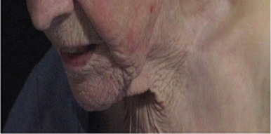
Intact type I collagen fibrils in the dermis provide mechanical stability and attachment sites for fibroblasts, which is critical for normal, balanced production of collagen and collagen-degrading enzymes. Without this attachment the fibroblasts collapse, and collapsed fibroblasts exacerbate the situation by producing more MMPs, advancing the aging process into a self-perpetuating, deleterious cycle. This phenomenon is well illustrated in Fig. 3.18 , showing both sun-protected and sun-exposed skin in an elderly female. Sunscreen, antioxidants, and retinoids help mitigate collagen loss . Microneedling, peels, and various energy devices work by inducing new collagen formation. There is some evidence that hyaluronic acid fillers may provide a “stretch effect” on fibroblasts and speculation on whether the collagen-stimulating fillers or stem cells from fat augmentation may play a similar role, leading to the skin improvement commonly seen after these procedures.
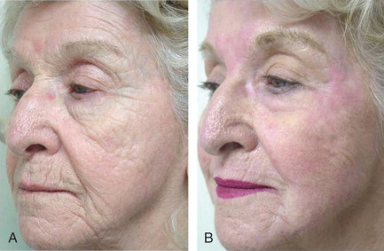
Be aware that it is very difficult to fill an elastotic outer skin envelope with volume augmentation of any kind, and these patients will require surgical intervention to both lift and fill. On the other hand, if the underlying craniofacial support and fat volume is good, and the skin still has a fair amount of collagen, any procedure that can effectively tighten skin will make an enormous difference, as seen in the patient in Fig. 3.19 who underwent a nonfractionated CO 2 laser resurfacing procedure as monotherapy.
Fat: Layer 2
The recent description of the superficial and deep fat compartments of the face by Rohrich and Pessa (utilizing dye sequestration in cadaveric dissections) and radiological confirmation by Gierloff et al. (utilizing radiopaque dye and computed tomography [CT]) has provoked great interest in both the role that these compartments play in the layered and spatial relationships existing in the face, as well as to what role this might contribute to the theory of facial deflation with aging. It should be noted that the theory of facial deflation is not universally accepted. It has been observed that inversion photographs of aging patients (either in a supine or Trendelenburg position) demonstrate an appearance consistent with that of photographs taken approximately 10 to 15 years prior . These authors feel that aging must, therefore, be partially gravitational: much of the volume in the face must be retained and not lost, because volumes drift back into position when the supine position is assumed.
The 3D technique employed by Gierloff et al. allowed these compartments to be simultaneously identified and measured, as well as viewed from several angles. This allowed the investigators to further define the deep subcutaneous fat of the midcheek into a medial and lateral compartment, as well as define the isolated buccal extension of the buccal fat pad. This finding—the discrete nature of buccal fat lobes—is of tremendous importance clinically to avoid a potential complication of augmentation of deep medial cheek fat (DMCF). Although injection of filler into the central lobe of buccal fat specifically augments that region alone, inadvertent injection of filler into the inferior lobe of buccal fat may lead to the creation of a more prominent jowl, and should therefore be avoided.
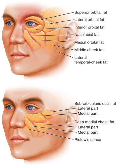
Schematics of the superficial and deep fat compartments of the midface (adapted from this first radiologic study) are shown in Fig. 3.20 .
Related posts:

Stay updated, free articles. Join our Telegram channel

Full access? Get Clinical Tree


