In some patients, the appearance of skin lesions may be so distinctive that the diagnosis is clear at a glance. In others, subjective symptoms and clinical signs alone are inadequate, and a complete history and laboratory examination, including a biopsy, are essential to arrive at a diagnosis.
The same disease may show variations under different conditions and in different individuals. The appearance of the lesions may have been modified by previous treatment or obscured by extraneous influences, such as scratching or secondary infection. Subjective symptoms may be the only evidence of a disease, as in pruritus, and the skin appearance may be generally unremarkable. Although history is important, the diagnosis in dermatology is most frequently made based on the objective physical characteristics and location or distribution of one or more lesions that can be seen or felt. Therefore careful physical examination of the skin is paramount in dermatologic diagnosis.
Cutaneous Signs
Typically, most skin diseases present with lesions that have distinct characteristics. They may be uniform or diverse in size, shape, and color or may be in different stages of evolution or involution. The original lesions are known as the primary lesions, and identification of such lesions is the most important aspect of the dermatologic physical examination. They may continue to full development or be modified by regression, trauma, or other extraneous factors, producing secondary lesions.
Primary Lesions
Primary lesions are of the following forms: macules (or patches), papules (or plaques), nodules, tumors, wheals, vesicles, bullae, and pustules.
Macules (Maculae, Spots)
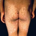
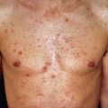
Macules are variously sized, circumscribed changes in skin color, without elevation or depression (nonpalpable) ( Fig. 2.1 ). They may be circular, oval, or irregular and may be distinct in outline or may fade into the surrounding skin. Macules may constitute the whole lesion or part of the eruption or may be merely an early phase. If the lesions become slightly raised, they are then designated papules or, in some cases, morbilliform eruptions.
Patches
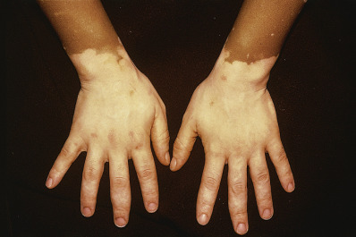
A patch is a large macule, 1 cm or greater in diameter, as may be seen in nevus flammeus or vitiligo.
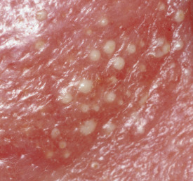
Papules
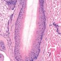
Papules are circumscribed, solid elevations with no visible fluid, varying in size from a pinhead to 1 cm ( Fig. 2.2 ). They may be acuminate, rounded, conical, flat topped, or umbilicated and may appear white (as in milium), red (eczema), yellowish (xanthoma), or black (melanoma).

Papules are generally centered in the dermis and may be concentrated at the orifices of the sweat ducts or at the hair follicles. They may be of soft or firm consistency. The surface may be smooth or rough. If capped by scales, they are known as squamous papules, and the eruption is called papulosquamous.
Some papules are discrete and irregularly distributed, as in papular urticaria, whereas others are grouped, as in lichen nitidus. Some persist as papules, whereas those of the inflammatory type may progress to vesicles or to pustules, or they may erode before regression takes place.
The term “maculopapular” should not be used. There is no such thing as a “maculopapule,” although there may be both macules and papules in an eruption. Typically, such eruptions are morbilliform.
Plaques
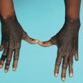
A plaque is a broad papule (or confluence of papules), 1 cm or more in diameter ( Fig. 2.3 ). It is generally flat but may be centrally depressed.
Nodules
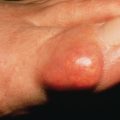
Nodules are morphologically similar to papules but are larger than 1 cm in diameter. Nodules most frequently are centered in the dermis or subcutaneous fat.
Tumors
Tumors are soft or firm, freely movable or fixed masses of various sizes and shapes, but usually are greater than 2 cm in diameter. General usage dictates that the word “tumor” means a neoplasm. They may be elevated or deep seated and in some cases are pedunculated (neurofibromas). Tumors have a tendency to be rounded. Their consistency depends on the constituents of the lesion. Some tumors remain stationary indefinitely, whereas others increase in size or break down.
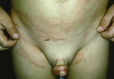
Wheals (Hives)
Wheals are evanescent, edematous, plateaulike elevations of various sizes ( Fig. 2.4 ). They are usually oval or of arcuate contours, pink to red, and surrounded by a “flare” of macular erythema. Wheals may be discrete or may coalesce. These lesions often develop quickly (minutes to hours). Because the wheal is the prototypic lesion of urticaria, diseases in which wheals are prominent are frequently described as “urticarial” (e.g., urticarial vasculitis). Dermatographism, or pressure-induced whealing, may be evident.
Vesicles (Blisters)
Vesicles are circumscribed, fluid-containing elevations 1–0 mm in size ( Fig. 2.5 ). They may be clear from serous exudate or red from serum mixed with blood. The apex may be rounded, acuminate, or umbilicated, as in eczema herpeticum. Vesicles may be discrete, irregularly scattered, grouped (e.g., herpes zoster), or linear, as in allergic contact dermatitis from urushiol (poison ivy/oak). Vesicles may arise directly or from a macule or papule and generally lose their identity in a short time. They may break spontaneously or develop into bullae through coalescence or enlargement. The inflammatory process may lead to pustule formation. When the contents are of a seropurulent character, the lesions are known as vesicopustules. Vesicles have either a single cavity (unilocular) or several compartments (multilocular).
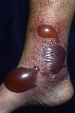
Bullae
Bullae are rounded or irregularly shaped blisters containing serous or serosanguineous fluid. They differ from vesicles only in size, being larger than 1 cm (see Fig. 2.5 ). They are usually unilocular but may be multilocular. Bullae may be located superficially in the epidermis, so their walls are flaccid and thin and subject to rupture spontaneously or from slight injury. After rupture, remnants of the thin walls may persist and, together with the exudate, may dry to form a thin crust. Alternatively the broken bleb may leave a raw and moist base, which may be covered with seropurulent or purulent exudate. Less frequently, irregular vegetations may appear on the base (as in pemphigus vegetans). When subepidermal, the bullae are tense, do not rupture easily, and are often present when the patient is examined.
Nikolsky sign refers to the diagnostic maneuver of putting lateral pressure on unblistered skin in a patient with a bullous eruption; a positive result occurs when the epithelium shears off. Asboe-Hansen sign refers to the extension of a blister to adjacent, unblistered skin when pressure is put on the top of the blister. Both these signs demonstrate the principle that in some diseases, the extent of microscopic vesiculation is more than what is evident by simple inspection. These findings are useful in evaluating the severity of pemphigus vulgaris and severe bullous drug reactions. Hemorrhagic bullae are common in pemphigus, herpes zoster, severe bullous drug reactions, and lichen sclerosus. The cellular contents of bullae may be useful in cytologically confirming the diagnosis of pemphigus, herpes zoster, and herpes simplex.
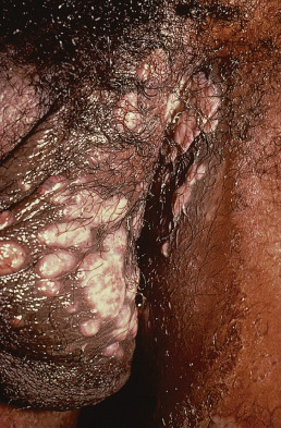
Pustules
Pustules are small elevations of the skin containing purulent material, usually necrotic inflammatory cells ( Fig. 2.6 ). They are similar to vesicles in shape and usually have an inflammatory areola. Pustules are usually white or yellow centrally but have a red tinge if they also contain blood. They may originate as pustules or may develop from papules or vesicles, passing through transitory early stages, during which they are known as papulopustules or vesicopustules.
Secondary Lesions
Secondary lesions are of many types; the most important are scales, crusts, erosions, ulcers, fissures, and scars.
Scales (Exfoliation)
Scales are dry or greasy, laminated masses of keratin. The body ordinarily is constantly shedding imperceptible tiny, thin fragments of stratum corneum. When the formation of epidermal cells is rapid or the process of normal keratinization is disturbed, pathologic exfoliation results, producing scales. These scales vary in size; some are fine, delicate, and branny, as in tinea versicolor, whereas others are coarser, as in eczema and ichthyosis, and still others are stratified, as in psoriasis. Scales vary in color from white-gray to yellow or brown from the admixture of dirt or melanin. Occasionally, they have a silvery sheen from trapping of air between their layers; these are micaceous scales, characteristic of psoriasis. When scaling occurs, it usually suggests a pathologic process in the epidermis, and parakeratosis is often present histologically.
Crusts (Scabs)
Crusts are dried serum, pus, or blood, usually mixed with epithelial and sometimes bacterial debris. When crusts become detached, the base may be dry or red and moist.
Excoriations and Abrasions (Scratch Marks)
An excoriation is a punctate or linear abrasion produced by mechanical means, usually involving only the epidermis but sometimes reaching the papillary layer of the dermis. Excoriations are caused by scratching with the fingernails in an effort to relieve itching. If the skin damage is the result of mechanical trauma or constant friction, the term “abrasion” may be used. Frequently, there is an inflammatory areola around the excoriation or a covering of yellowish dried serum or red dried blood. Excoriations may provide access for pyogenic microorganisms and the formation of crusts, pustules, or cellulitis, occasionally associated with enlargement of the neighboring lymphatic glands. In general, the longer and deeper the excoriations, the more severe is the pruritus that provoked them. Lichen planus is an exception, however, in which pruritus is severe, but excoriations are rare.
Fissures (Cracks, Clefts)
A fissure is a linear cleft through the epidermis or into the dermis. These lesions may be single or multiple and vary from microscopic to several centimeters in length with sharply defined margins. Fissures may be dry or moist, red, straight, curved, irregular, or branching. They occur most often when the skin is thickened and inelastic from inflammation and dryness, especially in regions subjected to frequent movement. Such areas are the tips and flexural creases of the thumbs, fingers, and palms; the edges of the heels; the clefts between the fingers and toes; at the angles of the mouth; the lips; and around the nares, auricles, and anus. When the skin is dry, exposure to cold, wind, water, and cleaning products (soap, detergents) may produce a stinging, burning sensation, indicating microscopic fissuring is present. This may be referred to as chapping, as in “chapped lips.” When fissuring is present, pain is often produced by movement of the parts, which opens or deepens the fissures or forms new ones.
Erosions
Loss of all or portions of the epidermis alone, as in impetigo, produces an erosion. It may or may not become crusted, but it heals without a scar.
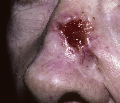
Ulcers
Ulcers are rounded or irregularly shaped excavations that result from complete loss of the epidermis plus some portion of the dermis. They vary in diameter from a few millimeters to several centimeters ( Fig. 2.7 ). Ulcers may be shallow, involving little beyond the epidermis, as in dystrophic epidermolysis bullosa, the base being formed by the papillary layer, or they may extend deeply into the dermis, subcutaneous tissues, or deeper, as with leg ulcers. Ulcers heal with scarring.