Contact Dermatitis
There are two types of dermatitis caused by substances coming in contact with the skin: irritant dermatitis and allergic contact dermatitis. Irritant dermatitis is an inflammatory reaction in the skin resulting from exposure to a substance that causes an eruption in most people who come in contact with it. Allergic contact dermatitis is an acquired sensitivity to various substances that produce inflammatory reactions only in those persons who have been previously sensitized to the allergen.
Irritant Contact Dermatitis
Many substances act as irritants that produce a nonspecific inflammatory reaction of the skin. This type of dermatitis may be induced in any person if there is contact with a sufficiently high concentration. No previous exposure is necessary, and the effect is evident within minutes, or a few hours at most. The concentration and type of toxic agent, duration of exposure, and condition of the skin at the time of exposure produce the variation in severity of the dermatitis from person to person, or from time to time in the same person. The skin may be more vulnerable because of maceration from excessive humidity or exposure to water, heat, cold, pressure, or friction. Dry skin, as opposed to wet skin, is less likely to react to contactants, although in chronic xerosis, as seen in elderly patients, increased sensitivity to irritants results. Thick skin is less reactive than thin skin. Atopic patients are predisposed to irritant hand dermatitis. Repeated exposure to some of the milder irritants may produce a hardening effect over time. This process makes the skin more resistant to the irritant effects of a given substance. Symptomatically, pain and burning are more common in irritant dermatitis, contrasting with the usual itch of allergic reactions. Avoidance, substitution of nonirritating agents when possible, and protection, most often by wearing gloves or using barrier creams, are the mainstays of treatment.
Alkalis
Irritant dermatitis is often produced by alkalis such as soaps, cement, detergents, bleaches, ammonia preparations, lye, drain pipe cleaners, and toilet bowl and oven cleansers ( Fig. 6.1 ). Alkalis penetrate and destroy deeply because they dissolve keratin. Strong solutions are corrosive, and immediate application of a weak acid such as vinegar, lemon juice, or 0.5% hydrochloric acid solution will lessen their effects.
The principal compounds are sodium, potassium, ammonium, and calcium hydroxides. Occupational exposure is frequent among workers in soap manufacturing. Sodium silicate (water glass) is a caustic used in soap manufacture and paper sizing and for the preservation of eggs. Alkalis in the form of soaps, bleaching agents, detergents, and most household cleansing agents figure prominently in the causes of hand eczema. Alkaline sulfides are used as depilatories. Calcium oxide (quicklime) forms slaked lime when water is added. Severe burns may be caused in plasterers.
Acids
The powerful acids are corrosive, whereas the weaker acids are astringent. Hydrochloric acid produces burns that are less deep and more liable to form blisters than injuries from sulfuric and nitric acids ( Fig. 6.2 ). Hydrochloric acid burns are encountered in those who handle or transport the product and in plumbers and those who work in galvanizing or tin-plate factories. Sulfuric acid produces a brownish charring of the skin, beneath which is an ulceration that heals slowly. Sulfuric acid is used more widely than any other acid in industry; it is handled principally by brass and iron workers and by those who work with copper or bronze. Nitric acid is a powerful oxidizing substance that causes deep burns; the tissue is stained yellow. Such injuries are observed in those who manufacture or handle the acid or use it in the making of explosives in laboratories. At times, nitric acid or formic acid is used in assaults secondary to interpersonal conflicts, resulting in scarring most prominently of the face, with the complication of renal failure present in a small number of cases.
Hydrofluoric acid is used widely in rust remover, in the semiconductor industry, and in germicides, dyes, plastics, and glass etching. It may act insidiously at first, starting with erythema and ending with vesiculation, ulceration, and finally necrosis of the tissue. Hydrofluoric acid is one of the strongest inorganic acids, capable of dissolving glass. Hypocalcemia, hypomagnesemia, hyperkalemia, and cardiac dysrhythmias may complicate hydrofluoric acid burns. Fluorine is best neutralized with hexafluorine solution, followed by 10% calcium gluconate solution or magnesium oxide.
Oxalic acid may produce paresthesia of the fingertips, with cyanosis and gangrene. The nails become discolored yellow. Oxalic acid is best neutralized with limewater or milk of magnesia to produce precipitation. Titanium hydrochloride is used in the manufacture of pigments. Application of water to the exposed part will produce severe burns. Therefore treatment consists only of wiping away the noxious substance.
Phenol (carbolic acid) is a protoplasmic poison that produces a white eschar on the surface of the skin. It can penetrate deep into the tissue. If a large surface of the skin is treated with phenol for cosmetic peeling effects, the absorbed phenol may produce glomerulonephritis and arrhythmias. Locally, temporary anesthesia may also occur. Phenol is readily neutralized with 65% ethyl or isopropyl alcohol.
Chromic acid burns, which may be seen in electroplating and dye production occupations, may result in extensive tissue necrosis and acute renal damage. Excision of affected skin down to the fascia should be accomplished rapidly, and hemodialysis to remove circulating chromium should start in the first 24 hours. Other strong acids that are irritants include acetic, trichloracetic, arsenious, chlorosulfonic, fluoroboric, hydriodic, hydrobromic, iodic, perchloric, phosphoric, salicylic, silicofluoric, sulfonic, sulfurous, tannic, and tungstic acids.
Treatment of acid burns consists of immediate rinsing with copious amounts of water and alkalization with sodium bicarbonate, calcium hydroxide (limewater), or soap solutions. Phosphorus burns should be rinsed off with water, followed by application of copper sulfate to produce a precipitate.
Airbag Dermatitis
Airbags are deployed as a safety feature on cars when rapid deceleration occurs. Activation of a sodium azide and cupric oxide propellant cartridge releases nitrogen gas, which expands the bag at speeds exceeding 160 km/hour (96 miles/hour). Talcum powder, sodium hydroxide, and sodium carbonate are released into the bag. Abrasions, thermal, friction, and chemical burns and an irritant contact dermatitis may result. Superficial erythema may respond well to topical steroids, but full-thickness burns may occur and require debridement and grafting.
Other Irritants
Metal salts that act as irritants include the cyanides of calcium, copper, mercury, nickel, silver, and zinc and the chlorides of calcium and zinc. Bromine, chlorine, fluorine, and iodine are also irritants. Occupational exposure to methyl bromide may produce erythema and vesicles in the axillary and inguinal areas. Insecticides, including 2,2-dichlorovinyl dimethyl phosphate used in roach powder and fly repellents and killers, can act as irritants.
Fiberglass Dermatitis
Fiberglass dermatitis is seen after occupational or inadvertent exposure. The small spicules of glass penetrate the skin and cause severe irritation with tiny erythematous papules, scratch marks, and intense pruritus. Usually, there is no delayed hypersensitivity reaction. Wearing clothes that have been washed together with fiberglass curtains, handling air conditioner filters, or working in the manufacture of fiberglass material may produce severe folliculitis, pruritus, and eruptions that may simulate scabies or insect bites. Fiberglass is also used in thermal and acoustic installation, the wind industry, padding, vibration isolation, curtains, draperies, insulation for automobile bodies, furniture, gasoline tanks, and spacecraft. Talcum powder dusted on the flexure surfaces of the arms before exposure makes the fibers slide off the skin. A thorough washing of the skin after handling fiberglass is helpful. Patch testing to epoxy resins should be done when evaluating workers in fiberglass and reinforced-plastics operations, because an allergic contact dermatitis may be difficult to discern from fiberglass dermatitis.
Dusts
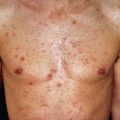
Some dusts and gases may irritate the skin in the presence of heat and moisture, such as perspiration. The dusts of lime, zinc, and arsenic may produce folliculitis. Dusts from various woods, such as teak, may incite itching and dermatitis. Dusts from cinchona bark, quinine, and pyrethrum produce widespread dermatitis. Tobacco dust in cigar factories, powdered orris root, lycopodium, and dusts of various nutshells may cause swelling of the eyelids and dermatitis of the face, neck, and upper extremities, the distribution of an airborne contact dermatitis. Dusts formed during the manufacture of high explosives may cause erythematous, vesicular, and eczematous dermatitis that may lead to generalized exfoliative dermatitis.
Capsaicin
Hand irritation produced by capsaicin in hot peppers used in Korean and North Chinese cuisine (Hunan hand) may be severe and prolonged, sometimes necessitating stellate ganglion blockade and gabapentin. Pepper spray, used by police in high concentrations and by civilians in less concentrated formulas, contains capsaicin and may produce severe burns. Cold water is not much help; capsaicin is insoluble in water. Acetic acid 5% (white vinegar) or antacids (Maalox) may completely relieve the burning, even if applied an hour or more after the contact. Application should be continued until the area can be dried without return of the discomfort.
Tear Gas Dermatitis
Lacrimators such as chloroacetophenone in concentrated form may cause dermatitis, with a delayed appearance about 24–72 hours after exposure. Irritation or sensitization, with erythema and severe vesiculation, may result. Treatment consists of lavage of the affected skin with sodium bicarbonate solution and instillation of boric acid solution into the eyes. Contaminated clothing should be removed.
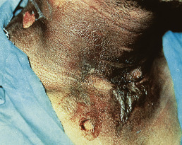
Sulfur mustard gas, also known as yperite (dichlorodiethyl sulfide), has been used in chemical warfare. Erythema, vesicles, and bullae result from mild to moderate exposure ( Fig. 6.3 ). Toxic epidermal necrolysis (TEN)–like appearance may follow more concentrated contact. The earliest and most frequently affected sites are areas covered by clothing and humidified by sweat, such as the groin, axillae, and genitalia.
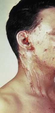
Mace is a mixture of tear gas (chloroacetophenone) in trichloroethane and various hydrocarbons resembling kerosene. It is available in a variety of self-defense sprays. Mace is a potent irritant and may cause allergic sensitization ( Fig. 6.4 ). Treatment consists of changing clothes, then washing with oil or milk, followed by washing with copious amounts of water.
Chloracne
Workers in the manufacture of chlorinated compounds may develop chloracne, with small, straw-colored follicular plugs and papules, chiefly on the malar crescent, retroauricular areas, earlobes, neck, shoulders, and scrotum. Histologically, there is a loss of sebaceous glands and the formation of cystic structures. The synthetic waxes chloronaphthalene and chlorodiphenyl, used in the manufacture of electric insulators and in paints, varnishes, and lacquers, predispose workers engaged in the manufacture of these synthetic waxes to chloracne. Exposure to 2,6-dichlorobenzonitrile during the manufacture of a herbicide, and to 3,4,3′,4′-tetrachloroazooxybenzene, which is an unwanted intermediate byproduct in the manufacture of a pesticide, may also produce chloracne.
A contaminant in the synthesis of herbicides and hexachlorophene, 2,3,7,8-tetracholorodibenzo- p -dioxin, produces a chemical burn in the acute stage, but chloracne, hyperpigmentation, hirsutism, and skin fragility (with or without criteria for porphyria cutanea tarda) are manifestations of chronic toxicity. Gastrointestinal tract cancer and malignancies of the lymphatic and hematopoietic systems are suspected to result. Although direct contact is the usual method of exposure, inhalation, ingestion, or contact with contaminated clothing may also result in chloracne. Chloracne may persist for long periods because dioxin is stored in the liver and released slowly into the circulation. Treatment is with medications used in acne vulgaris, including isotretinoin.
Hydrocarbons
Many hydrocarbons produce skin eruptions. Crude petroleum causes generalized itching, folliculitis, or acneiform eruptions. The irritant properties of petroleum derivatives are directly proportional to their fat-solvent properties and inversely proportional to their viscosity. Oils of the naphthalene series are more irritating than those of the paraffin series. Refined fractions from petroleum are less irritating than the unrefined products, although benzene, naphtha, and carbon disulfide may cause a mild dermatitis.
Lubricating and cutting oils are causes of similar cutaneous lesions. They represent a frequent cause of occupational dermatoses in machine tool operators, machinists, layout men, instrument makers, and setup men. Insoluble (neat) cutting oils are responsible for a follicular acneiform eruption on the dorsa of the hands, the forearms, face, thighs, and back of the neck. Hyperpigmentation, keratoses, and scrotal cancer have been found in those exposed to insoluble cutting oils. Soluble oils and synthetic fluids used in metalworking do not result in acne, but rather an eczematous dermatitis, usually of the dorsal forearms and hands. Approximately 50% of the time it is irritant and in the remainder it is allergic. Allergic contact dermatitis arises from various additives, such as biocides, coloring agents, and deodorizers.
Coal briquette makers develop dermatitis as a result of a tarry residue from petroleum used in their trade. Paraffin exposure leads to pustules, keratoses, and ulcerations. Shale oil workers develop an erythematous, follicular eruption that eventually leads to keratoses, which may become the sites of carcinoma. It is estimated that 50% of shale oil workers have skin problems.
Impure and low-grade paraffins and mineral oils cause similar skin eruptions. Initially, the skin changes are similar to those in chloracne. Over time, a diffuse erythema with dappled pigmentation develops. Gradually, keratoses appear, and after many years, some of these are the sites of carcinoma. Melanoderma may occur from exposure to mineral oils and lower-grade petroleum from creosote, asphalt, and other tar products. Photosensitization may play a role. Creosote is a contact irritant, sensitizer, and photosensitizer. Allergy is demonstrated by patch testing with 10% creosote in oil.
Petrolatum dermatitis may appear as a verrucous thickening of the skin caused by prolonged contact with impure petroleum jelly or, occasionally, lubricating oil. A follicular-centered process may occur in which erythematous horny nodules are present, usually on the anterior and inner aspects of the thighs. There are no comedones, and the lesions are separated by apparently normal skin.
Acne corne consists of follicular keratosis and pigmentation resulting from crude petroleum, tar oils, and paraffin. The dorsal aspects of the fingers and hands, the arms, legs, face, and thorax are the areas usually involved. The lesions are follicular horny papules, often black, and are associated at first with a follicular erythema and later with a dirty brownish or purplish spotty pigmentation, which in severe cases becomes widespread and is especially marked around the genitals. This syndrome may simulate pityriasis rubra pilaris or lichen spinulosus.
Coal tar and pitch and many of their derivatives produce photosensitization and an acneiform folliculitis of the forearms, legs, face, and scrotum. Follicular keratoses (pitch warts) may develop and later turn into carcinoma. Soot, lamp black, and the ash from peat fires produce dermatitis of a dry, scaly character, which over time forms warty outgrowths and cancer. Chimney sweep’s cancer occurs under a soot wart and is usually located on the scrotum, where soot, sebum, and dirt collect in the folds of the skin. This form of cancer has virtually disappeared.
Acquired perforating disease may occur in oil field workers who use drilling fluid containing calcium chloride. Patients develop tender, umbilicated papules of the forearms that microscopically show transepidermal elimination of calcium.
Solvents
The solvents cause approximately 10% of occupational dermatitis. When solvents are applied to the hands to cleanse them, the surface oil is dissolved, and a chronic fissured dermatitis results. Additionally, peripheral neuropathy and chemical lymphangitis may occur after the solvents are absorbed through the fissured skin. Solvent sniffers may develop an eczematous eruption around the mouth and nose; erythema and edema occur. This is a direct irritant dermatitis caused by the inhalation of the solvent placed on a handkerchief.
Trichloroethylene is a chlorinated hydrocarbon solvent and degreasing agent also used in the dry-cleaning and refrigeration industry. Inhalation may produce exfoliative erythroderma, mucous membrane erosions, eosinophilia, and hepatitis.
Allergic contact dermatitis caused by alcohol is rarely encountered with lower-aliphatic alcohols. A severe case of bullous and hemorrhagic dermatitis on the fingertips and deltoid region was caused by isopropyl alcohol. Although rare, ethyl alcohol dermatitis may also be encountered. Cetyl and stearyl alcohols may provoke contact urticaria.
Ale IS, et al: Irritant contact dermatitis. Rev Environ Health 2014; 29: 195.
Angelova-Fischer I: Irritants and skin barrier function. Curr Probl Dermatol 2016; 49: 80.
Bordel-Gomez MT, et al: Fiberglass dermatitis. Contact Dermatitis 2008; 59: 120.
Das KK, et al: Management of acid burns. Burns 2015; 41: 484.
Fartasch M: Wet work and barrier function. Curr Probl Dermatol 2016; 49: 144.
Flammiger A, et al: Sulfuric acid burns. Cutan Ocul Toxicol 2006; 25: 55.
Goon AT, et al: A case of trichloroethylene hypersensitivity syndrome. Arch Dermatol 2001; 137: 274.
Greenwood JE, et al: Alkalis and skin. J Burn Care Res 2016; 37: 135.
Herzemans-Boer M, et al: Skin lesions due to methyl bromide. Arch Dermatol 1988; 124: 917.
Jia X, et al: Adverse effects of gasoline on the skin of gasoline workers. Contact Dermatitis 2002; 46: 44.
Mostosi C, Simonart T: Effectiveness of barrier creams against irritant contact dermatitis. Dermatology 2016; 232: 353.
Patterson AT, et al: Skin diseases associated with Agent Orange and other organochlorine exposures. J Am Acad Dermatol 2016; 74: 143.
Saxena AK, et al: Multimodal approach for the management of Hunan hand syndrome. Pain Prac 2013; 13: 227.
Schep LJ, et al: Riot control agents. J R Army Med Corps 2015; 161: 94.
Steinritz D, et al: Medical documentation, bioanalytical evidence of an accidental human exposure to sulfur mustard and general therapy recommendations. Toxicol Lett 2016; 244: 112.
Tan CH, et al: Contact dermatitis. Clin Dermatol 2014; 32: 116.
Wang X, et al: A review of treatment strategies for hydrofluoric acid burns. Burns 2014; 40: 1447.
Wu JJ, et al: A case of air bag dermatitis. Arch Dermatol 2002; 138: 1383.
Yin S: Chemical and common burns in children. Clin Pediatr (Phila) 2017; 56: 8S.
Allergic Contact Dermatitis
Allergic contact dermatitis results when an allergen comes into contact with previously sensitized skin. It is caused by a specific acquired hypersensitivity of the delayed type, also known as cell-mediated (type IV) hypersensitivity. These sensitizers do not cause demonstrable skin changes on initial contact. Persons may be exposed to allergens for years before finally developing hypersensitivity. Genetic variability in the immunologic processes leading to sensitization and other factors, such as concentration of the allergen applied, its vehicle, timing and site of the exposure, presence of occlusion, age, gender, and race of the patient, and presence of other skin or systemic disorders, likely determine whether any given exposure will result in sensitization. Once sensitized, however, subsequent outbreaks may result from extremely slight exposure.
Childhood exposures do result in allergy, and the frequency of allergy in this age group is increasing. The most common relevant allergens in young children are nickel, cobalt, fragrance, lanolin, and neomycin. In adolescents potassium dichromate and Myroxylon pereirae become significant. Sensitivity is rarely lost over the years; older patients have similar rates of allergy as adults.
Occasionally, dermatitis may be induced when the allergen is taken internally by a patient first sensitized by topical application, as with substances such as cinnamon oil or various medications. The anamnestic response is termed systemic contact dermatitis. It may appear first at the site of the prior sensitization or past positive patch test, but may spread to a generalized morbilliform or eczematous eruption. Additional morphologic patterns include vesicular hand eczema, urticaria, erythema multiforme, vasculitis, or symmetric drug-related intertriginous and flexural exanthema (SDRIFE). Formerly called baboon syndrome, SDRIFE is a deep-red-violet eruption on the buttocks, genital area, inner thighs, and sometimes the axillae.
The most common causes of contact dermatitis in the United States are toxicodendrons (poison ivy, oak, or sumac), nickel, balsam of Peru (Myroxylon pereirae), neomycin, fragrance, formaldehyde and the formaldehyde-releasing preservatives, bacitracin, and rubber compounds. Frequent positive reactions to gold and thimerosal do not often correlate with the clinical exposure history. Gold reactions, which may be prolonged, can be correlated in some cases with oral gold exposure or occupational dermatitis, but in most cases, the relevance is questionable. Thimerosal reactions are probably related to its use as a preservative in common vaccines and skin-testing material. It also serves as a marker for piroxicam photosensitivity.
Eczematous delayed-type hypersensitivity reaction, as exemplified by allergic contact dermatitis and the patch test, must be distinguished from immediate-type hypersensitivity reaction. The latter presents within minutes of exposure with urticaria and is proved with a scratch test. It should be kept in mind, however, that persons who develop contact urticaria to a substance may concomitantly have a type IV delayed-type sensitization and eczema from the same allergen.
In some patients, impetigo, pustular folliculitis, and irritation or allergic reactions from applied medications are superimposed on the original dermatitis. A particularly vexing situation is when allergy to topical corticosteroids complicates an eczema, in which case the preexisting dermatitis usually does not flare, but simply does not heal as expected. The cutaneous reaction may also provoke a hypersusceptibility to various other, previously innocuous substances, which continues the eczematous inflammatory response indefinitely.
These eruptions resolve when the cause is identified and avoided. For acute generalized allergic contact dermatitis, treatment with systemic steroidal agents is effective, beginning with 40–60 mg/day of prednisone in a single oral dose, and tapering slowly to topical steroids. When the eruption is limited in extent and severity, local application of topical corticosteroid creams, lotions, or aerosol sprays is preferred.
Testing for Sensitivity
Patch Test.
The patch test is used to detect hypersensitivity to a substance that is in contact with the skin so that the allergen may be determined and corrective measures taken. So many allergens can cause allergic contact dermatitis that it is impossible to test a person for all of them. In addition, a good history and observation of the pattern of the dermatitis, its localization on the body, and its state of activity are helpful in determining the cause. The patch test is confirmatory and diagnostic, but only within the framework of the history and physical findings; it is rarely helpful if it must stand alone. Interpretation of the relevance of positive tests and the subsequent education of patients are challenging in some cases. The Contact Allergen Management Program (CAMP) provides names of alternative products that may be used by patients when an allergen is identified. This is available through the American Contact Dermatitis Society.
The patch test consists of application of substances suspected to be the cause of the dermatitis to intact uninflamed skin. Patch testing may be administered by the thin-layer rapid-use epicutaneous (TRUE) test or by individually prepared patches. The TRUE test has resulted in more screening for allergic contact dermatitis than in the past, but if it does not reveal the allergen for a highly suspect dermatitis, testing with an expanded series will on average yield relevant allergens in more than half of these patients. Dermatitis originating in the workplace will almost always require individualized testing.
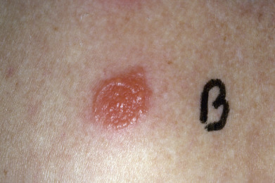
Test substances are applied usually to the upper back, although if only one or two are applied, the upper outer arm may be used. Each patch should be numbered to avoid confusion. The patches are removed after 48 hours (or sooner if severe itching or burning occurs at the site) and read. The patch sites need to be evaluated again at day 4 or 5 because positive reactions may not appear earlier. Some allergens may take up to day 7 to show a reaction, and the patient should be advised to return if such a delayed reaction occurs. Erythematous papules and vesicles with edema are indicative of allergy ( Fig. 6.5 ). Occasionally, patch tests for potassium iodide, nickel, or mercury will produce pustules at the site of the test application. Usually no erythema is produced; therefore the reaction has no clinical significance.
Strong patch test reactions may induce a state of hyperirritability (“excited skin syndrome”) in which adjacent tests that would otherwise be negative appear as weakly positive. Weakly positive tests in the presence of strong tests do not prove sensitivity. The skin and mucous membranes vary widely in the ability to react to antigens. The oral mucosa is more resistant to primary irritants and is less liable to be involved in allergic reactions. This may be because the keratin layer of the skin more readily combines with haptens to form allergens. Also, the oral mucosa is bathed in saliva, which cleanses and buffers the area and dilutes irritants. However, patch testing for various types of oral signs and symptoms, such as swelling, tingling and burning, perioral dermatitis, and the appearance of oral lichen planus, is useful in determining a cause in many cases.
Potent topical corticosteroids, ultraviolet (UV) light, prednisone, and the acquired immunodeficiency syndrome (AIDS) have been reported to interfere with the reliability of patch testing. Expert opinion regarding patch testing while on other immunosuppressants (e.g., methotrexate, azathioprine, biologics) is that these are less likely to produce unreliable testing. However, with all of these, false-negative reactions may result; the value of testing in such circumstances is that if a positive reaction occurs, a diagnosis may be made. Vitiliginous skin is less reactive than normally pigmented adjacent skin.
Provocative Use Test.
The provocative use test may be used to screen products used by the patient. Products that are made to stay on the skin once applied (as opposed to shampoos or soaps) are rubbed on normal skin of the inner aspect of the forearm several times daily for 5 days. A pink itchy patch will indicate the need to avoid the product. Further testing to its individual ingredients will help identify replacement products. This test may also confirm a positive closed patch test reaction to ingredients of the personal care product.
Photopatch Test.
The photopatch test is used to evaluate for contact photoallergy to such substances as sulfonamides, phenothiazines, p -aminobenzoic acid, oxybenzone, 6-methyl coumarin, musk ambrette, and tetrachlorosalicylanilide. A standard patch test is applied for 48 hours; this is then exposed to 5 to 15 J/m 2 of UVA and read after another 48 hours. To test for 6-methyl coumarin sensitivity, the patch is applied in the same manner but for only 30 minutes before light exposure, rather than for 48 hours. A duplicate set of nonirradiated patches is used in testing for the presence of routine delayed hypersensitivity reactions. Also, a site of normal skin is given an identical dose of UVA to test for increased sensitivity to light without prior exposure to chemicals. There is a steady increase in incidence of photoallergy to sunscreening agents and a decreasing incidence of such reactions to fragrance.
Regional Predilection
Familiarity with certain contactants and the typical dermatitis they elicit on specific parts of the body will assist in diagnosis of the etiologic agent.
Head and Neck.
The scalp is relatively resistant to the development of contact allergies; however, involvement may be caused by hair dye, hair spray, shampoo, or permanent wave solutions. The surrounding glabrous skin, including the ear rims and backs of the ears, may be much more inflamed and suggestive of the cause. Persistent otitis of the ear canal may be caused by sensitivity to neomycin, an ingredient of most aural medications. The eyelids are the most frequent site for nail polish dermatitis. Volatile gases, false-eyelash adhesive, fragrances, eye medications, preservatives, mascara, rubber in sponges used to apply cosmetics, and eye shadow are also frequently implicated ( Fig. 6.6 ). Perioral dermatitis and cheilitis may be caused by flavoring agents in dentifrices and gum, as well as fragrances, shellac, medicaments, and sunscreens in lipstick and lip balms. Perfume dermatitis may cause redness just under the ears or on the neck. Earlobe dermatitis is indicative of nickel sensitivity. Photocontact dermatitis may involve the entire face and may be sharply cut off at the collar line or extend down on to the sternum in a V shape. There is a typical clear area under the chin where there is little or no exposure to sunlight. The left cheek and left side of the neck (from sun exposure while driving) may be the first areas involved.
Trunk.
The trunk is an infrequent site; however, the dye or finish of clothing may cause dermatitis. The axilla may be the site of deodorant dermatitis and clothing-dye dermatitis; involvement of the axillary vault suggests the former; of the axillary folds, the latter. In women, brassieres cause dermatitis from the material itself, the elastic, or the metal snaps or underwires.
Arms.
The wrists may be involved because of jewelry or the backs of watches and clasps, all of which may contain nickel. Wristbands made of leather are a source of chrome dermatitis.
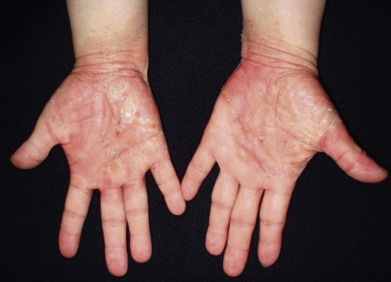
Hands.
Innumerable substances may cause allergic contact dermatitis of the hands, which typically occurs on the backs of the hands and spares the palms. Florists will often develop fingertip or palmar lesions. A hand dermatitis that changes from web spaces to fingertips or from palms to dorsal hands should trigger patch testing. Poison ivy and other plant dermatitides frequently occur on the hands and arms. Rubber glove sensitivity must be kept constantly in mind. Usually, irritancy is superimposed on allergic contact dermatitis of the hands, altering both the morphologic and histologic clues to the diagnosis.
Abdomen.
The abdomen, especially the waistline, may be the site of rubber dermatitis from the elastic in pants and undergarments. The metallic rivets in blue jeans may lead to periumbilical dermatitis in nickel-sensitive patients, as may piercings of the umbilicus.
Groin.
The groin is usually spared, but the buttocks and upper thighs may be sites of dermatitis caused by dyes. The penis is frequently involved in poison ivy dermatitis. Condom dermatitis may also occur. The perianal region may be involved from the “caine” medications in suppositories, as well as preservatives and fragrances in cleansing materials. Almost half of women with pruritus vulvae have one or more relevant allergens; often these are medicaments, fragrances, or preservatives.
Lower Extremities.
The shins may be the site of rubber dermatitis from elastic stockings. Feet are sites for shoe dermatitis, most often attributable to rubber sensitivity, chrome-tanned leather, dyes, or adhesives. Application of topical antibiotics to stasis ulcers frequently leads to sensitivity and allergic contact dermatitis.
Bangash HK, Petronic-Rosic V: Acral manifestations of contact dermatitis. Clin Dermatol 2017; 35: 9.
Bourke I, et al: Guidelines for the management of contact dermatitis. Br J Dermatol 2009; 160: 946.
Bryden AM, et al: Photopatch testing of 1155 patients. Br J Dermatol 2006; 155: 737.
Diepgen TL, et al: Guidelines for diagnosis, prevention and treatment of hand eczema—short version. J Dtsch Dermatol Ges 2015; 13: 77.
Feser A, et al: Periorbital dermatitis. Br J Dermatol 2008; 159: 858.
Jean SE, et al: Contact dermatitis in leg ulcer patients. J Cutan Med Surg 2009; 13: S38.
Kockentiet B, et al: Contact dermatitis in athletes. J Am Acad Dermatol 2007; 56: 1048.
Mahler V: Hand dermatitis—differential diagnoses, diagnostics, and treatment options. J Dtsch Dermatol Ges 2016; 14: 7.
Martin SF: Immunological mechanisms in allergic contact dermatitis. Curr Opin Allergy Clin Immunol 2015; 15: 124.
Mowad CM: Contact dermatitis. Dermatol Clin 2016; 34: 263.
Mowad CM, et al: Allergic contact dermatitis. J Am Acad Dermatol 2016; 74: 1029 and 1043.
Pelletier JL, et al: Contact dermatitis in pediatric. Pediatr Ann 2016; 45: e287.
Rashid RS, Shim TN: Contact dermatitis. BMJ 2016; 353: i3299.
Rodrigues DF, Goulart EM: Patch-test results in children and adolescents. An Bras Dermatol 2016; 91: 64.
Schalock PC, et al: American Contact Dermatitis Society Core Allergen Series. Dermatitis 2017; 28: 141.
Scheman A, et al: Contact allergy cross-reactions. Dermatitis 2017; 28: 128.
Schlosser BJ: Contact dermatitis of the vulva. Dermatol Clin 2010; 28: 697.
Thyssen JP, et al: The epidemiology of contact allergy in the general population. Contact Dermatitis 2007; 57: 287.
Torgerson RR, et al: Contact allergy in oral disease. J Am Acad Dermatol 2007; 57: 315.
Veien NK: Systemic contact dermatitis. Int J Dermatol 2011; 50: 1445.
Warshaw EM, et al: Positive patch test reactions in older individuals. J Am Acad Dermatol 2012; 66: 229.
Wentworth AB, Davis MD: Patch testing with the standard series when receiving immunosuppressive medications. Dermatitis 2014; 25: 195.
Winnicki M, et al: A systematic approach to systemic contact dermatitis and symmetric drug-related intertriginous and flexural exanthema (SDRIFE). Am J Clin Dermatol 2011; 12: 171.
Dermatitis Resulting From Plants
A large number of plants, including trees, grasses, flowers, vegetables, fruits, and weeds, are potential causes of dermatitis. Eruptions from them vary considerably in appearance but are usually vesicular and accompanied by marked edema. After previous exposure and sensitization to the active substance in the plant, the typical dermatitis results from reexposure. The onset is usually a few hours or days after contact. The characteristic linearly grouped lesions are probably produced by brushing the skin with a leaf edge or a broken twig or by carriage of the allergen under the nails. Contrary to popular belief, the contents of vesicles are not capable of producing new lesions.
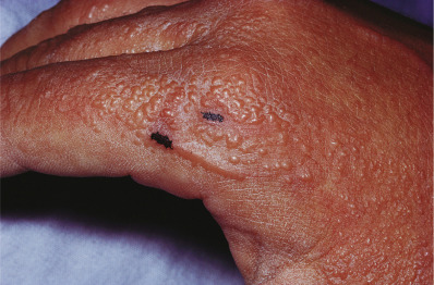
Toxicodendron (Poison Ivy).
Toxicodendron dermatitis includes dermatitis from members of the Anacardiaceae family of plants: poison ivy ( T. radicans, or Rhus radicans ), poison oak (T. diversilobum, R. diversaloba), poison sumac (T. vernix, R. vernix), Japanese lacquer tree, cashew nut tree (allergen in nutshell), mango (allergen in rind, leaves, or sap), Rengas tree, and Indian marking nut tree. The ginkgo (allergen in fruit pulp), spider flower or silver oak, Gluta species of trees and shrubs in Southeast Asia, Brazilian pepper tree, also known as Florida holly, and poisonwood tree contain almost identical antigens.
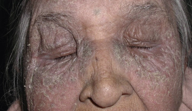
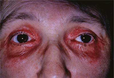
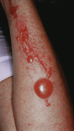
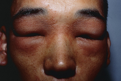
Toxicodendron dermatitis appears within 48 hours of exposure of a person previously sensitized to the plant. It usually begins on the backs of the fingers, interdigital spaces, wrists, and eyelids, although it may begin on the ankles or other parts that have been exposed. Marked pruritus is the first symptom; inflammation, vesicles, and bullae may then appear. The vesicles are usually grouped and often linear ( Fig. 6.7 ). Large bullae may be present, especially on the forearms and hands. The eyelids are puffy and worse in the morning, improving as the day progresses ( Fig. 6.8 ). Pruritus ani and involvement of the genital areas occur frequently. A black lacquer deposit may occur in which the sap of the plant has been oxidized after being bound to the stratum corneum ( Fig. 6.9 ). Untreated Toxicodendron dermatitis usually lasts 2–3 weeks.
The fingers transfer the allergen to other parts, especially the forearms and the male prepuce, which become greatly swollen. However, once the causative oil has been washed off, there is no spreading of the allergen. Some persons are so susceptible that direct contact is not necessary, the allergen apparently being carried by objects with prior exposure to the catechol. Occasionally, eating the allergen, as occurred in a patient who ingested raw cashew nuts in an imported pesto sauce, may result in SDRIFE (see earlier) or a systematized allergic contact dermatitis with the morphology of a generalized erythematous papular eruption.
The cause is an oleoresin known as urushiol, of which the active agent is a mixture of catechols. This and related resorcinol allergens are present in many plants and also in Philodendron species, wood from Persoonia elliptica, wheat bran, and marine brown algae.
The most striking diagnostic feature is the linearity of the lesions. It is rare to see vesicles arranged linearly except in plant-induced dermatitis. A history of exposure in the country or park to plants that have shiny leaves in groups of three, followed by the appearance of vesicular lesions within 2 days, usually establishes the diagnosis.
Eradication of plants having grouped “leaves of three” growing in frequented places is one easy preventive measure, as is recognition of the plants to avoid. An excellent resource is a pamphlet available from the American Academy of Dermatology. If the individual is exposed, washing with soap and water within 5 minutes may prevent an eruption. Protective barrier creams are available that are somewhat beneficial. Quaternium-18 bentonite has been shown to prevent or diminish experimentally produced poison ivy dermatitis.
Innumerable attempts have been made to immunize against poison ivy dermatitis by oral administration of the allergen or subcutaneous injections of oily extracts. To date, no accepted method of immunization is available. Repeated attacks do not confer immunity, although a single severe attack may achieve this by what has been called massive-dose desensitization.
When the diagnosis is clear and the eruption severe or extensive, systemic steroidal agents are effective, beginning with 40–60 mg of prednisone in a single oral dose daily, tapered off over a 3-week period. When the eruption is limited in extent and severity, local application of topical corticosteroid creams, lotions, or aerosol sprays is preferred. Time-honored calamine lotion without phenol is helpful and does no harm. Antihistaminic ointments should be avoided because of their sensitization potential. This also applies to the local application of the “caine” topical anesthetics.
Other Toxicodendron -Related Dermatitides.
Lacquer dermatitis is caused by a furniture lacquer made from the Japanese lacquer tree, used on furniture, jewelry, or bric-a-brac. Antique lacquer is harmless, but lacquer less than 1 or 2 years old is highly antigenic. Cashew oil is extracted from the nutshells of the cashew tree (Anacardium occidentale). This vesicant oil contains cardol, a phenol similar to urushiol in poison ivy. The liquid has many commercial applications, such as the manufacture of brake linings, varnish, synthetic glue, paint, and sealer for concrete.
Mango dermatitis is uncommon in natives of mango-growing countries (e.g., Philippines, Guam, Hawaii, Cuba) who have never been exposed to contact with Toxicodendron species. Many persons who have been so exposed, however, whether or not they had dermatitis from it, are sensitized by one or a few episodes of contact with the peel of the mango fruit. The palms carry the allergen, so the eyelids and the male prepuce are often early sites of involvement.
Ginkgo tree dermatitis simulates Toxicodendron dermatitis with its severe vesiculation, erythematous papules, and edema. The causative substances are ginkgolic acids from the fruit pulp of the ginkgo tree. Ingestion of the ginkgo fruit may result in perianal dermatitis. Ginkgo biloba given orally for cerebral disturbances is made from a leaf extract so it does not elicit a systemic contact allergy when ingested.
Flowers and Houseplants.
Among the more common houseplants, the velvety-leafed philodendron, Philodendron crystallinum (and its several variants), known in India as the “money plant,” is a frequent cause of contact dermatitis. The eruption is often seen on the face, especially the eyelids, carried there by hands that have watered or cared for the plant. English ivy follows philodendron in frequency of cases of occult contact dermatitis. Primrose dermatitis affects the fingers, eyelids, and neck with a punctate or diffuse erythema and edema. Formerly found most frequently in Europe, the primrose is now a common U.S. houseplant. Primin, a quinone, is the causative oleoresin abounding in the glandular hairs of the plant Primula obconica.
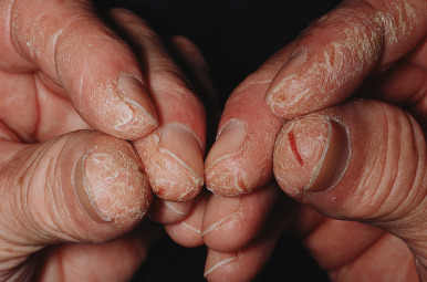
The popular cut flower, the Peruvian lily, is the most common cause of allergic contact dermatitis in florists. When handling flowers of the genus Alstroemeria, the florist uses the thumb and second and third digits of the dominant hand. Because it is chronic, fissured hyperkeratotic dermatitis results, identical to the “tulip fingers” seen among sensitized tulip workers (see Fig. 6.10 ). Testing is done with the allergen tuliposide A. It does not penetrate nitrile gloves. The geranium, scorpion flower ( Phacelia crenulata or P. campanularia ), hydrangea, creosote bush (Larvia tridentata), Heracula, daffodil, foxglove, pooja, lisianthus, lilac, lady slipper, magnolia, and tulip and narcissus bulbs are other flowers that commonly cause allergic reactions among florists. The poinsettia and oleander almost never cause dermatitis, despite their reputation for it, although they are toxic if ingested.
Chrysanthemums frequently cause dermatitis, with the hands and eyelids of florists most often affected. The α-methylene portion of the sesquiterpene lactone molecule is the antigenic site, as it is in the other genera of the Compositae family.
A severe inflammatory reaction with bulla formation may be caused by the prairie crocus ( Anemone patens L.), the floral emblem of the province of Manitoba. Several species of ornamental “bottle brush” from Queensland (Grevillea banksii, G. Robyn Gordon, G. robusta), may cause allergic contact dermatitis. It is exported to the United States and other Western countries. The allergen is a long-chain alkyl resorcinol. Cross-sensitivity to Toxicodendron has been demonstrated. Treatment of all these plant dermatitides is the same as that recommended for toxicodendron dermatitis.
Contact dermatitis may be caused by handling many other flowers, such as the geranium, scorpion flower ( Phacelia crenulata or P. campanularia ), hydrangea, creosote bush (Larvia tridentata), Heracula, daffodil, foxglove, lilac, lady slipper, magnolia, and tulip and narcissus bulbs. The poinsettia and oleander almost never cause dermatitis, despite their reputation for it, although they are toxic if ingested.
Parthenium hysterophorus is a photosensitizing weed. The well-deserved reputation for harmfulness of dieffenbachia, a common, glossy-leafed house plant, rests on the high content of calcium oxalate crystals in its sap, which burn the mouth and throat severely if any part of the plant is chewed or swallowed. Severe edema of the oral tissues may result in complete loss of voice, thus its common nickname, “dumb cane.” It does not appear to sensitize. The castor bean, the seed of Ricinus communis, contains ricin, a poisonous substance (phytotoxin). Its sap contains an antigen that may cause anaphylactic hypersensitivity and also dermatitis.
Fruit and Vegetables.
Many vegetables may cause contact dermatitis, including asparagus, carrot, celery, cow-parsnip, cucumber, garlic, Indian bean, mushroom, onion, parsley, tomato, and turnip. Onion and celery, among other vegetables, have been incriminated in the production of contact urticaria and even anaphylaxis. Several plants, including celery, fig, lime, and parsley, can cause a phototoxic dermatitis because of the presence of psoralens. Phototoxic contact dermatitis from plants is discussed more fully in Chapter 3 .
Trees.
Trees with timber and sawdust that may produce contact dermatitis include ash, birch, cedar, cocobolo, elm, Kentucky coffee tree, koa, mahogany, mango, maple, mesquite, milo, myrtle, pine, and teak. The latex of fig and rubber trees may also cause dermatitis, usually of the phototoxic type. Melaleuca oil (tea tree oil), which may be applied to the skin to treat a variety of maladies, can cause allergic contact dermatitis, primarily through the allergen D-limonene ( Fig. 6.11 ). The exotic woods, especially cocobolo and rosewood are prominent among allergens that may produce erythema multiforme (EM) after cutaneous exposure. Toxicodendron, tea tree oil, various medicaments, and a variety of other allergens may induce this reaction.
Tree-Associated Plants.
Foresters and lumber workers can be exposed to allergenic plants other than trees. Lichens are a group of plants composed of symbiotic algae and fungi. Foresters and wood choppers exposed to these lichens growing on trees may develop severe allergic contact dermatitis. Exposure to the lichens may also occur from firewood, funeral wreaths, and also fragrances added to aftershave lotions (oak moss and tree moss). Sensitization is produced by d -usnic acid and other lichen acids contained in lichens. The leafy liverwort (Frullania nisquallensis), a forest epiphyte growing on tree trunks, has produced allergic dermatitis in forest workers. The eruption is commonly called “cedar poisoning.” It resembles Toxicodendron dermatitis; its attacks are more severe during wet weather. The allergen is sesquiterpene lactone.
Pollens and Seeds.
The pollens in ragweed are composed of two antigens. The protein fraction causes the respiratory symptoms of asthma and hay fever, and the oil-soluble portion causes contact dermatitis. Ragweed oil dermatitis is a seasonal disturbance seen mainly during the ragweed growing season from spring to fall. Contact with the plant or with wind-blown fragments of the dried plant produces the typical dermatitis. The oil causes swelling and redness of the lids and entire face, and a red blotchy eruption on the forearms that, after several attacks, may become generalized, with lichenification. It closely resembles chronic atopic dermatitis, with lichenification of the face, neck, and major flexures, and severe pruritus. The distribution also mimics that of photodermatitis, with ragweed dermatitis differentiated by its involvement of the upper eyelids and the retroauricular and submental areas. Chronic cases may continue into the winter, although signs and symptoms are most severe at the height of the season. Sesquiterpene lactones are the cause. Coexistent sensitization to pyrethrum may account for prolongation of ragweed dermatitis. Men outnumber women in hypersensitivity reactions; farmers outnumber patients of all other occupations.
Marine Plants.
Numerous aquatic plants are toxic or produce contact dermatitis. Algae are the worst offenders. Freshwater plants are rarely of concern. Seaweed dermatitis is a type of swimmer’s eruption produced by contact with a marine blue-green alga, Lyngbya majuscula Gomont. The onset is within a few minutes of leaving the ocean, with severe itching and burning, followed by dermatitis, blisters, and deep, painful desquamation that affects the areas covered by the bathing suit, especially the scrotum, perineum, and perianal areas and occasionally the breasts in women). Patch tests with the alga are neither necessary nor helpful because it is a potent irritant. Bathing in fresh water within 10 or 15 minutes of leaving the ocean may prevent the dermatitis. The Bermuda fire sponge may produce contact erythema multiforme. Trawler fishermen in the Dogger Bank area of the North Sea develop allergic dermatitis after contact with Alcyonidium hirsutum. This seaweed-like animal colony becomes caught in nets and produces erythema, edema, and lichenification on the fishermen’s hands and wrists.
Plant-Associated Dermatitis.
The residua of various insecticides on plants may also produce dermatitis. This is especially true of sprays containing arsenic and malathion. Randox (2-chloro- N,N -diallyl-acetamide) has been reported as the cause of hemorrhagic bullae on the feet of farmers. Lawn care companies spray herbicides and fungicides throughout the spring, summer, and fall. Dryene, thiuram, carbamates, and chlorothalonil are potential sensitizers in these workers, whose clothing frequently becomes wetted while spraying.
Barbs, bristles, spines, thorns, spicules, and cactus needles are some of the mechanical accessories of plants that may produce dermatitis. Sabra dermatitis is an occupational dermatitis resembling scabies. It is seen among pickers of the prickly pear cactus plant. It also occurs in persons handling Indian figs in Israel, where the condition is seen from July to November. The penetration of minute, invisible thorns into the skin is the cause. Agave americana is a low-growing plant used for ornamental purposes in many southwestern U.S. communities. Trimming during landscaping can induce an irritant dermatitis caused by calcium oxalate crystals. The stinging nettle is a common weed that bears tiny spines with biologically active substances such as histamine that produce itching and urticaria within minutes of contact.
Plant Derivatives.
Sensitizing substances derived from plants are found in the oleoresin fractions that contain camphors, essential oils, phenols, resins, and terpenes. The chief sensitizers are the essential oils. These may be localized in certain parts of the plant, such as in the peel of citrus fruits, leaves of the eucalyptus tree, and bark of the cinnamon tree. Aromatherapy, an increasingly popular treatment for relief of stress, involves either inhaling or massaging with essential oils; this may cause allergic contact dermatitis in therapists or clients. Exposure to botanical extracts through many cosmetics and homeopathic remedies has resulted in an increasing number of reports of allergic contact sensitivity to individual ingredients, especially tea tree oil.
Cinnamon oil ( Cassia oil) is a common flavoring agent, especially in pastries. Hand dermatitis in pastry bakers is often caused by cinnamon. It is also used as a flavor for lipstick, bitters, alcoholic and nonalcoholic beverages, toothpaste, and chewing gum. Perioral dermatitis may be caused by cinnamon in chewing gum. A 5% cinnamon solution in olive oil is used for patch testing. Eugenol, clove oil, and eucalyptus oil are used by dentists, who may acquire contact dermatitis from them. Anise, peppermint, and spearmint oils may cause sensitization.
Nutmeg, paprika, and cloves are causes of spice allergy. Fragrance mix is a useful indicator allergen. Lemon oil from lemon peel or lemon wood may cause sensitization in the various handlers of these substances. Citric acid may cause dermatitis in bakers. Lime oil in lime-scented shaving cream or lotion may cause photoallergy. Myroxylon pereirae contains numerous substances, including essential oils similar to the oil of lemon peel. It is known to cross-react with vanilla, cinnamon, and many other substances. Vanillin is derived from the vanilla plant and frequently produces contact dermatitis, vanillism, in those connected with its production and use.
Turpentine frequently acts as an irritant and as an allergic sensitizer (carene). It is contained in paints, paint thinners, varnishes, and waxes.
Arberer W: Contact allergy and medicinal plants. J Dtsch Dermatol Ges 2008; 6: 15.
Crawford GH, et al: Use of aromatherapy products and increased risk of hand dermatitis in massage therapists. Arch Dermatol 2004; 140: 991.
de Groot AC, Schmidt E: Tea tree oil. Contact Dermatitis 2016; 75: 129.
Ferreira O, et al: Erythema multiforme–like lesions revealing allergic contact dermatitis to exotic woods. Cutan Ocul Toxicol 2012; 31: 61.
Foti C, et al: Occupational contact dermatitis caused by Eustoma exaltatum russellianum (lisianthus). Contact Dermatitis 2014; 71: 59.
Ghorpade A: Contact dermatitis caused by Indian marking nut juice used to relieve ankle pain. Int J Dermatol 2014; 53: 117.
Gladman AC: Toxicodendron dermatitis. Wilderness Environ Med 2006; 17: 120.
Hershko K, et al: Exploring the mango–poison ivy connection. Contact Dermatitis 2005; 52: 3.
Higgins C, et al: Eucalyptus oil. Contact Dermatitis 2015; 72: 344.
Jack AR, et al: Allergic contact dermatitis to plant extracts in cosmetics. Semin Cutan Med Surg 2013; 32: 140.
Lakshmi C: Fingertip eczema to pooja flowers. Indian J Dermatol Venereol Leprol 2015; 81: 514.
Linares T, et al: Phytodermatitis caused by Agave americana. Allergol Immunopathol (Madr) 2011; 39: 184.
McClanahan C, et al: Black spot poison ivy. Int J Dermatol 2014; 53: 752.
Sharma VK, et al: Parthenium dermatitis. Photochem Photobiol Sci 2013; 12: 85.
Swinnen I, et al: An update on airborne contact dermatitis: 2007–2011. Contact Dermatitis 2013; 68: 232.
Veien NK: Systemic contact dermatitis. Int J Dermatol 2011; 50: 1445.
Verma P, et al: Severe marking-nut dermatitis. Dermatitis 2012; 23: 293.
Dermatitis From Clothing
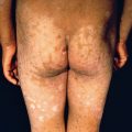
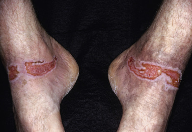
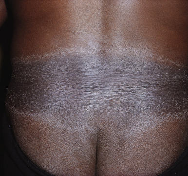
A predisposition to contact dermatitis from clothing occurs in persons who perspire freely or who are obese and wear clothing that tends to be tight. Depending on the offending substance, various regions of the body will be affected. Regional location is helpful in identifying the sensitizing substance. The axillary folds are often involved; the vaults of the axillae are usually spared. Sites of increased perspiration and sites where evaporation is impeded, such as the intertriginous areas, will tend to leach dyes from fabrics to produce dermatitis. Areas where the material is tight against the skin, such as the waistband or neck, are frequently involved ( Fig. 6.12 ). The thighs are affected when pants contain the offending allergen. The hands, face, and undergarment sites are usually spared, but otherwise these reactions may be scattered and generalized. Secondary changes of lichenification and infection occur frequently because of the chronicity of exposure.
Cotton, wool, linen, and silk fabrics were used exclusively before the advent of synthetic fabrics. Most materials are now blended in definite proportions with synthetics to produce superior lasting and esthetic properties. Dermatitis from cotton is virtually nonexistent. In most cases, there is no true sensitization to wool. Wool acts as an irritant because of the barbs on its fibers. These barbs may produce severe pruritus at points of contact with the skin, especially in the intertriginous areas. In persons with sensitive skin, such as those with atopic dermatitis, the wearing of wool is not advisable because of its mechanical irritative properties. Silk is a sensitizer, but rarely; the nature of the allergen is not known. Many patients believe their detergent is the source of a dermatitis, but this is rarely the case.
Numerous synthetic fibers are available for clothing and accessory manufacture, all of which again are remarkably free of sensitizing properties. Polyvinyl resins are the plastics used in such apparel as raincoats, rainhoods, wristbands, suspenders, plastic mittens, and gloves. These also are only infrequently found to be causes of contact dermatitis.
The most common causes of clothing dermatitis are the fabric finishers, dyes, and rubber additives. Fabric finishers are used to improve the durability, appearance, and feel of a material. Antiwrinkling and crease-holding chemicals are mostly resins, which are incorporated into the fibers as they are being manufactured or applied to the finished fabric. Fabrics are treated to make them less vulnerable to the effects of perspiration and ironing. Clothing may be treated with these substances to make it dry rapidly after washing. They are used to make clothing fabrics shrink resistant and water and stain repellent. When all these uses are taken into consideration, the low incidence of dermatitis from these formaldehyde resin materials is remarkable.
Ethylene urea melamine formaldehyde resin and dimethylol dihydroxyethylene urea formaldehyde resin are the best screening agents. Many persons also react to formaldehyde and the formaldehyde-releasing preservatives such as quaternium 15. Avoidance of exposure of the skin to formaldehyde resin is most difficult. New clothes should be thoroughly washed twice before wearing the first time. Even with this precaution, however, allergens may still be present in sufficient quantities to continue the dermatitis. T-shirts, sweat-shirts and pants, white underclothes suitable for bleaching, and garments of mixed synthetic fibers with cotton fibers added to make them drip-dry are most likely to cause problems in these patients.
Synthetic fabrics such as polyester and acetate liners in women’s clothing are also prime causes, and women are more affected than men. Even infants may be affected, however, with dyes in diapers accounting for some cases. Many patients do not react to paraphenylene diamine, but only to the disperse dye allergens. The best screening agents are disperse blue 106 and 124. Lymphomatoid contact allergy may result from clothing dye reactivity.
Suspected fabrics may be soaked in water for 15 minutes and applied under a patch for 72–96 hours. Jeans, Spandex, silk, 100% linen, 100% nylon, and 100% cotton that is not wrinkle resistant or colorfast are best tolerated. Spandex is a nonrubber (but elastic) polyurethane fiber. It is widely used for garments such as girdles, brassieres, and socks, but is generally safe in the United States because it is free of rubber additives.
Carlson RM, et al: Diagnosis and treatment of dermatitis due to formaldehyde resin in clothing. Dermatitis 2004; 15: 169.
Coman G, et al: Textile allergic contact dermatitis. Rev Environ Health 2014; 29: 163.
Donovan J, et al: Allergic contact dermatitis from formaldehyde textile resins in surgical uniforms and nonwoven textile masks. Dermatitis 2007; 18: 40.
Malinauskiene L, et al: Contact allergy from disperse dyes in textiles. Contact Dermatitis 2013; 68: 65.
Mobolaji-Lawal M, Nedorost S: The role of textiles in dermatitis. Curr Allergy Asthma Rep 2015; 15: 17.
Narganes LM, et al: Lymphomatoid dermatitis caused by contact with textile dyes. Contact Dermatitis 2013; 68: 62.
Reich HC, et al: Allergic contact dermatitis from formaldehyde textile resins. Dermatitis 2010; 21: 65.
Zug KA, et al: The value of patch testing patients with a scattered generalized distribution of dermatitis. J Am Acad Dermatol 2008; 59: 426.
Shoe Dermatitis
Footwear dermatitis may begin on the dorsal surfaces of the toes and may remain localized to that area indefinitely ( Fig. 6.13 ). There is erythema, lichenification, and, in severe cases, weeping and crusting. Secondary infection is frequent. In severe cases, an id reaction may be produced on the hands, similar to the reaction from fungal infection of the feet. A diagnostic point is the normal appearance of the skin between the toes, which has no contact with the offending substance. In fungal infections, the toe webs are usually involved. Another pattern seen is involvement of the sole with sparing of the instep and flexural creases of the toes. Also, purpuric reactions may occur to components of black rubber mix. Hyperhidrosis and atopy predispose to development of shoe allergy.
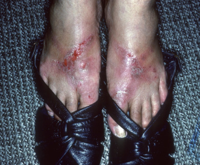
Shoe dermatitis is most frequently caused by the rubber accelerators mercaptobenzothiazole, carbamates, and tetramethylthiuram disulfide. Potassium dichromate in leather and the adhesives used in synthetic materials (especially p -tert-butylphenol formaldehyde resin) are also common shoe allergens. Diisocyanates are used in making foam rubber padding for athletic shoes and may cause allergy. Dimethyl fumarate is a preservative used in antihumidity sachets. It is a volatile substance and may deposit on shoes during its transport. Dimethyl fumarate is highly allergenic, and several outbreaks of shoe dermatitis in Europe have occurred secondary to this allergen. Other causative agents are felt, cork liners, formaldehyde, dyes, asphalt, and tar. Shoe refresher sprays may also induce allergy. Patch testing with pieces of various shoe parts may be done by soaking them for 15 minutes in water and applying them to the back for 72–96 hours. Once the allergen has been identified, selection of shoes without the offending substance will lead to resolution. Unfortunately, this is a difficult process, because most shoes are made in areas without mandatory labeling requirements, and plastic, wooden, or fabric shoes that contain fewer allergens are often impractical.
Castando-Tardan MP, et al: Allergic contact dermatitis to Crocs. Contact Dermatitis 2008; 58: 248.
Matthys E, et al: Shoe allergic contact dermatitis. Dermatitis 2014; 25: 163.
Mowitz M, Pontén A: Foot dermatitis caused by didecyldimethylammonium chloride in a shoe refresher spray. Contact Dermatitis 2015; 73: 374.
Švecová D, et al: Footwear contact dermatitis from dimethyl fumarate. Int J Dermatol 2013; 52: 803.
Washaw EM, et al: Shoe allergens. Dermatitis 2007; 18: 191.
Dermatitis From Metals and Metal Salts
Metal dermatitis is most frequently caused by nickel and chromates. Usually, with the exception of nickel, the pure metals generally do not cause hypersensitivity; only when they are incorporated into salts do they cause reactions. Most objects containing metal or metal salts are combinations of several metals, some of which may have been used to plate the surface, thereby enhancing its attractiveness, durability, or tensile strength. For this reason, suspected metal-caused dermatitis should be investigated by doing patch tests to several of the metal salts.
Patients have developed a variety of dermatoses, most often eczematous in type, after placement of an orthopedic, gynecologic, or dental implant or a pacemaker/defibrillator or endovascular device. When patients are symptomatic with an eczematous process after implantation, patch testing will allow evaluation of allergy by testing with an extended tray, metals, and bone cement. A positive diagnosis of allergy at a minimum requires the appearance of a chronic dermatitis after placement, no other cause, a positive patch test for the suspected metal (or with drug-eluting stents, the drug), and healing after removal. This scenario is exceedingly uncommon; the removal of the foreign material needs to be judged as necessary, reasonable, and safe, and no objective criteria exist to determine the necessity. Dental and gynecologic implants are more frequently replaced; some patients do improve. Patch testing before placement does not seem to predict complications and is therefore not recommended.
Black Dermatographism.
Black or greenish staining under rings, metal wristbands, bracelets, and clasps is caused by the abrasive effect of cosmetics or other powders containing zinc or titanium oxide on gold jewelry. This skin discoloration is black because of the deposit of metal particles on skin that has been powdered and that has metal, such as gold, silver, or platinum, rubbing on it. Abrasion of the metal results because some powders are hard (zinc oxide) and can abrade the metal.
Nickel.
Because we are all constantly exposed to nickel, nickel dermatitis is a frequent occurrence. Although still most common among women, sensitization is increasing among men. A direct relationship between prevalence of nickel allergy and number of pierced sites has been documented. Nickel produces more cases of allergic contact dermatitis than all other metals combined. Erythematous and eczematous eruptions, sometimes with lichenification, appear beneath earrings ( Fig. 6.14 ), bracelets, rings, wrist watches, clasps, and jeans buttons. The snaps on clothing have been implicated in producing allergy in children; nickel is the most common cause of allergic contact dermatitis in children as well as adults. Laptop computers, cell phones, electronic cigarettes, and microneedling devices are newer products capable of causing nickel dermatitis. Metals, including nickel, are increasingly being recognized as a cause of cosmetic allergies. Nickel ranks highly on lists of occupationally induced allergic contact dermatitis.
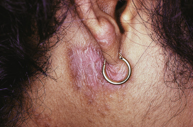
Nickel dermatitis is seen most frequently on the earlobes. Piercing the earlobes with nickel-plated instruments or wearing nickel-plated jewelry readily induces nickel sensitivity. Earlobes should be pierced only with stainless steel instruments, and only stainless steel earrings should be worn until the ears have healed. Exposure to the metal may not be readily apparent most of the time. Even with gold jewelry, the clasps and solder may contain nickel. Nickel objects may be plated with chrome but may still cause nickel dermatitis through the leaching of some of the nickel through the small pores of the chromium plating.
Nickel oxides in green paints may produce nickel dermatitis. Homeopathic and complementary medicaments may also contain enough nickel to produce a contact allergy. Sweat containing sodium chloride may combine with nickel to form nickel chloride. This affects the degree of nickel dermatitis, being more severe in persons who perspire profusely.
The diagnosis is established by a positive patch test reaction to nickel sulfate. Nickel may be detected by applying dimethylglyoxime solution to the test object. In the presence of nickel, the cotton swab used to apply the solution will turn orange-pink. A positive test always means that nickel is present, but a negative test does not rule out its presence. Sweat, blood, or saline may leach nickel from stainless steel.
Prophylactic measures should include the reduction of perspiration in those sensitive to nickel. Topical corticosteroids applied before exposure to nickel, such as before putting on a wristband, may be successful. Clasps and other objects are available in plastic material so that some of the exposure to nickel may be decreased. Polyurethane varathane 91 applied in three coats will give protection for several months. Treatment of nickel dermatitis consists of the application of topical corticosteroids. In Europe, laws regulating the maximum content of nickel in jewelry have led to a marked decrease in sensitization. Dr. Sharon Jacob is leading an effort to have a similar law passed in the United States.
Hand eczema and pompholyx in nickel-sensitive or cobalt-sensitive patients have rarely been aggravated by ingested metals in the diet. In severe, treatment-resistant dermatitis, a specific diet low in nickel and cobalt may be tried.
Chromium.
The chromates are strongly corrosive and irritating to the skin and may act as primary irritants or as sensitizers to produce allergic contact dermatitis. Besides affecting employees in chromate works, chrome dermatitis is encountered among tanners, painters, dyers, photographers, polishers, welders, aircraft workers, diesel engine workers, and those involved with the bleaching of crude oils, tallows, and fats. Traces of dichromates in shoe leather and gloves may cause eczema of the feet and hands. Many zippers are chromium plated, and the nickel underneath may be the causative agent. Chromium metal and stainless steel do not produce contact dermatitis.
Zinc chromate paint is a source of dermatitis. Matches, hide glues, chrome alloys, cigarette lighters, and leather hatbands, sandals, or camera cases may cause chrome dermatitis. Anticorrosion solutions used for refrigeration and other recirculation systems often contain chromates that produce dermatitis. Most workers in the cement industry who have cement eczema show positive patch tests to dichromates. Cement eczema is often a primary irritant dermatitis complicated by allergic contact dermatitis to the hexavalent chromates. The incidence of cement dermatitis has decreased significantly over the years, believed to be the result of the addition of ferrous sulfate, delivery of premixed cement to the job site, and improved education.
The skin changes are multiform, ranging from a mild follicular dermatitis to widespread nodular and crusted eruptions, all being worse on exposed parts. Often the eruptions are slow to clear up, lasting from a few weeks to 6 months after contact has ceased. Heavy exposure of industrial workers to chromates may produce chrome ulcers on the backs of the hands and forearms, usually beginning around a hair follicle, or in the creases of the knuckles or finger webs. The hole begins as a small abrasion that deepens and widens as its edges grow thick, eventually forming a conical, indolent ulceration. Chrome ulcers may also arise on—and perforate—the nasal septum. Arsenic exposure may result in similar ulcers.
Diagnosis of chrome sensitivity is made by a positive patch test to potassium dichromate in petrolatum. The hexavalent chrome compounds are the most frequent cause of chrome dermatitis because these penetrate the skin more easily than the trivalent form. Both forms are sensitizers. Even with avoidance of chromate-containing materials, chromate-induced dermatitis is often persistent.
Mercury.
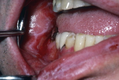
The mercurials may act not only as irritants but also as sensitizers. Thimerosal is a mercuric-containing preservative; it is an allergen that is rarely relevant. Allergy to this compound is likely to have been caused by exposure during childhood vaccinations and to tincture of merthiolate antiseptic. In general, these patients tolerate repeated vaccinations well. Most individuals are sensitized to the ethyl mercuric component of thimerosal; however, those who react to the thiosalicylic acid portion develop photodermatitis to piroxicam. Mercury in amalgam dental fillings has been shown in multiple large studies to cause oral lichenoid eruptions. The relationship is especially strong when the oral lesion, often with a painful erosion present, is apposed to a gold or amalgam filling. In many cases, when sensitivity is proved by patch testing and fillings are replaced, involution of the oral findings occurs.
Cobalt.
Cobalt is frequently combined with nickel as a contaminant, and patients allergic to cobalt typically are also allergic to nickel. The metals have similar properties but do not produce cross-reactions. Cobalt dermatitis may occur in those involved in the manufacture of polyester resins and paints, hard metals used for cutting and drilling tools, and cement. Cobalt dermatitis may also occur in producers of pottery, ceramics, metal alloys, glass, carbides, and pigments. Individuals may be exposed to cobalt in hair dye, flypaper, and vitamin B 12 . Blue tattoo pigment contains cobalt oxide. Rarely, cobalt chloride may cause nonimmunologic local release of vasoreactive materials, with a local urticarial response.
Gold.
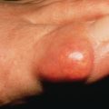
Gold dermatitis may rarely occur from the wearing of gold jewelry. A predisposing factor in such patients is the presence of dental gold. Oral lichenoid eruptions have also been reported with gold, similar to the situation with mercury-containing amalgams ( Fig. 6.15 ). It is not uncommon to see positive reactions to gold when patch-testing patients with facial, eyelid, or widespread dermatitis of unknown cause. Although it is difficult to make a direct clinical correlation with any one piece of jewelry, occasionally patients will clear if they stop wearing all gold jewelry. One possible explanation is that titanium dioxide in sunscreen products may liberate gold particles from jewelry. In most patients, however, there is a lack of relevance. Gold reactions on patch testing may be delayed in onset and remain inflamed for months.
Other Metals.
Most other common metals are not important in causing contact dermatitis. Platinum dermatitis may occur from exposure to platinum salts and sprays in industry. Platinum rings, earrings, white gold spectacles, clasps, and other jewelry cause eruptions resembling those caused by nickel. Zinc, aluminum, copper sulfate, titanium, and antimony dermatitis rarely occur, although these metals may act as irritants.
Atanaskova Mesinkovska N, et al: The effect of patch testing on surgical practices and outcomes in orthopedic patients with metal implants. Arch Dermatol 2012; 148: 687.
Borowska S, Brzóska MM: Metals in cosmetics. J Appl Toxicol 2015; 35: 551.
Bregnbak D, et al: Chromium allergy and dermatitis. Contact Dermatitis 2015; 73: 261.
Chen JK, Lampel HP: Gold contact allergy. Dermatitis 2015; 26: 69.
Crépy MN: Skin diseases in musicians. Eur J Dermatol 2015; 25: 375.
Fage SW, et al: Copper hypersensitivity. Contact Dermatitis 2014; 71: 191.
Fage SW, et al: Titanium. Contact Dermatitis 2016; 74: 323.
Fowler JF Jr: Cobalt. Dermatitis 2016; 27: 3.
Goldenberg A, et al: Nickel allergy in adults in the U.S. Dermatitis 2015; 26: 216.
Gölz L, et al: Nickel hypersensitivity and orthodontic treatment. Contact Dermatitis 2015; 73: 1.
Honari G, et al: Hypersensitivity reactions associated with endovascular devices. Contact Dermatitis 2008; 59: 7.
Jacob SE, et al: Nickel allergy and our children’s health. Pediatr Dermatol 2015; 32: 779.
Maridet C, et al: The electronic cigarette. Contact Dermatitis 2015; 73: 49.
Mislankar M, et al: Low nickel diet scoring system for systemic nickel allergy. Dermatitis 2013; 24: 190.
Pacheco KA: Allergy to surgical implants. J Allergy Clin Immunol Pract 2015; 3: 683.
Stuckert J, et al: Low cobalt diet for dyshidrotic eczema. Contact Dermatitis 2008; 59: 361.
Teo Wendy ZW, Schalock PC: Hypersensitivity reactions to implanted metal devices. J Investig Allergol Clin Immunol 2016; 26: 279.
Thomas P, et al: DKG statement on the use of metal alloy discs for patch testing in suspected intolerance to metal implants. J Dtsch Dermatol Ges 2015; 13: 1001.
Thyssen JP, et al: Patch test reactivity to metal allergens following regulatory intervention. Contact Dermatitis 2010; 63: 102.
Warshaw EM, et al: Body piercing and metal allergic contact sensitivity. Dermatitis 2014; 25: 255.
Contact Stomatitis
The role of contact allergy in oral symptomatology is significant. Approximately 30% of patients with oral symptoms will have relevant allergens, most frequently metals used in dental fillings, food additives (flavorings and antioxidants), and dental products (acrylic monomers, epoxy resins, hardeners used in prosthodontics and dental impression materials). Chewing gums and dentifrices may also produce contact stomatitis. Ingredients responsible for this are hexylresorcinol, thymol, dichlorophen, oil of cinnamon, and mint.
Clinical signs may be bright erythema of the tongue and buccal mucosa with scattered erosions. Angular cheilitis may also develop. Oral lichenoid lesions may be caused by sensitization to metals in dental fillings and gold caps or crowns.
Batchelor JM, et al: Allergic contact stomatitis caused by a polyether dental impression material. Contact Dermatitis 2010; 63: 296.
de Groot A: Contact allergy to (ingredients of) toothpastes. Dermatitis 2017; 28: 95.
Isaac-Renton M, et al: Cinnamon spice and everything not nice: many features of intraoral allergy to cinnamic aldehyde. Dermatitis 2015; 26: 116.
Johns DA, et al: Allergic contact stomatitis from bisphenol-a-glycidyl dimethacrylate during application of composite restorations. Indian J Dent Res 2014; 25: 266.
Krishen C, et al: Dental allergic contact dermatitis. Dermatitis 2012; 23: 222.
O’Gorman SM, Torgerson RR: Contact allergy in cheilitis. Int J Dermatol 2016; 55: e386.
Sharma R, et al: Role of dental restoration materials in oral mucosal lichenoid lesions. Indian J Dermatol Venereol Leprol 2015; 81: 478.
Torgerson RR, et al: Contact allergy in oral disease. J Am Acad Dermatol 2007; 57: 315.
Zug KA, et al: Patch-testing North American lip dermatitis patients. Dermatitis 2008; 19: 202.
Rubber Dermatitis
Rubber dermatitis generally occurs on the hands from wearing rubber gloves, as by surgeons, nurses, and homemakers. The eruption is usually sharply limited to the gloved area but may spread up the forearms. Rubber dermatitis also develops from exposure to condoms, diaphragms, swim goggles ( Fig. 6.16 ), caps and scuba masks, wet suits, bandages for chronic leg ulcers, respirators, gas masks, rubber sheets, and cosmetic sponges. Shoe dermatitis may be caused by rubber allergy to insoles or sneakers.
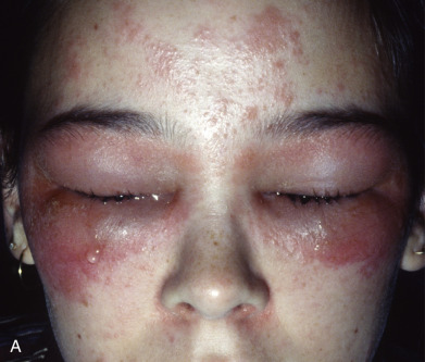
Natural and synthetic rubbers are used separately or in combination to make the final rubber product. The chemicals added in the rubber manufacturing process, most importantly the accelerators and antioxidants, are the common causes of allergic contact dermatitis. A similar list of additives is present in neoprene, a synthetic rubber, but also includes the dialkyl thioureas. These are not in the standard patch trays and thus may escape detection unless applied as a supplemental allergen. Elastic in underwear is chemically transformed by laundry bleach into a potent sensitizing substance. The allergen is permanent and cannot be removed by washing. The offending garments must be thrown out and the use of bleaches interdicted.
Accelerators.
During the manufacturing process, chemicals are used to hasten the vulcanization of rubber. Among the numerous chemicals available, tetramethylthiuram disulfide, mercaptobenzothiazole, and diphenylguanidine are frequently used. Tetramethylthiuram disulfide and its analogs, known as disulfiram and thiuram, may produce contact dermatitis when moist skin is exposed to the finished rubber product. In a 10-year study of 636 cases of allergy to rubber additives, thiuram mix was by far the most common sensitizer. Mercaptobenzothiazole is most often the cause in shoe allergy and thiuram in glove allergy.
Antioxidants.
Antioxidants are used to preserve rubber. Amine antioxidants, such as phenyl-α-naphthylamine, are most effective. Hydroquinone antioxidants may cause depigmentation of the skin, as well as allergic contact dermatitis. A frequent antioxidant sensitizer, propyl p -phenylenediamine, is used in tires, heavy-duty rubber goods, boots, and elastic underwear.
Bendewald MJ, et al: An 8-year retrospective review of patch testing with rubber allergens. Dermatitis 2010; 21:33.
Cao LY, et al: Allergic contact dermatitis to synthetic rubber gloves. Arch Dermatol 2010; 146: 1001.
Higgins CL, et al: Occupational skin disease among Australian healthcare workers. Contact Dermatitis 2016; 75: 213.
Ibler KS, et al: Prevalence of delayed-type and immediate-type hypersensitivity in healthcare workers with hand eczema. Contact Dermatitis 2016; 75: 223.
Schwensen JF, et al: Contact allergy to rubber accelerators remains prevalent. J Eur Acad Dermatol Venereol 2016; 30: 1768.
Warshaw EM, et al: Positive patch-test reactions to mixed dialkyl thioureas. Dermatitis 2008; 19: 190.
Adhesive Dermatitis
Cements, glues, and gums may cause adhesive dermatitis. Formaldehyde resin adhesives contain free formaldehyde, naphtha, glue, and disinfectants. Synthetic resin adhesives contain plasticizers; hide glues may contain chromates from the tanned leather, and other glues incorporate preservatives such as formaldehyde. Dental bonding adhesives may contain acrylic monomers and epoxy resins and hardeners. Pressure-sensitive adhesives contain rubber and acrylates, and anaerobic adhesives have primarily acrylates.
Vegetable gums, such as gum tragacanth, gum arabic, and karaya, may be used in denture adhesives, hair wave lotions, topical medications, toothpastes, and depilatories, and many cause contact dermatitis. Resins are used in adhesive tapes and in various adhesives such as tincture of benzoin. Turpentine is frequently found in rosin; abietic acid in the rosin is the causative sensitizer.
Adhesive tape reactions are frequently irritant in nature. Allergic reactions to adhesive tape itself are caused by the rubber components, accelerators, antioxidants, and various resins or turpentine. Some adhesive tapes contain acrylate polymers rather than rubber adhesives. These acrylates may cause allergic contact dermatitis. Pressure-sensitive adhesives are increasingly used. Allergens present in these adhesives include rosin, rubber accelerators, antioxidants, acrylates, hydroquinones, lanolin, thiourea compounds, and N -dodecylmaleamic compounds.
Bennike, NH, et al: Please, label the label; a case report of occupational allergic contact dermatitis caused by methylisothiazolinone in adhesive labels. Contact Dermatitis 2016; 75: 314.
Bhargava K, et al: Eyelid allergic contact dermatitis caused by ethyl cyanoacrylate–containing eyelash adhesive. Contact Dermatitis 2012; 67: 306.
Corazza M, et al: Allergic contact dermatitis in a volleyball player due to protective adhesive taping. Eur J Dermatol 2011; 21: 430.
Davis MD, Stuart MJ: Severe allergic contact dermatitis to Dermabond Prineo, a topical skin adhesive of 2-octyl cyanoacrylate increasingly used in surgeries to close wounds. Dermatitis 2016; 27: 75.
Jacobs M, et al: Peristomal allergic contact dermatitis caused by stoma adhesive paste containing N-butyl monoester of polymethyl vinyl ether maleic acid. Contact Dermatitis 2015; 73: 314.
Lau YN, et al: Use of skin adhesive in dermatological surgery. Clin Exp Dermatol 2014; 39: 773.
Özkaya E, Kavlak Bozkurt P: Allergic contact dermatitis caused by self-adhesive electrocardiography electrodes. Contact Dermatitis 2014; 70: 121.
Schwensen JF, et al: Sensitization to cyanoacrylates caused by prolonged exposure to a glucose sensor set in a diabetic child. Contact Dermatitis 2016; 74: 124.
Synthetic Resin Dermatitis
The many varieties of synthetic resins preclude adequate discussion of each. The reactions during the manufacture of these substances are more common than those in their finished state.
Epoxy Resins.
The epoxy resins in their liquid (noncured, monomer) form may produce severe dermatitis, especially during the manufacturing process. The fully polymerized or cured product is nonsensitizing. Nonindustrial exposure is usually to epoxy resin glues, nail lacquers, and artificial nails. Epoxy resins are used in the home as glues and paints (bathtub and refrigerator). Artists and sculptors frequently use epoxy resins.
Epoxy resins consist of two or more components, the resin and the curing agent. Approximately 90% of allergic reactions are to the resin and 10% to the hardener. The numerous curing agents include the amines, phenolic compounds, peroxides, and polyamides. These may be irritants and/or allergens. The resin, based on an acetone and phenol compound known as bisphenol A, in its raw state may cause allergic contact dermatitis. BIS-GMA, a combination of bisphenol A and glycidyl methacrylate, is the main allergen in dental bonding agents. Epoxy resins are used also as stabilizers and plasticizers. Their use in the manufacture of polyvinyl chloride (plastic) film has caused dermatitis from plastic handbags, beads, gloves, and panties.
Polyester Resins.
Ordinarily, completely cured or polymerized resins are not sensitizers. The unsaturated polyester resins are dissolved and later copolymerized with vinyl monomers. Such polyester resins are used for polyester plasticizers, polyester fibers (Dacron), and polyester film (Mylar). The unsaturated polyester resins, on the other hand, will produce primary irritation in their fabrication or among sculptors. The dermatitis occurs typically as an eczematous eruption on the back of the hands, wrists, and forearms. Polyester resins are incorporated into other plastic material as laminates to give them strength; applications include boat hulls, automobile body putty, safety helmets, fuel tanks, lampshades, and skylights.
Acrylic Monomers.
Cyanoacrylates are used widely as adhesives in a variety of home and commercial products. They are generally a rare cause of contact dermatitis. Multifunctional acrylic monomers may produce allergic or irritant contact dermatitis. Pentaerythritol triacrylate, trimethylolpropane triacrylate, and hexanediol diacrylate are widely used acrylic monomers. Printers handling multifunctional acrylic monomers in printing inks and acrylic printing plates may present with an erythematous, pruritic eruption, mainly of the hands and arms, swelling of the face, and involvement of the eyelids.
Orthopedic surgeons experience contact dermatitis from the use of acrylic bone cement (methyl methacrylate monomer) used in mending hip joints. Dentists and dental technicians are exposed when applying this to teeth. The sensitizer passes through rubber and polyvinyl gloves and may additionally cause paresthesias. In patients who are allergic to their acrylate dental prosthesis, coating this with UV light–cured acrylate lacquer may allow it to be worn without adverse effects.
Benzoyl peroxide is a popular acne remedy. It is also used for bleaching flour and edible oils and for curing plastics, such as acrylic dentures. Infrequently, an allergic contact dermatitis may result.
Aalto-Korte K, et al: Contact allergy to epoxy hardeners. Contact Dermatitis 2014; 71: 145.
Aalto-Korte K, et al: Occupational allergic contact dermatitis caused by epoxy chemicals. Contact Dermatitis 2015; 73: 336.
Bowen C, et al: Allergic contact dermatitis to 2-octyl cyanoacrylate. Cutis 2014; 94: 183.
Kocak O, Gul U: Patch test results of the dental personnel with contact dermatitis. Cutan Ocul Toxicol 2014; 33: 299.
Shmidt E, et al: Patch-testing with plastics and glues series allergens. Dermatitis 2010; 21: 269.
Strazzula L, et al: Fingertip purpura in a dental student. JAMA Dermatol 2014; 150: 784.
Cosmetic Dermatitis
Cutaneous reactions to cosmetics may be divided into irritant, allergic, and photosensitivity reactions. More than half the reactions occur on the face and are caused primarily by skin care products, nail cosmetics, shaving preparations, and deodorants. The leading cause of allergic contact dermatitis associated with cosmetics is from fragrance. A close second is preservatives, such as methylchloroisothiazolinone/methyllisothiazolinone, 2-bromo-2-nitropropane-1,3-diol, quaternium 15, methyldibromo glutaronitrile, and imidazolidinyl urea. The third leading cause is p -phenylenediamine in hair dye. It is recommended that patch testing with the patient’s own product, as long as it is applied to the skin as a leave-on product, be part of the evaluation.
Fragrances.
Almost all cosmetic preparations, skin care products, and many medications contain fragrance; even those labeled “nonscented” often contain a masking fragrance that may be a sensitizer. Even “fragrance-free” products have been documented to contain the raw fragrance ingredients, such as rose oil in “all-natural” products. Fragrances are the most common cosmetic ingredient causing allergic contact dermatitis. Photodermatitis, irritation, contact urticaria, and dyspigmentation are other types of reactions that fragrances may produce.
The most common individual allergens identified are cinnamic alcohol, oak moss, cinnamic aldehyde, hydroxy citronellal, musk ambrette, isoeugenol, geraniol, coumarin, lyral ( Fig. 6.17 ), and eugenol. Hyperperoxides of linalool and oxidized limonene are newer important fragrance allergens. Frequently, unspecified allergens are the cause, because they are not listed on labels, and fragrances are combinations of many different ingredients. Myroxylon pereirae (balsam of Peru) will identify approximately half of those often unsuspected cases of allergic dermatitis, and additional testing with the fragrance mixes will identify over 90%. Additionally, a natural fragrance mixture of jasmine absolute, ylang-ylang oil, narcissus absolute, spearmint oil, and sandalwood oil is recommended. Other essential oils may be missed if not individually tested. Fragrance mixes should be prepared the day of application as evaporation may compromise their reliability. New products should be tested for tolerance in patients with a history of fragrance sensitivity.
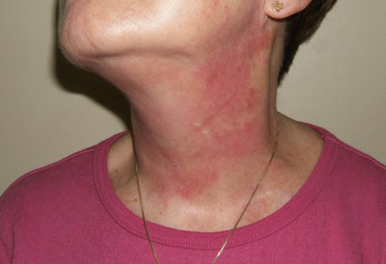
At least 1% of the population have fragrance sensitivity. Women still outnumber men, but as the frequency of fragrance contact reactions has increased over the years, men have shown a steeper increase in sensitivity. Fragrance is one allergen that may be transferred by skin-to-skin contact to a sensitive person, causing connubial contact dermatitis. Ingestion of balsam-related foods, such as tomatoes, citrus fruits, and spices, may cause a flare in some sensitive patients. In particularly difficult-to-treat patients, balsam-restricted diets may be beneficial but are not easy to follow.
Hair Dyes.
Permanent hair dyes incorporate p -phenylenediamine (PPDA), a popular but potent sensitizer that may cross-react with many chemicals. In rinses and tints, the azo dyes, acid violet 6B, water-soluble nigrosine, and ammonium carbonate may sensitize and cross-react with PPDA. Workers in the manufacture of PPDA, furriers, hairdressers, and those in the photographic and rubber vulcanization industries develop eruptions first on the back of the hands, wrists, forearms, eyelids, and nose, consisting of an eczematous, erythematous, oozing dermatitis. It will penetrate most protective gloves, but not nitrile gloves. Lichenification and scaling are seen in the chronic type. In those with dyed hair, sensitivity is manifested by itching, redness, and puffiness of the upper eyelids, tops of the ears, temples, and back of the neck ( Fig. 6.18 ). Beard/mustache dermatitis may be caused by coloring of the facial hair and eyelid dermatitis by dying eyelashes. PPDA added to temporary henna tattoos to make them darker has resulted in acute vesicular allergic reactions, some with scarring and hyperpigmentation. Kumkum is a common cosmetic in India, primarily smeared on the forehead of women to denote their marital status; one of many reported allergens in the product is PPDA.
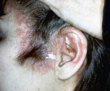
For those sensitive to this type of hair dye, use of semipermanent or temporary dyes might be the solution. In the case of sensitivity to these alternatives, vegetable dyes such as henna may be tried. Metallic dyes are usually not favored by women but are frequently used by men as “hair color restorers.” The metallic hair dyes may contain nickel, cobalt, chromium, or lead. Hair dyes containing FD&C and D&C dyes often do not cross-react with PPDA and 70% of PPDA-sensitive patients exposed to a dye containing the less allergenic PPDA derivative 2-methoxymehtyl-PPD tolerated it.
Other Hair Products.
Hair bleach products incorporate peroxides, persulfates, and ammonia, which may act as primary irritants. Hair bleaches that contain ammonium persulfate, a primary irritant, may produce a local urticarial and a generalized histamine reaction.
Several types of permanent wave preparations exist. The alkaline permanent wave preparations, which use ammonium thioglycolate, are rarely if ever sensitizers and usually cause only hair breakage and irritant reactions. The hot type, or acid perm, is a common sensitizer, the allergen being glyceryl monothioglycolate. Cosmetologists are at risk for development of hand dermatitis. The glyceryl monothioglycolate persists in the hair for at least 3 months after application and may cause a long-lasting dermatitis. It readily penetrates rubber and vinyl gloves. A more neutral pH permanent wave solution is less allergenic than the acid perms; however, allergy to cysteamine hydrochloride found in neutral permanent wave products may occur. This allergen does not penetrate household-weight latex gloves, and hair waved with it does not produce allergic reactions in sensitized individuals. Also, it is an amine salt, not a thioglycolate, so cross-reactivity is unlikely.
Hair straighteners using greases and gums are not sensitizers; however, the perfume incorporated in these preparations can be sensitizing. Thioglycolates are also used, and hair breakage may occur with these products.
Hair sprays may contain shellac, gum arabic, sunscreens, and synthetic resins as sensitizers, and allergic reactions occur infrequently. Lanolin is frequently incorporated into aerosol sprays.
Chemical depilatories containing calcium thioglycolate and the sulfides and sulfhydrates may cause primary irritant dermatitis. Mechanical hair removers include the mercaptans, waxes, and resins; resins may produce allergic dermatitis.
Hair tonics and lotions with tincture of cinchona produce allergic sensitization; tincture of cantharidin and salicylic acid cause primary irritation. Resorcin, quinine sulfate, and perfumes such as bay rum are also sensitizers.
Nail Products.
Nail lacquers may contain tosylamide/formaldehyde resin and are a frequent cause of eyelid and neck dermatitis. Polishes free of this resin are available. Nail polish removers are solvents such as acetone, which can cause nail brittleness. The acrylic monomers in artificial nails, as well as the ethyl cyanoacrylate glue required to attach the prosthetic nail, may produce allergic sensitivity. Photoinitiating agents, such as benzophenone, used in photo-bonded acrylic sculptured nails are other potential allergens.
Lipsticks.
Various R and C dyes, sunscreens, shellac, flavoring agents, preservative, and lipstick perfumes may cause sensitization reactions. Lipsticks are tested as is. Lip plumpers may cause contact urticaria in those being kissed. Propolis is found in many so-called natural products, including lip balms, toothpastes, lotions, shampoos, and other cosmetics. Its main allergens are two types of caffeates.
Eye Makeup.
In mascara, eye shadow, and eyeliners, preservatives, shellac, metals, base wax, and perfumes are the components that may produce sensitization, but this occurs rarely. False-positive reactions to some mascaras occur when a closed patch test is used. This is caused by the irritative qualities of the solvents. An open or nonocclusive patch test is recommended. A provocative use test in the antecubital fossae may ultimately be necessary. The rubber sponges used to apply eye makeup, nickel in the tools used, or cocamidopropylbetaine in eye makeup remover also cause eyelid dermatitis.
Sunscreens.
p -Aminobenzoic acid (PABA) and its derivatives (e.g., padimate O, padimate A, glycerol PABA), dibenzoylmethanes, salicylates, cinnamates, and benzophenones are photosensitizers as well as sensitizers. The benzophenones (most often benzophenone-3) and dibenzoylmethanes are the most common sunscreen allergens and photoallergens. These agents frequently are found in various other cosmetic products. The physical blockers titanium dioxide and zinc oxide do not cause allergy. If allergy to PABA exists, its derivatives should be avoided, and the patient should be aware that thiazides, sulfonylurea antidiabetic medication, azo dyes, p -aminosalicylic acid, benzocaine, and PPDA all may cause dermatitis from cross-reactions.
Bleaching Creams.
Hydroquinones are occasional sensitizers. Ammoniated mercury is a sensitizing agent formerly used in bleaching creams.
Lanolin.
The fatty alcohol lanolin is rarely a sensitizer on normal skin, so it usually does not produce dermatitis when exposure is with cosmetic and skin care products. It provokes allergic reactions more frequently in therapeutic agents used by atopic patients and in emollient products that may be used postsurgically.
Dentifrices and Mouthwashes.
Dentifrices and mouthwashes contain sensitizers, such as the essential oils used as flavoring agents, preservatives, formalin, antibiotics, and antiseptics. Circumoral dermatitis and cheilitis may be caused by tartar-control types of dentifrice.
Axillary Antiperspirants.
Aluminum salts, such as aluminum chloride and chlorhydroxide, and zinc salts, such as zinc chloride, act as primary irritants and may rarely produce a folliculitis. Aluminum chlorhydrate is considered to be the least irritating antiperspirant. Zirconium salt preparations, now removed from all antiperspirants, produced a granulomatous reaction. Zirconium-aluminum complexes, however, are often used as the active ingredient in topical antiperspirants and may produce granulomas. Quaternary ammonium compounds in some roll-on deodorants may produce allergic contact dermatitis.
Axillary Deodorants and Feminine Hygiene Sprays.
Fragrances, bacteriostats, and propellants cause the majority of the reactions seen with these products. Deodorants that contain cinnamic aldehyde can induce irritation on axillary skin even when tolerated on healthy skin in other sites.
Cosmetic Intolerance Syndrome.
Occasionally, a patient will complain of intense burning or stinging after applying any cosmetic. The patient usually has only subjective symptoms, but objective inflammation may also be present. The underlying cause may be difficult to document, even after thorough patch, photopatch, and contact urticaria testing. Endogenous disease, such as seborrheic dermatitis, rosacea, or atopic dermatitis, may complicate the assessment. Avoidance of all cosmetics, with only glycerin being allowed, for 6–12 months is often necessary to calm the reactive state. Adding back cosmetics one at a time, no more frequently than one a week, may then be tolerated.
Antelmi A, et al: Are gloves sufficiently protective when hairdressers are exposed to permanent hair dyes? An in vivo study. Contact Dermatitis 2015; 72: 229.
Beleznay K, et al: Analysis of the prevalent of allergic contact dermatitis to sunscreen. J Cutan Med Surg 2014; 18: 15.
Bourgeois P, Goossens A: Allergic contact cheilitis caused by menthol in toothpaste and throat medication. Contact Dermatitis 2016; 75: 113.
Bråred Christensson J, et al: Positive patch test reactions to oxidized limonene. Contact Dermatitis 2014; 71: 264.
Cheng J, Zug KA: Fragrance allergic contact dermatitis. Dermatitis 2014; 25: 232.
Das S, et al: Shellac. Dermatitis 2011; 22: 220.
de Groot AC: Propolis. Dermatitis 2013; 24: 263.
de Groot AC, Roberts DW: Contact and photocontact allergy to octocrylene. Contact Dermatitis 2014; 70: 193.
Diepgen TL, et al: Prevalence of fragrance contact allergy in the general population of five European countries. Br J Dermatol 2015; 173: 1411.
Elliott JF, et al: Severe intractable eyelid dermatitis probably caused by exposure to hydroperoxides of linalool in a heavily fragranced shampoo. Contact Dermatitis 2017; 76: 114.
Ghazavi MK, et al: Photo-allergic contact dermatitis caused by isoamyl P-methoxycinnamate in an “organic” sunscreen. Contact Dermatitis 2011; 64: 115.
González-Muñoz P, et al: Allergic contact dermatitis caused by cosmetic products. Actas Dermosifiliogr 2014; 105: 822.
Haylett AK, et al: Sunscreen photopatch testing. Br J Dermatol 2014; 171: 370.
Heurung AR, et al: Adverse reactions to sunscreen agents. Dermatitis 2014; 25: 289.
Jacob SE, et al: Nickel ferrule applicators. Pediatr Dermatol 2015; 32: e62.
Jefferson J, et al: Update on nail cosmetics. Dermatol Ther 2012; 25: 481.
Kock M, et al: Continuous usage of a hair dye product containing 2-methoxymethyl-para-phenylenediamine by hair-dye-allergic individuals. Br J Dermatol 2016; 174: 1042.
Landers MC, et al: Permanent wave dermatitis. Am J Contact Dermat 2003; 14: 157.
Le Q, et al: The rising trend in allergic contact dermatitis to acrylic nail products. Australas J Dermatol 2015; 56: 221
Leventhal JS, et al: Crystal deodorant-induced axillary granulomatous dermatitis. Int J Dermatol 2014; 53: e59.
Lev-Tov H, et al: The sensitive skin syndrome. Indian J Dermatol 2012; 57: 419.
Lopez IE, et al: Clues to diagnosis of connubial contact dermatitis to paraphenylenediamine. Dermatitis 2014; 25: 32.
Miest RY, et al: Diagnosis and prevalence of lanolin allergy. Dermatitis 2013; 24: 119.
Montgomery R, et al: Contact allergy resulting from the use of acrylate nails is increasing in both users and those who are occupationally exposed. Contact Dermatitis 2016; 74: 120.
Mowitz M, et al: Fragrance patch tests prepared in advance may give false-negative reactions. Contact Dermatitis 2014; 71: 289.
Nardelli A, et al: Results of patch testing with fragrance mix 1, fragrance mix 2, and their ingredients, and Myroxylon pereirae and colophonium, over a 21-year period. Contact Dermatitis 2013; 68: 307.
Panasoff J: Cheilitis caused by to mint-containing toothpastes. Contact Dermatitis 2016; 75: 260.
Panfili E, et al: Temporary black henna tattoos and sensitization to para-phenylenediamine (PPD). Int J Environ Res Public Health 2017; 14: E421.
Park ME, Zippin JH: Allergic contact dermatitis to cosmetics. Dermatol Clin 2014; 32: 1.
Sabroe RA, et al: Contact allergy to essential oils cannot always be predicted from allergy to fragrance markers in the baseline series. Contact Dermatitis 2016; 74: 236.
Scheman A, et al: Balsam of Peru. Dermatitis 2013; 24: 153.
Uter W, et al: Coupled exposure to ingredients of cosmetic products. Contact Dermatitis 2014; 70: 219.
Preservatives
Preservatives are added to any preparation that contains water to kill microorganisms and prevent spoilage. Such products include moist materials such as baby wipes, which when used in either infants or adults can produce reactions caused by preservatives. The most important class is formaldehyde and the formaldehyde-releasing compounds, including quaternium 15 (the leading preservative sensitizer in the United States), imidazolidinyl urea, diazolidinyl urea, DMDM hydantoin, and 2-bromo-2-nitropropane-1,3-diol.
Kathon CG, or methylchloroisothiazolinone/methylisothiazolinone (MCI/MI), and Euxyl K 400 (methyldibromoglutaronitrile and phenoxyethanol in 1 : 4 ratio) are other important preservative allergens. MCI/MI is widely used in cosmetics and personal care products such as baby wipes and moist towelettes, but may be found in medical devices, and in industrial products such as cutting oils, household detergents, paints, and glues. MCI is a stronger allergen, however use of MI alone in high concentrations has led to a increase in primary sensitization to it. Testing should be to each chemical separately. In Euxyl K 400, the methyldibromoglutaronitrile component produces the allergic response. This preservative may produce false-negative results on testing, so repeat open testing is indicated if a specific leave-on product is suspected of causing allergy. European regulations limit exposure to methyldibromoglutaronitrile. As with similar laws regulating nickel in Europe, allergy to this preservative is also lowering in incidence over time.
Tea tree oil is an additive to some natural products that may serve as an antimicrobial. It is a frequent sensitizer as many products include this oil as a “natural” antimicrobial agent. Sorbic acid is a rare sensitizer among the preservatives; however, it is a cause of facial flushing and stinging through its action as an inducer of nonimmunologic contact urticaria. Benzalkonium chloride is widely used but a rare sensitizer. Triclosan and benzyl alcohol are weak sensitizers. Thimerosal is discussed earlier.
Formaldehyde and Formaldehyde-Releasing Agents.
Formaldehyde is used rarely, primarily in shampoos. Because it is quickly diluted and washed away, sensitization through this exposure is rare. Formaldehyde releasers are polymers of formaldehyde that may release small amounts of formaldehyde under certain conditions. Allergy may be to the formaldehyde-releasing preservatives (which act as antibacterial and antifungal agents in their own right) or to the released formaldehyde. Cross-reactivity among them is common, so when allergy is proved to one compound and avoidance does not clear the eruption, screening for clinically relevant reactions to the others is indicated. This may be done by repetitive open application testing to the leave-on product or by extended patch testing.
Parabens.
Allergic contact dermatitis may develop from parabens, which are used in cosmetics, foods, drugs, dentifrices, and suppositories. The paraben esters (methyl, ethyl, propyl, and butyl p -hydroxybenzoates) are used in low concentrations in cosmetics and rarely cause dermatitis. They are found in higher concentration in topical medicaments and may be the cause of allergic reactions. Parabens, which are frequently used as bacteriostatic agents, are capable of producing immunologically mediated, immediate systemic hypersensitivity reactions. Cross-reactivity to p -phenylenediamine and benzocaine occurs in some individuals.
Other Important Preservatives.
Iodopropynyl butylcarbamate is a wood and paint preservative that is being used more commonly in cosmetics and personal care products. This accounts for its reported higher importance among preservative allergens. The hands are the most frequent area sensitized. Gallates are antioxidant preservatives that are also emerging as important allergens; propyl gallate is the most commonly reported gallate allergen. It usually produces hand and facial eruptions as it is most commonly encountered by patients in cosmetics and personal care products. p -Chloro-metaxylenol (PCMX), a chlorinated phenol antiseptic is used in many over-the-counter products with the disinfectant properties of p -chloro-metacresol to which it is cross-reactive.
Aerts O, et al: Contact allergy caused by methylisothiazolinone. Eur J Dermatol 2015; 25: 228.
Cheng S, et al: Contact sensitivity to preservatives in Singapore. Dermatitis 2014; 25: 77.
Chow ET, et al: Frequency of positive patch test reactions to preservatives. Australas J Dermatol 2013; 54: 31.
Curry EJ, et al: Benzyl alcohol allergy. Dermatitis 2005; 16: 203.
Hauksson I, et al: Formaldehyde in cosmetics in patch tested dermatitis patients with and without contact allergy to formaldehyde. Contact Dermatitis 2016; 74: 145.
Holcomb ZE, et al: Gallate contact dermatitis. Dermatitis 2017; 28: 115.
Murad A, Marren P: Prevalence of methylchloroisothiazolinone and methylisothiazolinone contact allergy in facial dermatitis. J Eur Acad Dermatol Venereol 2016; 30: 60.
Pontén A, Bruze M: Formaldehyde. Dermatitis 2015; 26: 3.
Warshaw EM, et al: North American contact dermatitis group patch test results. Dermatitis 2015; 26: 49.
Wilson M, et al: Chloroxylenol. Dermatitis 2007; 18: 120.
Yim E, et al: Contact dermatitis caused by preservatives. Dermatitis 2014; 25: 215.
Yu SH, et al: Patch testing for methylisothiazolinone and methylchloroisothiazolinone-methylisothiazolinone contact allergy. JAMA Dermatol 2016; 152: 67.
Vehicles
Formulation of topically applied products is complex, and additives are blended to make a pleasant base for carriage of the active ingredient to the skin. Various emulsifiers, humectants, stabilizers, surfactants, and surface active agents are used to make esthetically pleasing preparations. These may cause irritation, erythema, and allergy. The surfactant cocamidopropyl betaine produces dermatitis of the head and neck in consumers and the hands in hairdressers, often from its presence in shampoos. Sorbitan sesquioleate is an emulsifier in the fragrance I mix. It may produce false-positive results to fragrance in the few patients sensitive to it. Propolis and lanolin are discussed previously under “Cosmetic Dermatitis.”
Propylene Glycol.
Propylene glycol is widely used as a vehicle for topical medications, cosmetics (especially antiperspirants), and various emollient lotions. It is used in the manufacture of automobile brake fluid and alkyd resins, as a lubricant for food machinery, and as an additive for food colors and flavoring agents. Propylene glycol must be considered as a sensitizer able to produce contact dermatitis, and it can cause a flare of the contact dermatitis when ingested. It is tested as a 4% aqueous solution, but irritant reactions or false-negative results are common. A use test of the implicated propylene glycol–containing products may be required.
Ethylenediamine.
Ethylenediamine is used as a stabilizer in medicated creams. It may cause contact dermatitis and cross-react with internally taken aminophylline, which consists of theophylline and ethylenediamine. Hydroxyzine is a piperazine derivative that is structurally based on a dimer of ethylenediamine, to which patients sensitive to the stabilizer may develop a generalized itchy, red eruption that recurs each time hydroxyzine is taken orally.
Bennike NH, Johansen JD: Sorbitan sesquioleate; a rare cause of contact allergy in consecutively patch tested dermatitis patients. Contact Dermatitis 2016; 74: 242.
Goossens RA: Allergic contact dermatitis from the vehicle components of topical pharmaceutical products. Immunol Allergy Clin North Am 2014; 34: 663.
Jacob SE, et al: Cocamidopropyl betaine. Dermatitis 2008; 19: 157.
Reid N, et al: Worsening of contact dermatitis by oral hydroxyzine: a case report. Dermatol Online J 2013; 19: 4.
Warshaw EM, et al: Positive patch-test reactions to propylene glycol. Dermatitis 2009; 20: 14.
Welling JD, et al: Chronic eyelid dermatitis secondary to cocamidopropyl betaine allergy in a patient using baby shampoo eyelid scrubs. JAMA Ophthalmol 2014: 132: 357.
Topical Drug Contact Dermatitis
Drugs, in addition to their pharmacologic and possible toxic action, also possess sensitizing properties. Sensitization may occur not only from topical application ( Fig. 6.19 ) but also from ingestion, injection, or inhalation. Some drugs, such as the antihistamines, including topical doxepin, sensitize much more frequently when applied topically than when taken orally. With the advent of transdermal patches for delivery of medications (e.g., nitroglycerin, hormones, nicotine, clonidine, fentanyl, lidocaine, and scopolamine) reports of sensitization have been increasing. Clonidine induces the highest rate of allergic reactions. At times, EM-like reactions may occur with transdermally applied drugs.