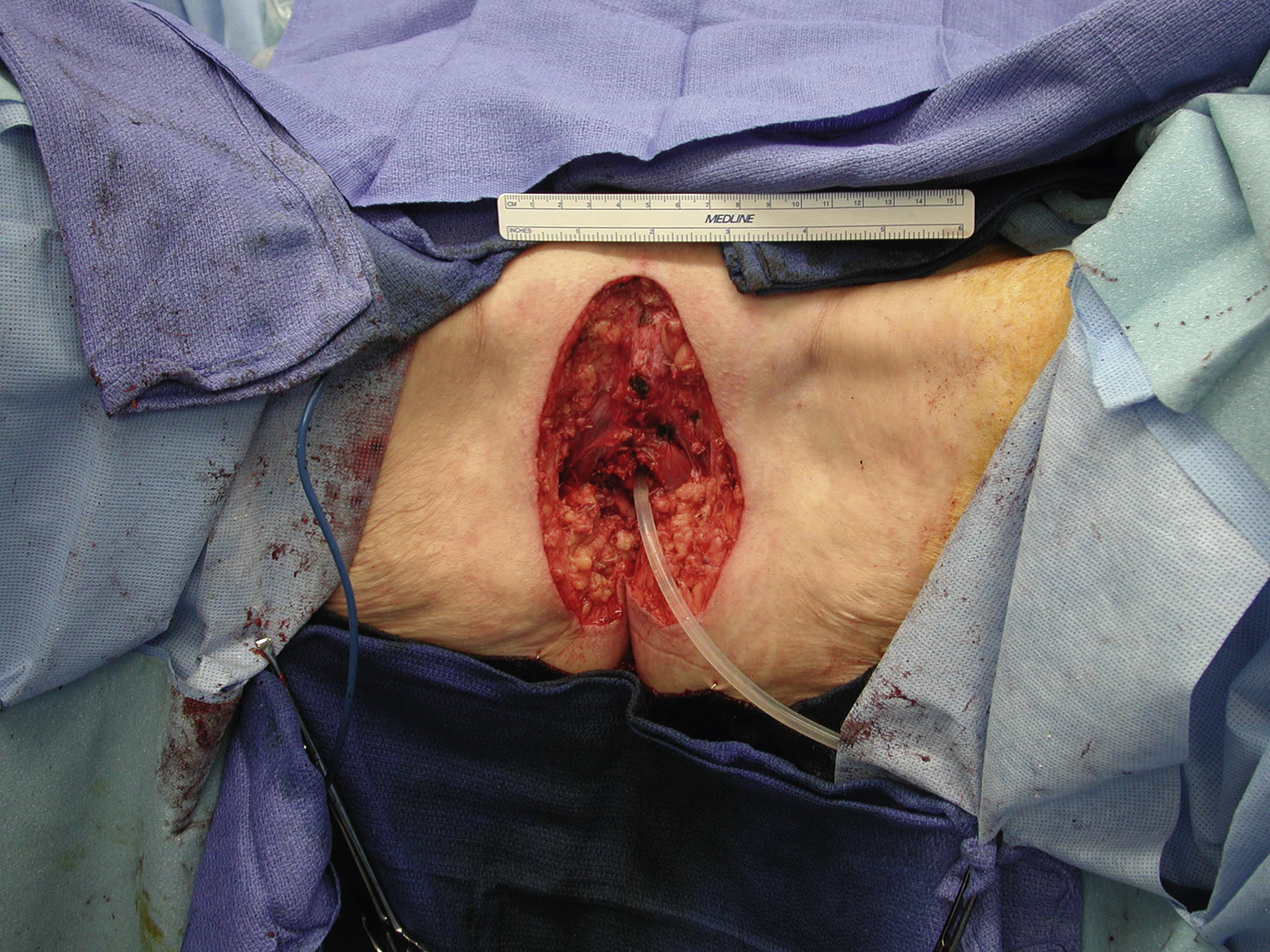
Clinical Presentation
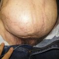
An 86-year-old White female underwent a wide local excision (WLE) for a vulvar squamous cell cancer (SCC) by the gynecologic oncology service. A 15 × 8-cm large soft tissue defect remained over the vulva down to the deep muscles and tissues. The plastic surgery service was asked to help to close this large vulvar wound once the WLE was confirmed to be adequate for a vulvar cancer surgical excision. Therefore, the definitive soft tissue reconstruction could be performed in the same setting immediately after oncological WLE of the vulvar SCC cancer ( Fig. 36.1 ).
Operative Plan and Special Considerations
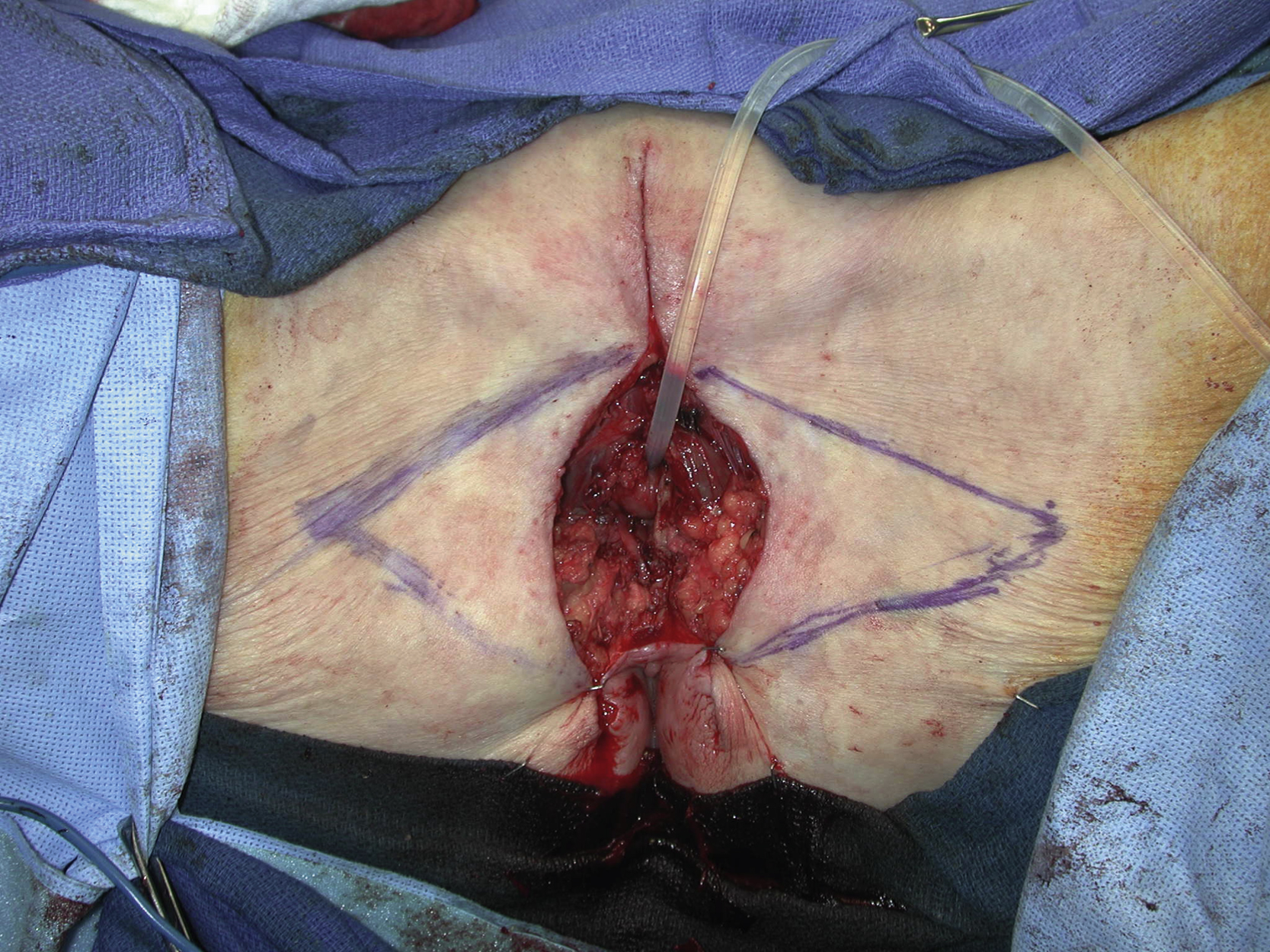
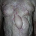
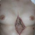
The shape and size of this vulvar soft tissue defect and availability of the adjacent normal perineal and thigh skin determine that bilateral V-to-Y skin advancement flaps could be designed to close the defect after complex closure for direct approximation for both superior and inferior aspects of the defect based on an intraoperative decision. The closure with such bilateral V-to-Y advancement flaps may provide a relatively simple reconstruction with almost no donor site problems. It would be a better option than a skin graft procedure for durable vulvar soft tissue reconstruction. Wound separation after any flap reconstruction is common in this location and the patient should be well informed about such a complication.
Operative Procedures
Under general anesthesia, the patient was placed in the lithotomy position and the vulva soft tissue defect was assessed. A Folly catheter was placed prior to surgical excision. The superior and inferior areas of the defect could be closed after simple skin undermining. Bilateral skin V-to-Y advancement flaps were designed and the skin marking was extended to the proximal thigh. The extent of the flap dissection was outlined for each side ( Fig. 36.2 ).
Related posts:


Stay updated, free articles. Join our Telegram channel

Full access? Get Clinical Tree


