Clinical Presentation
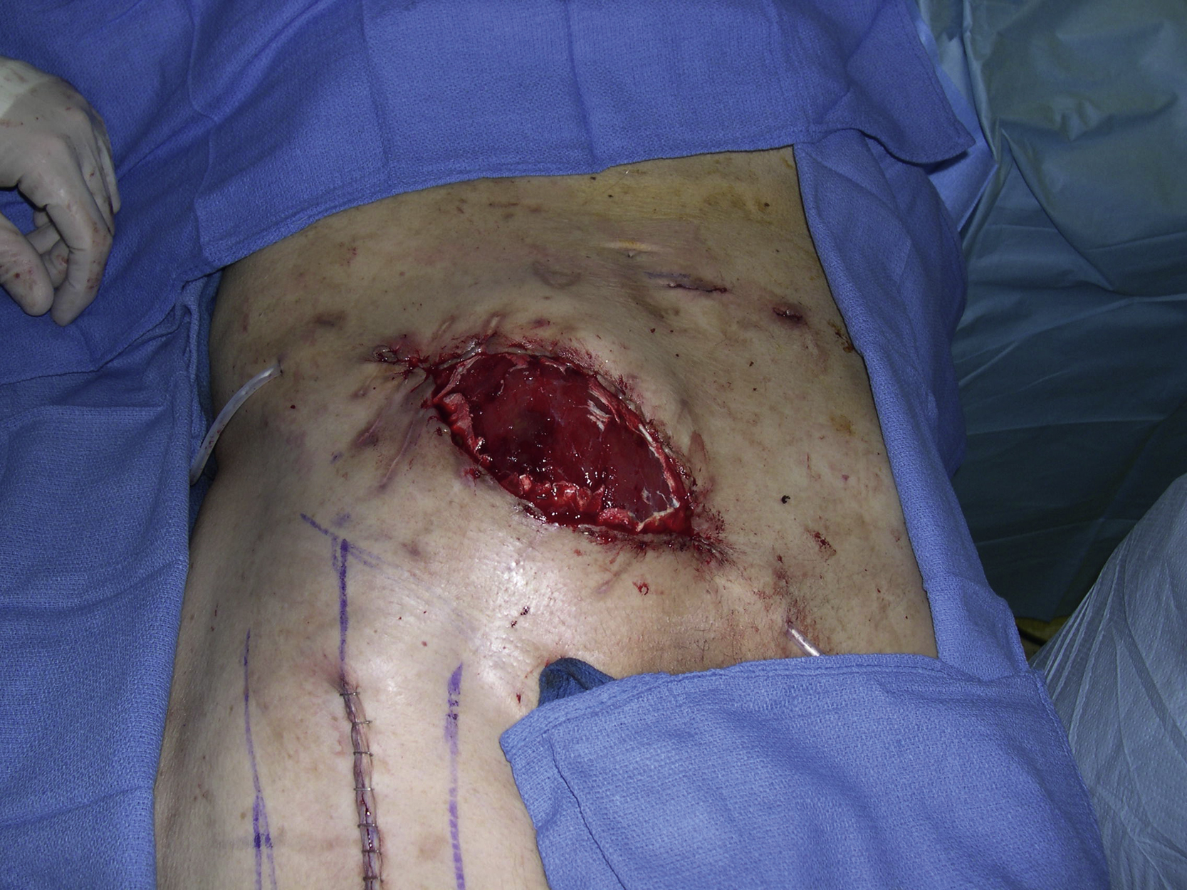
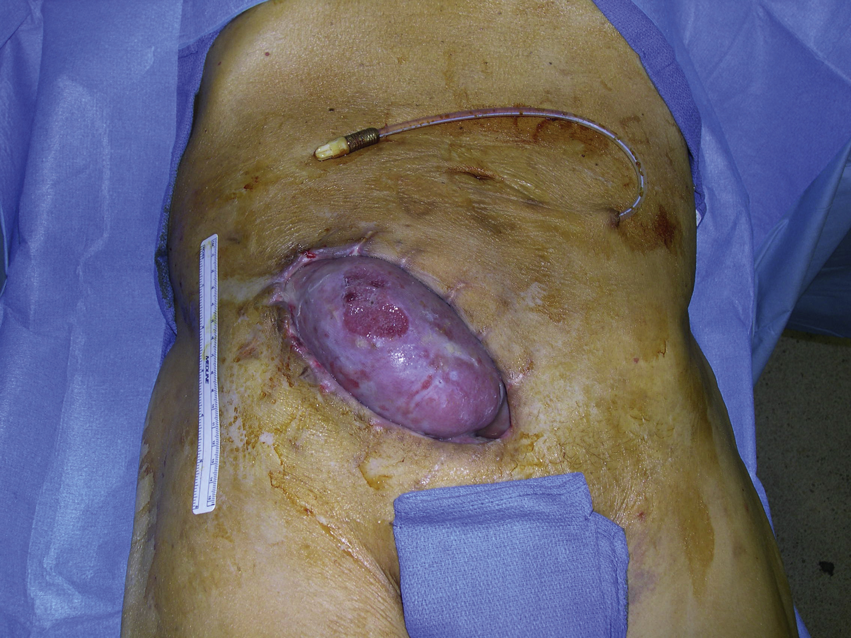
A 66-year-old White male had a renal transplantation for end-stage renal disease secondary to polycystic kidney disease by the transplant surgery service. He also had 50 pack-year of smoking history. He had chronic hypoalbuminemia caused by peritoneal dialysis. Although he underwent successful deceased-donor transplantation, he required peritoneal dialysis because his early allograft function was delayed. One week after surgery, he was found to have an enlarging perigraft hematoma. He was taken to the operating room for hematoma evacuation. Over the following days, he continued to require dialysis and developed a superficial infection of lower abdominal incision, which was opened at the bedside and packed with gauze. He was discharged home with a vacuum-assisted closure dressing and continued ongoing peritoneal dialysis. One week later, it was noted that the superior pole of the transplanted kidney was extruding from the wound and the plastic surgery service was consulted for a soft tissue reconstruction of wound closure ( Fig. 27.1 ).
Operative Plan and Special Considerations
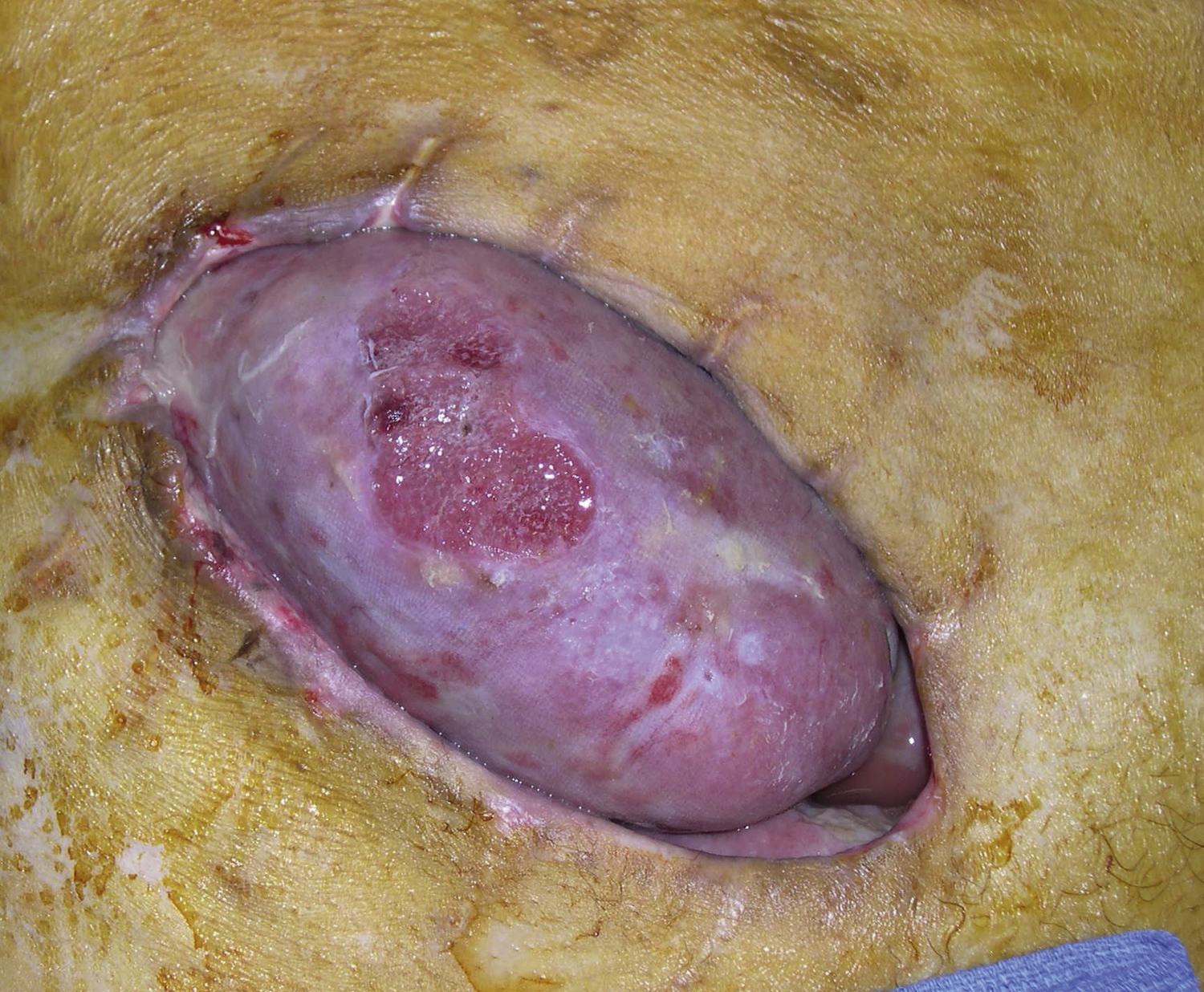
After examining the open wound over the right lower quadrant of the abdomen, it appeared that the entire incision for the kidney transplant was dehisced. The surrounding wound edges were not healthy but the transplanted kidney, confirmed by biopsy, was still functioning ( Fig. 27.2 ). A pedicled right rectus femoris muscle flap with a skin graft was selected for reconstruction of this right lower quadrant soft tissue wound. The flap has type II circulation pattern and receives blood supply dominantly from the descending branch of the lateral circumflex femoral artery and venae comitantes. It can be used as an alternative option for lower abdominal wall reconstruction. It is not an expandable muscle but approximation of the distal edges of the vastus medialis and lateralis muscles after the flap elevation may help to minimize impairment of full leg extension.
Operative Procedures
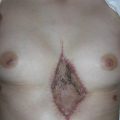
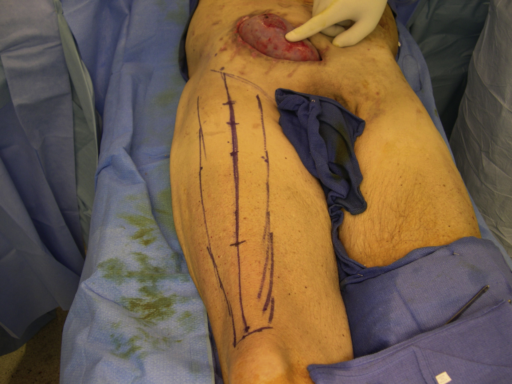
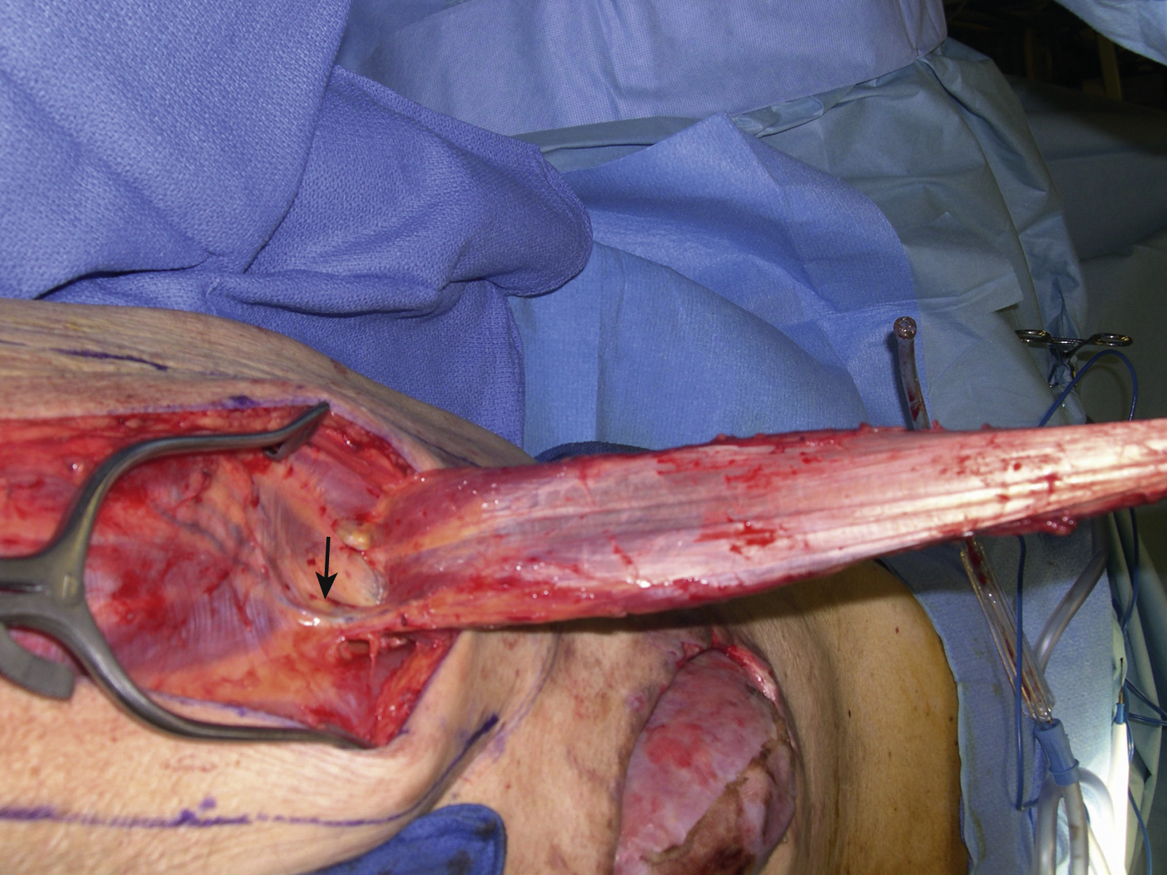
Under general anesthesia with the patient in the supine position, surgical debridement was performed to remove all necrotic, infected, or colonized tissues within the wound including the surface of the exposed transplanted kidney. The outline of the rectus femoris muscle flap and the planned incision for the flap elevation were marked in the right thigh ( Fig. 27.3 ). The rectus femoris muscle was identified first after the skin incision had been made down to the fascia. The surgical dissection could be done quickly once the vastus lateralis and medialis muscles were dissected free from the rectus femoris muscle. The distal insertion of the muscle to the patella was divided and the muscle could be quickly elevated ( Fig. 27.4 ). Once the pedicle had been identified, the proximal insertion was divided. The flap was now completely dissected free and tunneled to the right lower abdominal wound.
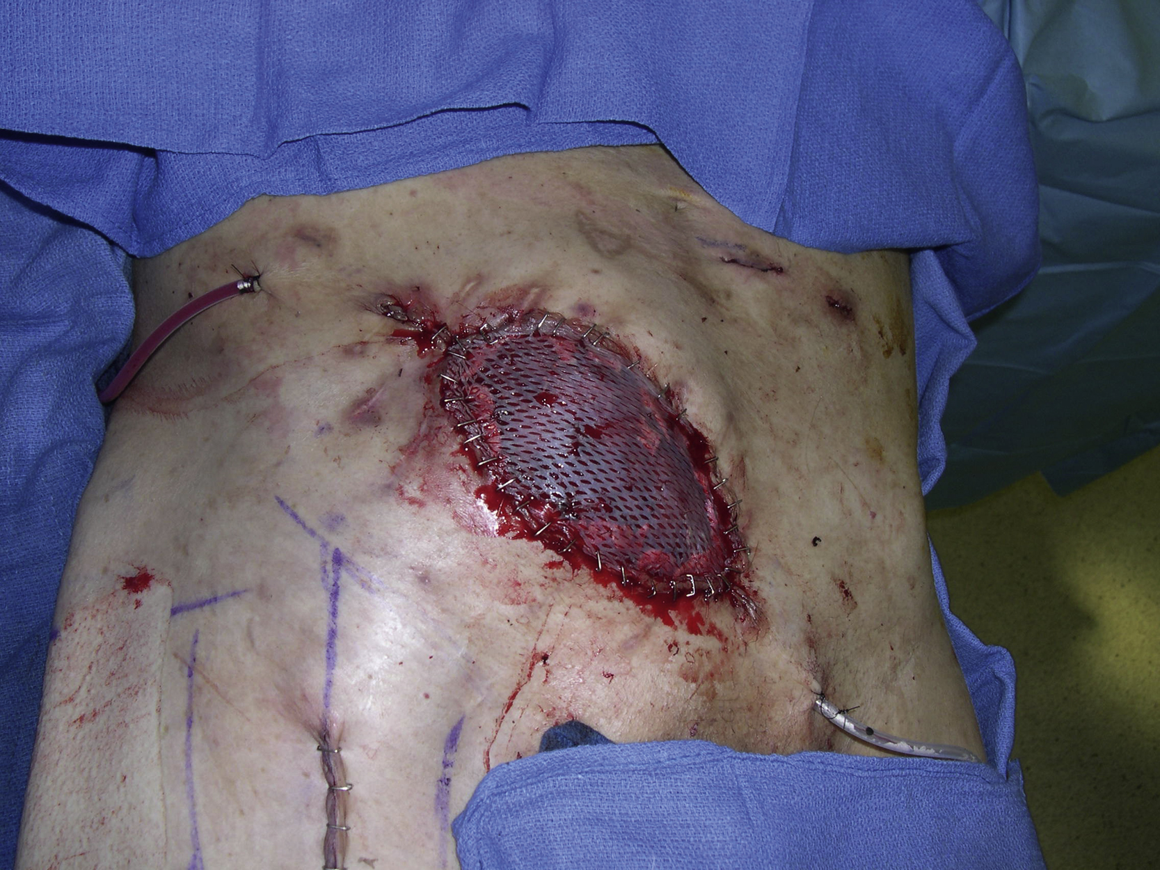
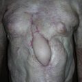
The flap was then placed onto the exposed kidney and approximated to the adjacent skin edges of the open wound with interrupted 3-0 Monocryl sutures in half-buried horizontal mattress fashion after drain placements ( Fig. 27.5 ). A split-thickness skin graft was harvested from his right thigh and meshed to 1 to 1.5 ratio. It was placed over the muscle flap and secured with skin staples ( Fig. 27.6 ). The donor site incision was closed in two layers over a drain ( Fig. 27.7 ).