Clinical Presentation
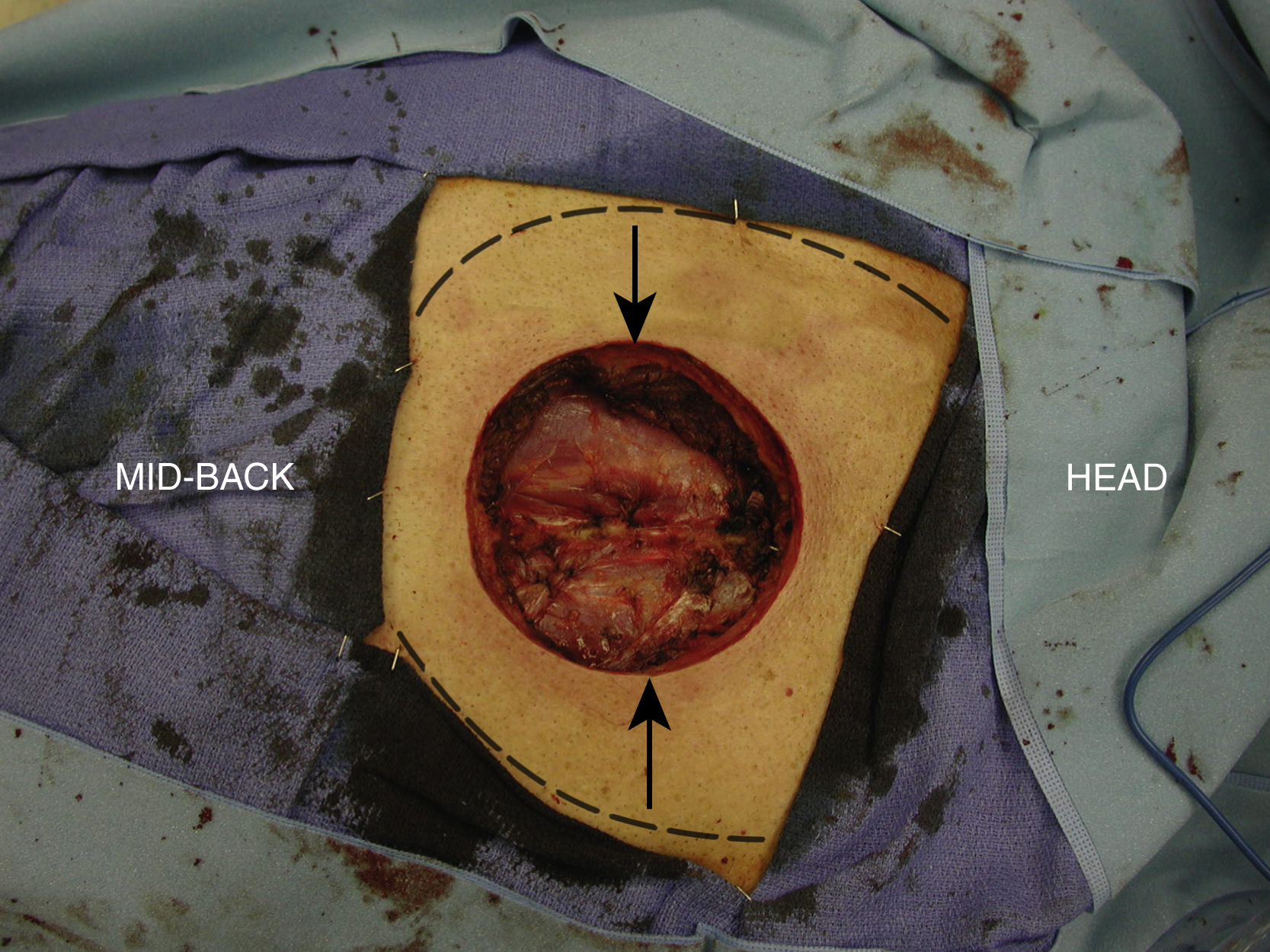
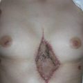
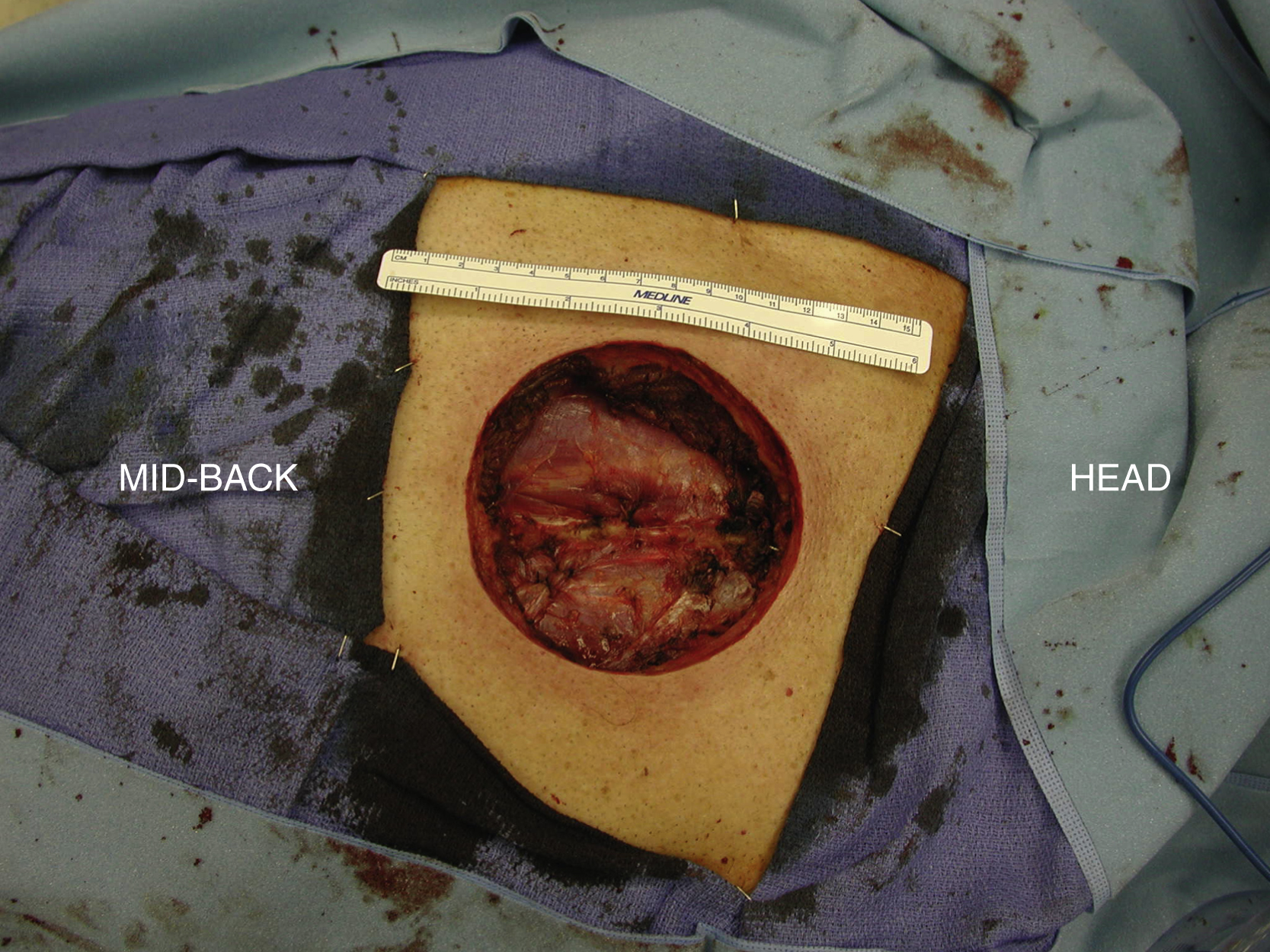
A 63-year-old White male underwent a wide local excision of his upper back soft tissue sarcoma by the surgical oncology service, which left a 12 × 12 cm large soft tissue defect down to the deep muscle. The plastic surgery service was asked to help to close this wound after the wide local excision had been made for the primary sarcoma resection leaving an adequate margin. Thus, the definitive soft tissue reconstruction would be performed in the same setting immediately after oncological resection of the primary tumor ( Fig. 28.1 ).
Operative Plan and Special Considerations
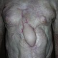
Because the location of this soft tissue defect was primarily in the upper back and the trapezius muscle in the adjacent area remained intact after the wide local excision, bilateral trapezius myocutaneous advancement flaps could be used to close this soft tissue defect. The flap receives blood supply from the transverse cervical artery and can be approximated in the midline of the upper back. Its attachment to the thoracic vertebrae could be released. Such a reconstruction could eventually provide a durable soft tissue coverage, best possible cosmetic outcome, and almost no donor site healing issues.
Operative Procedures
Under general anesthesia, the patient was placed in the prone position. Once the wide local excision had been completed by the surgical oncology service, the soft tissue defect was inspected. The paraspinal muscles were exposed at the base of the wound. Within the upper back wound, each side of the medial trapezius muscle edge was identified. The extent of the flap dissection was outlined for each side ( Fig. 28.2 ). The flap elevation was started by detaching its muscle attachment to the thoracic vertebrae. Once such a surgical dissection had been performed adequately, submuscular dissection was performed so that the muscle and its overlying subcutaneous tissue and skin were elevated as a single unit. Such a dissection could be easily performed with the aid of a mammary light retractor and electrical cautery. During the dissection, the main pedicle vessels was visualized under the muscle and should be protected.