Single-Bone Forearm Reconstruction
Carley Vuillermin
Operative Indications
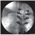
Severely maligned forearm with dislocated radial head proximally and distal radioulnar joint (DRUJ) at wrist (Figure 12.1)
Most commonly secondary to multiple exostosis/osteochondromatoses, but also with Ollier multiple enchondromatoses, neuropathic conditions such as brachial plexopathy and cerebral palsy, and congenital dislocations
This operation is highly effective for the severe deformity, especially in the young, as a single-stage reconstruction with low chance of future surgery
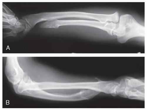 Figure 12-1 ▪ Anteroposterior (AP) (A) and lateral (B) radiographs of chronic forearm deformity from osteochondromatosis with dislocated radial head at elbow and distal radial-ulnar joint at wrist. |
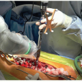
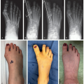
 Figure 12-2 ▪ Photograph of dislocated radial head and malaligned forearm in a 2-year-old child for single-bone reconstruction at early age. (Courtesy of Children’s Orthopaedic Surgery Foundation.) |
Alternative Treatments
Natural history
Skeletal realignment with corrective osteotomies, bone lengthening, and joint reduction of radiocapitellar joint, proximal radioulnar joint, and distal radioulnar joint (DRUJ) in similar cases of severe skeletal imbalance and joint dislocations
Frequent repeat surgeries throughout growth
Equipment
Fluoroscopy
Radiolucent arm and hand table
Tourniquet
Power drill
Power saw
Osteotomes
Smooth C-wires
Standard dissecting kit
AO small fragment and modular hand set (depending on the size of forearm)
Corticocancellous allograft
Casting materials and cast saw
Positioning
Supine with radiolucent arm and hand table
Surgical Approach
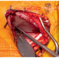
Extensile exposure for the entire length of forearm from elbow to wrist (Figure 12.3)
Proximal curvilinear aspect of incision allows for anconeus posterolateral approach to radiocapitellar joint
Incision straightens out and extends distally along the ulnar border to the level of DRUJ
Elevation of periosteum for the entire length of ulna, with circumferential diaphyseal exposure
Proximal Kocher exposure of dislocated radial head
Protect posterior interosseous nerve
Distal exposure to ulnar head protecting triangular fibrocartilage complex (TFCC)
Protect dorsal ulnar sensory nerve
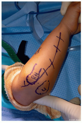 Figure 12-3 ▪ Extensile incision outlined for complete exposure for proximal radial head resection; diaphyseal realignment, bone grafting, and fixation; and distal ulna resection. (Courtesy of Children’s Orthopaedic Surgery Foundation.) |
Technique in Steps ( Video)
Video)Resect Radial Head
Enter elbow joint and debride pulvinar and synovitis with a rongeur
Expose dislocated radial head completely while protecting more distal
Posterior interosseous nerve (PIN)
Biceps tendon insertionRelated posts:

Stay updated, free articles. Join our Telegram channel

Full access? Get Clinical Tree