and Frank Hölzle2
(1)
Department of Oral and Maxillofacial Surgery, Klinikum rechts der Isar, Technische Universität Munich, Munich, Germany
(2)
Department of Oral and Maxillofacial Surgery, University Hospital of RWTH Aachen University, Aachen, Germany
Electronic supplementary material
The online version of this chapter (doi:10.1007/978-3-319-53670-5_8) contains supplementary material, which is available to authorized users.
8.1 Development and Indications
The subscapular vascular system and its suitability for flap harvesting first was investigated in an anatomic study by Saijo in 1978 [459]. Two years later, Dos Santos made use of these previous anatomical findings [122]. He described the scapular flap as a lipocutaneous flap, nourished by a transverse septocutaneous branch from the circumflex scapular artery. This flap, the axis of which was oriented inferior and parallel to the scapular spine, was successfully transferred by Gilbert already in 1979 [167]. Following further, more detailed anatomical studies [168, 353, 541], a number of clinical series were reported using this flap, which was soon accepted as another useful tool for covering soft tissue defects [35, 171, 196, 547, 555]. A variation of this flap was described in 1982 by Nassif and coworkers, who proposed to use the descending septocutaneous branch of the circumflex scapular artery as the nourishing skin vessel [398]. Thus they designed the skin paddle of this parascapular flap along the lateral border of the scapula. Already in 1981, Teot and coworkers published that from an anatomical point of view all preconditions are fulfilled to build purely osseous flap from the scapula bone [541]. Nevertheless, it was not earlier than 1986 when it was popularized to harvest osteocutaneous flaps by including the lateral border of the scapula [499, 521]. Since that time, the indications of flaps raised from the scapular donor site have been considerably expanded [31, 132, 499, 521]. Due to the fact that the vascular pedicle develops from the same source artery like the latissimus dorsi flap, both flaps can be combined using only one set of anastomoses at the subscapular vessels [397]. The indicational spectrum of flaps raised from the scapular region thus in the head and neck area reaches from contour augmentations using de-epithelialized adipo-fascial flaps to the closure of extended perforating composite defects with simultaneous mandible reconstruction using osteomyocutaneous scapular and latissimus dorsi flaps [31, 101, 102, 261, 432, 437, 554]. Moreover, a number of useful applications soon were described for defect coverage at the upper [66, 150, 224] or lower extremities [79, 106, 168, 295, 488, 555].
8.2 Anatomy
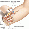
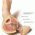
The circumflex scapular artery represents one of the two main branches of the subscapularis artery, which has a diameter of 3–4 mm at its origin from the distal third of the axillary artery [333]. During its course to the scapula region, the artery, which is accompanied by two comitant veins, has to penetrate the posterior triangle. This triangle is built by the teres muscles and the long head of the triceps. After giving off small branches to the surrounding muscles, the circumflex scapula artery (CSA) divides into a deep and superficial branch, the first of which is running underneath the teres major muscle and dividing into terminal branches to reach the periosteum of the lateral border of the scapula bone. Blood supply to the periosteum of scapula was investigated by Coleman and Sultan [101]. According to their findings, an angular branch, nourishing the tip of the scapula, arose from the thoracodorsal artery just proximal to the serratus branch in 58% of all cases, so that the tip of the scapula can be transferred on the thoracodorsal vessels, too. This angular branch first was described by Deraemacher et al., who reported on the possibility to transfer the tip of scapula together with the serratus anterior muscle on the thoracodorsal artery [119]. In a detailed anatomical study, the angular branch was found to travel between the serratus, subscapular, and teres major muscles to the inferior angle of the scapula. The second main branch, the superficial branch of the circumflex scapular artery, divides into the transverse and descending cutaneous branches to perfuse the scapular and the parascapular skin flaps, respectively.
Whereas in more than 100 cadaver dissections the transverse branch has been found to be always present, having a diameter of 1.5–2.5 mm [123, 168, 555], Godina was unable to identify this cutaneous vessel in 3 of 28 clinical cases [171]. When raising the skin paddle as a scapular flap, flap axis is outlined below and parallel to the spine of scapula. According to Urbaniak et al., the limits of the skin paddle should be 2 cm below the scapular spine, 2 cm above the angle and 2 cm lateral to the midline [555]. Although in an anatomical dissection it was shown that the vascular tree passes the midline and reaches the contralateral acromion [240], Hamilton pointed out that the maximum length of a scapula skin paddle should not have more than 24 cm and may not reach the midline because of the risk of necrosis of the tip of the skin island [196]. By additionally anastomosing the flap to the contralateral circumflex scapular artery, a 50 × 10 cm large bi-scapular flap can be harvested [36, 140]. When planning a parascapular skin island, the flap axis is oriented above the lateral scapular border, which can have a length of 25 cm [92] or even 30 cm [492]. Because the terminal branches to the skin are forming a dense network of anastomoses, building a subdermal and epifascial vascular plexus, fascio-subcutaneous and deep subcutaneous flap compartments can be transferred separately and used for contour augmentations [153, 554]. The length of the vascular pedicle depends on the extent of proximal dissection. If the vascular pedicle is limited to the circumflex scapular artery, its maximum length will be 7–10 cm. Lengthening of the pedicle up to 11–14 cm is possible by including the subscapular vessels, transecting these vessels at their point of takeoff from the axillary artery and vein [398]. The circumflex scapular artery is joined by two comitant veins, having sizes between 2.5 and 4 mm. In the majority of cases, these veins unify with the thoracodorsal vein or, which is the case in 10%, enter the axillary vein separately [123].
8.3 Advantages and Disadvantages
The major advantages of the scapular skin flap become obvious when comparing it with the osteocutaneous flaps of other donor sites: the skin of the scapular flap mostly is hairless and similar to the facial skin in texture and color. It is carrying only a thin layer of adipose tissue, and primary closure of the donor site is possible up to a flap width of 8–10 cm. Moreover, the vascular pedicle can be dissected to an acceptable length and is having a high caliber. There are only few anatomical variations concerning the vascular pedicle, and flap design can be variable. The possibility to simultaneously raise latissimus dorsi [397] or parascapular flaps, which can be left pedicled at the same source artery, further enlarges the indicational spectrum [499, 521]. To lengthen the pedicle, raising of reverse flow flaps is possible, performing anastomoses to the distal end of the thoracodorsal artery and subscapular vein, respectively [630]. Up to four flap components can be created, each of them offering the possibility for free and independent positioning [165, 416, 437]. Because of the specific architecture of the scapula, reconstructions of the maxilla can be performed using the blade of the bone to replace the hard palate [261, 437, 559, 629], but also specific indications for mandible reconstructions using the scapular tip have been described [624]. Primary closure of the donor-site defect mostly is possible even following harvesting of wide flaps, but unacceptable broad scars can result, if tensionless wound closure is impossible [333]. Raising of combined scapular and parascapular flaps is always limited by the ability to achieve direct closure [437]. To avoid the application of skin grafts, pretransfer expansion of the skin has been performed in suitable cases [554]. By prelaminating the bone with dermis and simultaneous insertion of endosseous implants, reconstructions of the alveolar ridge with a mucosa-like surface are possible [476], and raising of an additional skin flap can be avoided. Even after harvest of osteocutaneous flaps, which makes transection of the teres muscles necessary, disability of the shoulder is reported to be low [102, 416, 559]. This is even more the case when raising scapular tip flaps only, leading to very low shoulder morbidity [146]. Postoperative care includes immobilization of the arm for 3–4 days and physiotherapy to strengthen the muscles of the shoulder girdle, starting about 2–3 weeks after surgery.
The main disadvantage of the scapular donor site is given by the fact that simultaneous flap raising is impossible when tumor resections in the head and neck area have to be carried out. In these cases, flap harvesting cannot be started until tumor resection is completed, so that a considerable time loss will result; additionally, new positioning and re-prepping of the patient is time consuming, too. Whereas identification of the cutaneous branches normally is achieved quickly, dissection of the vascular pedicle by working through the posterior triangle can become difficult in the raising of cutaneous flaps, especially if a long pedicle is needed [131]. To simplify dissection of the vascular pedicle, Gahhos et al. proposed to perform a second incision at the axilla, which facilitates identification of the subscapular vessels [155]. Then, the flap can be pulled toward the axilla to obtain maximum length of the pedicle.
8.4 Patient Positioning
Flap elevation is performed with the patient in prone or lateral decubitus position. Shoulder, back, lateral thorax, and upper arm are circularly prepped to allow for movement of the extremity and exposure of the subscapular system from an axillary approach, if needed. In the lateral decubitus position, bags are used to stabilize the patient and to protect the dependent shoulder. Preoperatively, the location of the lateral circumflex scapular artery (CSA) is identified using a Doppler in the triangular space at the lateral rim of the scapula. Due to constant anatomy, angiography is only necessary if operative procedures at the donor site (axillary lymphadenectomy) have been performed previously.
8.5 Flap Design
Skin paddles can be elevated along the axis of the transverse (scapular flap) or descending branch (parascapular flap) of the CSA. In the standard situation, a scapular flap is outlined, keeping the upper, lower, and medial margins of the flap at least 2 cm away from the scapular spine, inferior angle, and posterior midline. Angle, spine, and lateral border of the scapula have to be palpated before outlining the flap. For both the scapular and the parascapular flaps, it is critical to outline the lateral part of the skin paddle safely above the triangular space, where the CSA runs along the fascial septum between the teres major and minor muscles to enter the posterior thoracic fascia and the skin. This triangular space is found either by palpation of the muscular groove lateral to the scapular bone or, more exactly, by preoperative mapping of the artery using a Doppler. The width of the flap may not exceed 8–10 cm to allow for direct closure. The bone segment is harvested from the lateral scapular border, inferior to the glenohumeral joint and mostly including the inferior angle (◘ Figs. 8.1, 8.2, 8.3 and 8.4).
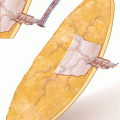
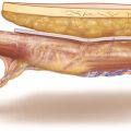
Circumcision of Medial Pole of Scapular Skin Paddle to the Deep Fascia
Starting medially, skin and subcutaneous fatty tissue are incised to the deep fascia overlying the infraspinatus muscle. The fascia, which consists of multiple layers, is included at the undersurface of the flap, but the deepest layer of fascia, directly covering the muscle fibers, is left intact (◘ Fig. 8.5).
Identification of Superficial Branch of the Circumflex Scapular Artery
The dissection proceeds to the lateral direction by bluntly separating the fasciocutaneous flap from the infraspinatus and teres minor muscle, until the posterior muscle triangle is reached. Here, the position of the CSA has already been marked at the skin preoperatively using a Doppler. The pulsation of the cutaneous branch, which is enveloped in the fascia, can now be seen and palpated easily. After the cutaneous branch has been exposed, the skin paddle is peritomized at its lateral portion and completely elevated (◘ Fig. 8.6).
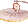
Dissection into Triangular Space, Identification of Deep Branch of CSA
Now, the CSA is traced proximally, and the fascial space between the teres minor and major muscles is opened. The lateral margin of scapula is identified by retracting the teres minor medially to expose the perforators to the bone, branching off from the deep segment of the CSA. A vessel loop is placed around the CSA proximal to the bone feeders, which are carefully protected during further flap raising (◘ Fig. 8.7).
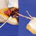
Identification of Bone Feeders
In the close-up view, the deep segment of the CSA is visible, giving off three branches to the proximal lateral border of the scapula. The cutaneous branch is coursing directly into the undersurface of the skin paddle, where it divides into the horizontal (scapula skin paddle) and the descending branch (parascapula skin paddle) (◘ Figs. 8.8 and 8.9).
Incision of Infraspinatus and Teres Minor Muscles to Gain Access to Scapular Bone
To gain access to the scapular bone, the infraspinatus muscle is incised 3 cm parallel to the lateral border of scapula, leaving a muscle cuff attached to the bone. The muscle is transected completely, starting at the inferior angle of scapula and ending cranially to the bone feeders (◘ Fig. 8.10).
Transection of Muscle Fibers Cranially
Cranial to the branches of the CSA to the bone, the teres minor and infraspinatus muscles are transected perpendicular to the muscle fibers to prepare for the osteotomy (◘ Fig. 8.11).
Undermining the Inferior Angle of Scapula
The teres major, which originates from the inferior angle and lateral rim of scapula, is separated from the latissimus dorsi at the inferior angle, and the caudal portion of scapula is undermined (◘ Fig. 8.12).
Undermining and Elevation of Teres Major Muscle
The teres major now is elevated and undermined, so that the angular branch of the thoracodorsal artery becomes visible. Although this vessel contributes to the blood supply of the tip region, it can be transected without endangering the viability of the scapular bone flap. If an isolated angular bone segment is planned, the vascular pedicle should include the thoracodorsal artery, and the angular branch is left intact (◘ Fig. 8.13).
Related posts:
Stay updated, free articles. Join our Telegram channel

Full access? Get Clinical Tree


