and Frank Hölzle2
(1)
Department of Oral and Maxillofacial Surgery, Klinikum rechts der Isar, Technische Universität Munich, Munich, Germany
(2)
Department of Oral and Maxillofacial Surgery, University Hospital of RWTH Aachen University, Aachen, Germany
Electronic supplementary material
The online version of this chapter (doi:10.1007/978-3-319-53670-5_9) contains supplementary material, which is available to authorized users.
9.1 Development and Indications
The first microvascular bone transfer was performed by Taylor and coworkers, who used a vascularized myoosseous segment of the fibula to treat a posttraumatic defect of the tibia in 1975 [539]. Since this first description, the primary indications for the fibular bone flap have been reconstructions of extended bone defects in the extremities by using a posterior approach for flap harvesting, which was originally described by Taylor et al. [539].
Whereas these first transfers of the fibula have been performed without including a skin paddle, Chen and Yan were the first to report about an osteocutaneous fibula flap in 1983 [88]. This extension of flap raising became possible following the proposal of Gilbert to use a lateral approach for harvesting the bone flap, which was easier to perform and allowed for visualization of the cutaneous branches of the peroneal artery [167]. A valuable extension of the indicational spectrum of the fibular flap was achieved by Hidalgo, who in 1989 performed the first lower jaw reconstruction using osteotomies to mimic the shape of nearly a whole mandible [217]. Since that time, the fibula flap has proven to be a valuable method for mandible reconstruction, especially in extended defects, exceeding the length of half a mandible [88, 151, 217, 327, 468, 491, 583, 596]. By inclusion of the soleus muscle, which than was connected to motor branches at the recipient site, restoration of motor function was achieved [96]. Because of the bone length and the potential to vary the position of the skin paddle, it is possible to combine bone segments and skin islands from different parts of the flap, allowing more flexibility in flap design [586, 596, 608]. Moreover, two separate skin paddles can be harvested and used for closure of through and through defects of the cheek, simultaneously reconstructing the mandible using the fibula bone [151, 217, 586, 596]. To overcome the limited height of the fibula, Jones introduced the possibility to fold two osteotomized bone segments over itself [257]. This “double barreled” fibular flap was first used for reconstruction of segmental defects of the femur, until this method was adopted to mandible reconstruction. To restore sensation of the skin paddle, Hayden and O’Leary harvested the sural cutaneous nerve together with the skin island and anastomosed this nerve to sensible nerves of the oral cavity [210]. Sensate fibular flaps later were used for penile reconstruction too [457]. Flap combinations were performed by anastomosing a second free flap to the distal peroneal artery and vein, which do not reduce in caliber significantly and thus additionally can serve as “donor vessels” at the recipient site [583].
9.2 Anatomy
The dominant vascular pedicle of the fibular flap is the peroneal artery which develops from the posterior tibial artery. Together with the tibial anterior artery, it represents one of the three main branches of the popliteal artery. Accompanied by two veins, the lateral of which is usually larger [190], the peroneal artery travels distally between the flexor hallucis longus and tibialis posterior muscles and, besides the muscular branches, gives off several periosteal and medullary branches to the fibula bone as well as a number of cutaneous perforators, running along the posterior intermuscular septum to the skin of the lateral calf. Normally, the peroneal artery does not contribute to the blood supply of the foot significantly, but due to a number of anatomic variations concerning the tibial anterior or posterior vessels, the peroneal artery can become a dominant nutrient vessel for the food. According to the anatomic literature, the tibial anterior and posterior vessels can only be rudimentally present or completely missing [156, 163, 177, 216, 229, 237, 241, 251, 312, 383, 421], so a preoperative angiography or magnetic resonance tomography has to be performed to evaluate the vascular anatomy of the donor site [228, 539, 596, 608, 628]. If one of the three main arteries is significantly reduced in caliber or missing, no flap raising should be performed at this leg. Additionally, atherosclerotic changes will lead to an increased risk for flap loss, and possibly, long-term ischemic complications at the donor site will occur, so in these cases, another donor site should be considered. Although the venous anatomy was found to be unique in every individual, no contraindications were found from a venous standpoint to raise fibula flaps. The two venae comitantes do not necessarily coalesce into a single common peroneal vein, but in 66%, they unify to a common peroneal vein and can be lengthened up to the confluence with the popliteal vein. Nevertheless, because of some anatomical exceptions, the choice of donor vein and venous pedicle length should be made dependent on the intraoperative presenting anatomy [190].
Although the fibular bone is non weight-bearing, 7–8 cm segments must be preserved at the proximal and distal ends during flap raising to protect the common peroneal nerve at the fibular neck and prevent instability of the ankle. Despite these limitations, up to 25 cm of bone length can be harvested, which are enough to restore subtotal or even total mandibular defects [167, 217]. For osteocutaneous transfer of the fibular flap, location and course of the cutaneous perforators are of particular interest. According to clinical experience and anatomical studies which have been performed to evaluate the reliability of blood supply to the skin, the cutaneous perforators of the peroneal artery vary in location, course, size and number. Thus, different survival rates of the skin paddle have been reported, and different proposals have been made to improve reliability of the fibular flap skin island. Hidalgo, who performed five osteocutaneous fibula transfers in his first series of 12 patients, reported of four complete or partial losses and only one complete survival of the skin paddle [217]. To increase the number of cutaneous perforators and thus the safety of skin perfusion, he, therefore, suggested to always include the whole posterior intermuscular septum, independent of the size of the skin paddle [219]. Because of the anatomical variations of the perforating vessels, Urken [556] considered loss rates between 7% and 10% to be unevitable [556]. In an antomic study on 52 cadavers, Chen et al. found 4–7 cutaneous branches, most of them having a myocutaneous course and perforating the soleus muscle [88]. Another description of the cutaneous arteries was given by Yoshimura et al., who differentiated myocutaneous perforators, travelling through the peroneal muscles; septomyocutaneous perforators, running between the peroneus and soleus muscles and giving off further muscular branches; and purely septocutaneous vessels [627]. A different classification was proposed by Wei and coworkers, who only made a distinction of septocutaneous perforators, traversing the whole intermuscular posterior septum, and musculocutaneous perforators, additionally coursing through either the peroneus, tibialis posterior or soleus muscle [583]. In a later publication, these authors reported about a 100% survival rate of the skin paddle in more than 100 patients by centering the skin paddle over the transition of the middle and distal third of the fibula [586]. In contradiction to the findings of Wei, Carriquiry could only identify septocutaneous perforators in their ten anatomical dissections [70]; this was supported by Carr and coworkers [67], who stated that the cutaneous perfusion exclusively is maintained by septocutaneous vessels. In an extensive anatomical dissection on 80 cadavers and supported by their clinical experience on 18 patients, Schusterman et al. could find 3.7 cutaneous perforating vessels from the peroneal artery in average, 1.3 of these having a septocutaneous, 1.9 a myocutaneous course, and 0.6 showed a direct adhesion on the muscle fascia without penetrating the muscles [482]. Because of this variability, the authors proposed always to include a cuff of tibialis posterior and soleus on either side of the septum for safety reasons. A similar suggestion already has been made in 1986 by Harrison, who could improve his success rate with the skin paddle using this method [203]. Despite this, Van Twisk considered the inclusion of a muscle cuff to be necessary only if no septocutaneous vessels could be visualized [568]. Yoshimura, who gave the first description on the peronal flap [625], which is nourished by the same cutaneous vessels like the osteocutaneous fibula flap, in his anatomical study on 80 cadavers could find 4.8 cutaneous vessels in average, 71% of these having a myocutaneous course to the skin [626]. Whereas he believed that the skin paddle should be designed at the junction of the middle and distal third of the fibula, other authors proposed to center the flap 2 cm superior to the midpoint between the fibula head and ankle [88, 333, 479, 518] and subfascial incision [151]. If no septocutaneous vessels are identified, part of the soleus muscle has to be included to capture the myocutaneous perforators [151]. As a result of an anatomical study by Wolff, 4.2 cutaneous perforators were found, most of them having a myocutaneous course through the tibialis posterior and soleus muscles at the proximal and a septocutaneous course at the distal lower leg [608]. The most reliable region to build a skin paddle turned out to be 8–12 cm proximal to the ankle, because here a strong perforator, mostly having a septocutaneous pattern, was found in all of the 50 cadavers. As a consequence of these anatomical findings, the author proposed to routinely design the skin paddle at the junction between the medial and the distal third of the fibula, additionally offering the possibility to dissect a long vascular pedicle. Depending on the needed length of the bone segment and the level on which the peroneal artery unifies with the tibial posterior vessels, the vascular pedicle can reach up to 15 cm, if the skin paddle is raised distally [590, 596, 608]. To obtain a long pedicle, Hidalgo proposed to remove the longest fibula segment possible and then to discharge the proximal bone segment after having dissected the pedicle together with the surrounding soft tissues in a subperiosteal plane [218]. Blood supply to the distal fibula segment is not altered by this maneuver. Dye injections have been carried out to determine the territory of skin available for the osteocutaneous fibula transfer. When injecting proximally into the peroneal artery, a skin territory of about 10 cm in width and 20 cm in length is stained, allowing to transfer nearly the whole skin of the lateral calf. Raising such a large skin paddle was considered as problematic because of the extensive donor site skin defect. Therefore, other authors propose to raise large skin flaps from an additional donor site or to use another osteocutaneous flap [327, 586]. Selective injection studies have shown that a skin territory of about 12 × 7 cm is safely perfused by a single perforating vessel [590], giving the anatomical basis to build two separate skin paddles not only by deepithelization [151] but also by complete transection of the flap between both perforators. To facilitate identification of the perforators, preoperative mapping using an audible Doppler is strongly advisable. Direct closure of the skin is achieved up to a flap width of 6–7 cm in the upper and middle third of the lower leg, whereas distally, skin grafts have to be used for wound coverage in most cases. After flap raising, the patient is immobilized for 3–4 days and then allowed to ambulate with physiotherapeutic assistance.
9.3 Advantages and Disadvantages
The fibula is the longest bone flap available and can be transferred as a bone flap or in combination with one or two skin paddles. Its indicational spectrum, therefore, reaches from bony reconstruction at the extremities to replacement of the whole mandible, including closure of large perforating defects of the oral cavity. Flap raising can be carried out using the two team approach, making this donor site attractive especially for primary reconstructions in the head and neck area. The quality of the thin and pliable skin paddle is comparable to the radial forearm skin, and the 3–5 cm broad septum provides good flexibility to the skin island, which can be brought into the oral cavity for lining without tension. Thus, the osteocutaneous fibula flap is perfectly suited for reconstruction of composite defects of the mandible [227]. If the intraoral soft tissue defect is limited, myofascial lining can also be performed, reducing the need of raising a skin paddle and debulking the flap secondarily [138]. The flap possesses a sufficiently long and high-caliber vascular pedicle, making microsurgical anastomoses easy to perform. Although the vertical dimension of the fibula is limited to the half of a toothed mandible, endosseous dental implants can be regularly inserted, reaching a high primary stability due to the high amount of cortical bone [230, 285]. The limited height of the fibula is not a problem in patients already having an atrophy of the alveolar process because there are no considerable differences in the height between the fibula and the atrophied mandible. In nonatrophied, toothed mandibles, the application of a double fibula transplant was suggested to compensate for the narrowness of the transplant in order to create better prerequisites for prosthetic management. The majority of authors were able to show, however, that prosthetic rehabilitation is also possible without using a double transplant [218, 249, 327, 586]. However, thinning of the skin flap around the implant is always necessary before prosthetic rehabilitation can be performed. The accuracy of mandible reconstruction can be improved by three-dimensional guided techniques using patient-specific planning tools. These techniques allow for exactly matching the osteotomies of the mandible to the fibular flap, which can be designed with multiple segments according to the configuration of the defect [375, 548, 587]. Despite the numerous advantages attributed to skin quality, bone length and vascular pedicle, for isolated bone defects of the mandible not exceeding the midline, the iliac crest should be preferred due to its better suitability to mimic a natural shape of the lower jaw.
The frequency of arteriosclerotic changes in the lower leg vessels is a well-known clinical fact and must be taken into consideration in the choice of flaps. Although some authors [121, 218, 334] considered a routine angiography in cases of clinically normal findings at the foot pulses to be not justified, the majority of authors perform assessment of donor site vascular anatomy and status of vascular integrity by preoperative measures like angiography or magnetic resonance imaging (MRI) [323, 344, 539, 596, 627]. According to clinical experience, one out of five candidates has to be excluded from fibula transfer due to severe arteriosclerotic damage or venous insufficiency of the lower leg vessels [596]. The reliability of skin supply was another criticism of the osteocutaneous fibula transplant and has been the reason for numerous studies, in which particularly the variability of the cutaneous perforating vessels and the limited size of the skin island have been reported as disadvantageous [67, 151, 217, 218, 249, 482, 586, 626]. According to the reports by Hidalgo [219] and Schusterman [482], a loss of the skin island must be considered in 7–9% of the cases. On the basis of anatomical studies and clinical experience of other authors [257, 559, 583, 586, 590, 596, 608], the transition of the middle and the distal third of the fibula represents a reliable donor site for the fibula skin paddle, which at this location is supplied by septocutaneous peroneal perforators. Having a survival rate of at least 95%, the safety of this skin paddle does not differ from other proven transplants [257, 559, 586, 596]. The possibility of forming two isolated skin islands, which was already demonstrated by Yoshimura, extends its indicational spectrum [627]. Nevertheless, Yokoo and coworkers pointed out that the perforator of the skin paddle as a variation can branch off from the tibialis posterior instead from the peroneal vessels [623]. In these cases, it is necessary to directly anastomose the perforating vessel or, if no other peroneal cutaneous perforator is available, to use a second skin flap. A neurocutaneous reinnervation by connecting the sural nerve, as suggested by Sadove et al. [457] and Wei et al. [586], is not an absolute requirement for sensory innervation of the flap; rather, it seems in at least some cases that there is spontaneous neurocutaneous reinnervation due to sprouting of sensory fibers from the periphery.
Some authors report the length of the vascular pedicle as ranging from 4 to a maximum of 8 cm so that in many of their cases vein grafts were necessary [151, 327, 568]. The dissectible vascular pedicle length is, however, much longer, if the transplant is raised from the distal third of the lower leg, where not only the skin supply is more reliable via septocutaneous perforating vessels but the fibula is better perfused via periostal branches [608]. Vein grafts may only become necessary if long bone segments are required for subtotal mandible reconstruction because, in these cases, proximal lengthening by separation from the fibula is only possible to a limited extent. A short vascular pedicle can also result, if the exit of the peroneal artery lies more distally, which can, however, be recognized preoperatively by angiography.
According to reports of the literature, donor site morbidity of the fibula flap generally is low. Apart from hypesthesia at the lateral malleolus, slight initial pain and tendency towards edema can be found, and the flexing or stretching function of the large toe or ankle joint is objectively reduced but hardly perceived subjectively [104, 173, 218]. Nevertheless, some patients report about pain and weakness on ambulation for several months after surgery [52, 362, 559], and a lower preferred velocity on walking was found compared with control subjects [52]. Instability of the ankle joint was not found in any of the patients [104, 313]. Radiological signs of osteoporosis can occur in the distal fibula segment after several years, but it does not lead to any disability on ambulation or shape of the ankle joint [313]. Hematomas can occur due to oozing from the resection margins of the bone at the donor site, and care must be taken to prevent development of a compartment syndrome [99]. Primary closure of the donor site defect should only be undertaken if this can be accomplished with no tension, because according to a study of Shindo et al., primary closure otherwise tends to a higher rate of complications compared with split-thickness skin grafting [498]. In order to ensure optimal healing of split-thickness skin grafts, a tie-over dressing should be applied and the lower leg should be immobilized for about 1 week.
9.4 Flap Raising
9.4.1 Preoperative Management
Due to possible variations of the tibial posterior and anterior vessels and the prevalence of arteriosclerotic damage in the lower extremities, conventional angiography or, which is less invasive, magnetic resonance angiography is mandatory before raising the fibula flap. Patients showing clinical signs of vascular damage (varicosis, missing foot pulses, pain on ambulation) should primarily be excluded. Marking of the cutaneous perforators along the posterior intermuscular septum using a Doppler facilitates intraoperative exposure of these vessels.
9.4.2 Patient Positioning
The leg is bent into the knee joint and brought in a prone position to get better access to the lateral and posterior aspect of the calf. This is facilitated by supporting the hip with a beanbag. The entire lower extremity is prepped circumferentially, and the foot is draped, leaving the pulses accessible. No tourniquet is used, because identification of the pulsating perforators is easier in the perfused leg. With consequent hemostasis and careful dissection, flap raising is possible without any significant bleeding, and the risk for diffuse oozing after release of the tourniquet and postoperative hematoma formation (compartment phenomenon) is reduced.
9.4.3 Flap Design
Despite mapping of the perforators and thus positioning of the skin island is possible preoperatively using a Doppler, designing the skin paddle should not be performed until the cutaneous branches are clearly seen intraoperatively. In the standard situation, the skin paddle is centered vertically along the septum with its center at the junction between the middle and lower third of the fibula. If only one perforator is enclosed, the flap size should not exceed 6 × 10 cm. A distance of 8 cm from the lower osteotomy to the ankle must be kept for stability of the malleolar joint; proximally, a 6-cm bone segment is maintained to protect the peroneal nerve (◘ Figs. 9.1, 9.2 and 9.3).
Incision of Skin and Fascia
Skin incision is made along the peroneus longus muscle, keeping a distance of 2 cm to the posterior intermuscular septum, which easily can be palpated posterior to the muscle. According to the location of the perforator found by preoperative mapping, the incision is slightly curved anteriorly in the region of the skin paddle. The strong crural fascia is incised according to the skin incision (◘ Fig. 9.4).
Identification of Perforator
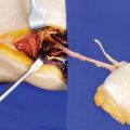
The perforator is visualized by careful separating the fascia from the peroneal muscles and blunt dissection in posterior direction. The posterior intermuscular septum, which covers the perforator from both sides, is exposed and must always be left intact in the region of the osteocutaneous flap. Once the perforator is identified, the peroneal muscles are retracted anteriorly, and the lateral margin of fibula is palpated (◘ Fig. 9.5).
Exposure of Lateral Margin of Fibula
Proximal to the skin paddle, the posterior intermuscular septum is incised sharply along the lateral margin of the fibula. To obtain better access to the deep flexor space, the peroneal muscles are retracted anteriorly and the soleus muscle is retracted posteriorly using sharp hooks. Dorsal to the fibula bone, the flexor hallucis longus muscle becomes visible (◘ Fig. 9.6a, b).
Related posts:
Stay updated, free articles. Join our Telegram channel

Full access? Get Clinical Tree


