and Frank Hölzle2
(1)
Department of Oral and Maxillofacial Surgery, Klinikum rechts der Isar, Technische Universität Munich, Munich, Germany
(2)
Department of Oral and Maxillofacial Surgery, University Hospital of RWTH Aachen University, Aachen, Germany
Electronic supplementary material
The online version of this chapter (doi:10.1007/978-3-319-53670-5_1) contains supplementary material, which is available to authorized users.
1.1 Development and Indications
In 1978, a fasciocutaneous free flap from the volar aspect of the forearm and pedicled on the radial artery was first used in China. When this so-called “Chinese flap” was originally described by Yang et al. in 1981 [619] and Song et al. in 1982 [506], both groups already had performed more than 100 successful flap transfers. Shortly after, this technique was popularized by different European surgeons, who visited their colleagues in China. In 1981, Mühlbauer was the first to describe the advantages of the radial forearm flap (RFF) in the European literature, especially its excellent pliability, thinness, the ease of flap raising, as well as the constant anatomy and the long and high caliber vascular pedicle [386, 388]. Very soon, many authors favored this flap for reconstructions in the head and neck region and for intraoral lining. In a number of publications, Soutar and coworkers reported on different indications of the radial forearm flap for reconstructions of the oral cavity and the hand [510–513], and Cheng used this flap for tongue reconstruction [89]. Hatoko et al. and Chen et al. favored the forearm flap for defect coverage at the hard and soft palate and thus proposed this flap for the rehabilitation of the cleft lip and palate patient [80, 207]. Apart from a reliable closure of oroantral fistulas, they were able to resurface the alveolar ridge and build a vestibule for reliable fitting of dentures. Moreover, the forearm flap was used as a tubed flap to reestablish the ability of phonation or deglutition by inserting it in defects of the hypopharynx, trachea, or esophagus [83, 200, 617]. By including a bony segment of the radius, an osteocutaneous flap can be raised, which was proposed for mandible reconstruction [386, 513, 516]. Because of the rich vascularization, two or more isolated skin paddles can be built which are suitable for the closure of perforating defects of the oral cavity [55]. Niranjan and Watson described a technique for cheek reconstruction using the tendon of the palmaris longus muscle to elevate the denervated angle of the mouth [402]. Lip reconstructions were performed by incorporating a segment of the brachioradialis muscle into the radial flap, which then was reinnervated by a branch of the facial nerve and sutured to the ends of the resected orbicularis muscle [456, 525]. As another variation, vascularized fascial flaps from the forearm were placed into the oral cavity to allow for reepithelialization and thus to achieve a mucosal surface [340]. When covering the fascia with a skin graft prior to flap raising, ultrathin flaps can be prefabricated which show less shrinkage compared to pure fascial flaps. Moreover, the appearance of the donor site is improved by linear closure of the forearm skin, which is not used for flap raising [597]. Although sensory recovery of the radial forearm flap may be facilitated by anastomosing a branch of the antebrachial cutaneous nerve to a sensory nerve of the recipient site [565], according to clinical experience, sensation will at least partially be reestablished spontaneously after years even without neurocutaneous anastomoses, probably by nerve sprouting.
Apart from these many indications in the head and neck area, the radial forearm flap is a workhorse flap in traumatology of the extremities and trunk and may be used in many other reconstructive procedures.
1.2 Anatomy
The radial artery, which forms the deep palmar arch of the hand, is located in the lateral intermuscular septum between the brachioradialis and flexor carpi radialis muscles, giving off 9–17 branches to the forearm fascia [559], most of them existing in the distal third of the forearm. The strongest of these branches, the inferior cubital artery, is located proximally at the forearm [492, 559]. These numerous fascial branches form a dense fascial plexus, which provides perfusion of the entire forearm skin. Because of this, the forearm flap is a fasciocutaneous flap. Although the radial artery, which terminates in the deep palmar arch, is the main source vessel for the cutaneous branches of the forearm, the ulnar artery as well as the anterior and posterior interosseous arteries contribute to the blood supply of the forearm skin and the hand, too [97, 99]. According to a clinical study of Kerawala, the mean arterial backflow pressure of the distal stump of the radial artery is 40 mmHg in average [266]. Thus, the vascular supply to the hand is normally maintained, and ischemia of the hand after raising the radial flap [237] as well as vascular anomalies, such as duplication of the radial artery [338, 466] or other irregularities [503], are described extremely seldom. A number of unnamed branches of the radial artery to the skin, muscles, and periosteum enable the transfer of different flap types with a great variation in design and flap components. Keeping in mind that the whole skin of an amputated forearm can safely be transferred at the radial artery alone [559], the size of the flap can vary considerably. Song and Gao pointed out that all the cutaneous vessels are traveling along with the antebrachial fascia, most of them between the brachioradialis and flexor carpi radialis muscles at the distal third of the forearm [506]. Because of this, the forearm fascia must be left attached to the undersurface of the skin during flap raising. Nutrition of the bone is provided by periosteal and direct medullar as well as indirect vascular branches, which perforate the flexor hallucis longus muscle and anastomose with the medullary vascular system. Alternatively, the forearm skin can be transferred at the ulnar or inferior cubital artery, designing the skin paddle over the ulnar side of the forearm. Due to the fact that the ulnar skin is less hair-bearing, the ulnar forearm flap was considered to be of higher skin quality [310], leading to a lower donor site morbidity when raised at the proximal part of the forearm [324]. An analysis of more than 300 ulnar flaps was provided by Hakim and coworkers, showing that despite the need to dissect the ulnar nerve, the harvest of the flap is safe, with only low donor site morbidity [183]. When comparing the donor site morbidities, the ulnaris flap had less reduction of pressure and cold perception in the hand, and there was no reduced strength in the donor hand [214]. A disadvantage of the ulnar forearm flap is the fact that it carries a significant smaller number of cutaneous branches. According to Morisson, cutaneous branches from the ulnar artery can be missing completely [384].
Venous drainage of the forearm flap is established either by the deep radial veins or by the superficial venous system which forms multiple anastomoses between each other. Because of the different branching patterns of the deep and the superficial venous systems, and the variability of the size and the course of the subcutaneous veins [543], the decision of whether anastomosing a superficial or deep vein has to be made depending on the individual situation. Although the large caliber of the subcutaneous veins permits an easier anastomosis, venous drainage by the superficial system can become unreliable in small flaps and after occult damage of the intima, for example, caused by repeated catheterization of the vein. Flow volumes of the superficial and deep veins were measured using Doppler ultrasonography and showed a significantly higher blood flow through the deep veins compared to the superficial veins in the early stage of flap transfer [242, 243]. Despite the presence of valves in the deep and superficial system, a retrograde flow is possible via the numerous interconnecting veins, allowing to raise distally based radial forearm flaps [135, 320, 337, 544], which may be useful as pedicled flaps for defect coverage of the hand [255].
1.3 Advantages and Disadvantages
The radial forearm flap is a thin, pliable, and mostly hairless fasciocutaneous flap, having great value for reconstructions in the head and neck region, especially in the oral cavity. The high caliber of the vessels (artery 2–3 mm, cephalic vein 3–4 mm, deep veins: 1–3 mm) and the long vascular pedicle as well as the variability in flap perfusion (ortho- and retrograde flow, venous drainage via the superficial or deep system) considerably facilitate to perform the anastomoses. Flap raising is possible simultaneous to tumor resection in the head and neck area and can be carried out quickly. Because of the ease of flap elevation, the radial forearm flap is recommended for beginners in free flap surgery.
Besides these advantages, some disadvantages have to be pointed out concerning the donor site of the forearm flap. Since harvesting the flap always leads to complete interruption of the radial artery, perfusion of the hand must be maintained by the ulnar artery and the remaining anterior and posterior interosseous vessels. In an anatomic investigation on 750 cadavers, the radial and ulnar arteries were found to be always present, and the dominant vessel for hand perfusion was regularly found to be the ulnar artery, which terminates in the superficial palmar arch [354]. Despite this, blood supply to the thumb and index can totally depend on the integrity of the radial artery, if two anatomical variations are coexisting: first, if there are no branches of the superficial palmar arch to the index and thumb, and second, if there is no anastomosis between the deep and superficial palmar arch [103, 383]. To prevent postoperative ischemia of the hand, the Allen’s test or, if still in doubt, an angiography has to be carried out to prove the reliability of hand perfusion via the ulnar artery. An absence of the radial artery was described by Porter, who found that the arterial supply of the forearm to be based on codominant median and ulnar arteries [418]. An aberrant ulnar artery with a superficial direct subcutaneous position was found in 1.5% of cases [183].
A considerable disadvantage is the appearance of the donor site, which is located in an esthetically exposed region. A number of publications can be found reporting on complications at the donor site, the frequence of which being 30–50%, mostly caused by the poor transplant bed for the split thickness skin graft [32, 54, 133, 145, 184, 186, 187, 343, 359, 510, 512, 520, 545]. Compared to other donor sites, such as the anterolateral thigh (ALT) flap, the short-term donor site morbidity of the radial forearm flap is significantly worse, with tendon exposure in 14% [286], and the disability of the whole upper extremity was found to be significantly higher [139]. To reduce donor site morbidity, different techniques have been developed to achieve direct wound closure, such as the VY-plastik [133], the transposition flap [32], the use of tissue expanders [184, 343], or prelamination of the forearm fascia [597]. Full-thickness skin grafts can be taken from the abdomen, groin region, forearm, or neck when doing a neck dissection [197], as long as tensionless primary closure is possible. According to McGregor, take of the skin graft can be improved by bringing the wrist in an extended position for 20 days [359]. To achieve protection of the flexor carpi radialis tendon, it was proposed to cover this tendon by oversewing it with the flexor [145] muscles or to provide a well vascularized bed for the split thickness skin graft by approximating the flexor digitorum muscle to the flexor and abductor pollicis longus muscles [287]. To enable primary donor site closure, the flap can be designed as a long and narrow ellipse (“snake flap”), if appropriate for the defect [157]. Apart from these problems concerning the healing of the donor site, other complications like edema formation, reduced strength and extension of the hand [214], loss of sensation due to injury of the superficial branches of the radial nerve, and cold intolerance are reported [545]. Following harvest of an osteocutaneous forearm flap, the arm has to be immobilized for about 6 weeks but nevertheless, fractures are common [545], unless the donor arm is primarily stabilized by rigid fixation using plates [572]. Using the tibia of sheeps, Meland and coworkers found a considerable weakness and loss of stability of the bone even if only small amounts of the cortical bone had been removed [365]. Therefore, and because of other flaps available providing much more bone material to be raised, the osteocutaneous forearm flap cannot be considered a method of the first choice for mandible reconstruction. Finally, there is a tendency of edema formation in the flap, probably by changing the perfusion from a “flow through” to a “terminal flow” pattern. This edema sometimes can cause functional restrictions, especially in the oral cavity, but within a few weeks, it will dissolve spontaneously [56]. Nevertheless, development of sleep apnea was described after tongue reconstruction using this flap [166]. Although the radial forearm flap still is a workhorse flap especially in the head and neck area, these disadvantages may reduce its acceptance among surgeons and patients considerably [333].
1.4 Flap Raising
1.4.1 Preoperative Management
The Allen test has to be performed to assess the adequacy of the circulation of the hand (especially of the thumb) through the ulnar artery alone after sacrifice of the radial artery. Flap raising is carried out on the nondominant arm (mostly on the left side). The use of a tourniquet is not mandatory, because with consequent hemostasis, the operating field can be held absolutely dry even in the perfused arm.
1.4.2 Patient Positioning
The arm is brought in an abducted and supine position so that the volar aspect of the whole forearm can be used for flap elevation. Circular disinfection is necessary from the fingers up to the axilla.
1.4.3 Standard Flap Design
The distal flap border is placed 3 cm proximal to the wrist, and the ulnar margin of the flap is outlined over the flexor carpi ulnaris muscle. If the cephalic vein, which is variable in size and course and can be missing completely, is not used for venous drainage, the radial flap margin is placed over the brachioradialis muscle. The flap should not be extended to the dorsal aspect of the arm for esthetic reasons. Drainage through the deep comitant veins alone is always reliable and sufficient. The position of the proximal margin depends on the flap size needed. For exposure of the proximal vascular pedicle, a wave-line incision helps to reduce postoperative scar shrinking (◘ Figs. 1.1, 1.2, 1.3, 1.4, and 1.5).
Ulnar Skin Incision and Undermining of Forearm Fascia
The skin is incised at the ulnar border through the subcutaneous fatty tissue until the forearm fascia is reached. The fascia, which has a dense and tight structure, is bluntly undermined above the flexor carpi ulnaris tendon (◘ Fig. 1.6).
Incision of Fascia and Exposure of Flexor Carpi Ulnaris
The fascia is incised and elevated, until the tendon of the flexor carpi ulnaris muscle is exposed. The paratenon which envelopes the tendon is left untouched. The cut margin of the fascia is clearly visible (◘ Fig. 1.7).
Distal Skin Incision, Subfascial Dissection
The incision at the distal margin is made through the skin and the fascia in the same fashion. The flap, containing skin, subcutaneous tissue, and fascia, can now be elevated. The further dissection is performed strictly underneath the fascia, and the tendons of the flexor digitorum and palmaris longus muscles become visible. The fibrous attachments between the undersurface of the forearm fascia and the paratenon are carefully transected. The paratenon itself is not removed. If, like in this case, the palmaris longus tendon is hypoplastic, it is transected and left attached to the fascia (◘ Fig. 1.8).
Exposure of the Flexor Carpi Radialis Tendon
Now, the strong tendon of the flexor carpi radialis muscle is reached and subsequently isolated from the forearm fascia in its distal portion (◘ Fig. 1.9).
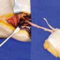
Identification of Radial Vessels and Superficial Radial Nerve at Distal Flap Margin
Directly radial to this tendon, the radial artery is palpated, which is running into the septum between the flexor carpi radialis and brachioradialis muscle. At the most distal point, this septum is opened and a short segment of the radial artery is exposed. Before ligating the artery, which is always accompanied by two comitant veins, the superficial branch of the radial nerve is identified over the tendon of the brachioradialis muscle. The nerve is carefully preserved during further dissection (◘ Fig. 1.10).
Ligation of Radial Vessels at Distal Flap Border
The radial artery is divided at the distal border of the flap. In the perfused arm, the pulsation of the distal stump of the radial artery, caused by the intact circulation through the palmar vessel arches, is visible (◘ Fig. 1.11).
Radial Skin Incision
Now, the skin incision is made 1 cm radial to the artery down to the forearm fascia. The cephalic vein and the superficial branches of the radial nerve are left intact. If the cephalic vein is included and used for venous drainage, the flap is extended toward the dorsal aspect of the forearm, and the cephalic vein is divided distally (◘ Fig. 1.12).
Dissecting the Pedicle along Brachioradialis Muscle
The fascia is incised keeping a safe distance to the radial artery, and the tendon of the brachioradialis muscle is exposed and retracted laterally. Having the superficial branch of the radial nerve identified, the intermuscular septum which contains the radial artery is separated from the brachioradialis muscle. The artery is carefully elevated together with the flap and remains firmly connected to the forearm fascia. Numerous small branches to the deep muscles and the radial bone have to be cauterized or clipped in this area. The deep dissection plane during this step of flap raising is above the flexor pollicis brevis muscle (◘ Fig. 1.13).
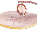
Complete Subfascial Flap Elevation
It is clearly visible that the undersurface of the flap is built by the forearm fascia and that the vascular bundle is securely attached to the fascia by the intermuscular septum. In the distal third of the forearm, where the radial artery is not covered by muscle bellies, the septum contains the highest number of cutaneous perforators. Because these perforators are first reaching the fascia to form a dense vascular network before they enter the skin, the radial forearm flap is a fasciocutaneous flap. The hypoplastic tendon of the palmaris longus muscle is left attached to the flap fascia and is removed from the forearm for a better take of the skin graft (◘ Fig. 1.14).
Related posts:
Stay updated, free articles. Join our Telegram channel

Full access? Get Clinical Tree


