Synopsis
- ▪
Macromastia, or mammary hypertrophy, is a disease process which can result in physical and psychological symptoms.
- ▪
Macromastia symptoms rarely improve without surgical intervention, which typically results in significant improvement in the patient’s quality of life.
- ▪
Reduction mammaplasty techniques have evolved over millennia, with particularly great strides made in the last 100 years.
- ▪
Currently, there exist several well-designed techniques based on sound surgical principles to address macromastia via reduction mammaplasty.
Keywords
Breast, Reduction, Mammaplasty, Gynecomastia
Synopsis
- ▪
Macromastia, or mammary hypertrophy, is a disease process which can result in physical and psychological symptoms.
- ▪
Macromastia symptoms rarely improve without surgical intervention, which typically results in significant improvement in the patient’s quality of life.
- ▪
Reduction mammaplasty techniques have evolved over millennia, with particularly great strides made in the last 100 years.
- ▪
Currently, there exist several well-designed techniques based on sound surgical principles to address macromastia via reduction mammaplasty.
Brief introduction
- ▪
Patients with mammary hypertrophy can present with a variety of symptoms.
- ▪
Physical complaints include neck and back pain, shoulder grooving from bra straps indenting the skin, headaches, difficulty finding well-fitted clothes and limited ability to exercise, intertriginous skin maceration, and rashes.
- ▪
Psychosocial issues include embarrassment, especially teenagers and elderly women.
- ▪
Though the symptomatic improvement of patients suffering from mammary hypertrophy is the primary goal of reduction mammaplasty, there is another goal that is nearly as equally important – creating a more aesthetic breast.
- ▪
Spear describes the reduction mammaplasty as “the clearest example of the interface between reconstructive plastic surgery and aesthetic plastic surgery”.
- ▪
This chapter seeks to demonstrate the most popular techniques for reduction mammaplasty. The key point for choosing the reduction mammoplasty technique is finding what works for you, the surgeon, and what gives your patients the best results.
- ▪
As breast reduction procedures have evolved, certain goals have been consistent:
- •
Aesthetic, natural breast shape.
- •
Maintenance of shape long term.
- •
Reducing scar length.
- •
- ▪
Like macromastia, gynecomastia can result in many of the same symptoms, complaints, and anatomical concerns.
Preoperative considerations
- ▪
Careful consideration of the factors that compel patients to seek reduction mammaplasty are key in the plastic surgeon’s assessment of patients with mammary hypertrophy.
- ▪
A thorough history of symptoms associated with mammary hyperplasia should be recorded.
- ▪
A personal and family history of breast disease and surgery should be recorded, and the results of any testing, such as mammography, breast ultrasound or MRI, and BRCA testing, should be obtained prior to surgical intervention.
- ▪
In addition to screening, a thorough physical examination should be performed as well, noting any relevant points of the patient’s general condition as well as an examination of the breast. In the United States, only some breast reduction procedures are considered medically necessary.
- ▪
Such salient general points relate to the patient’s height, weight, and habitus, and these measures are often mandatory for insurers to calculate the amount of breast tissue they require for the reduction procedure to be covered.
- ▪
This amount of tissue can, however, vary from state to state and from insurer to insurer.
- ▪
A focal breast exam is mandatory as well, evaluating for any masses of the breast, axilla, and supra- and infraclavicular fossae. The nipple–areolar complex should be assessed for changes or discharge, as well as its preoperative sensitivity.
- ▪
Some women have decreased sensitivity due to prior surgery, but often there is decreased sensitivity due to the excess weight of the breast causing traction injury to the cutaneous innervation of the nipple–areolar complex.
- ▪
The skin of the breast should be scrutinized to assess for stigmata of previous operations or physiologic changes, such as scars or striae, which should be pointed out to the patient preoperatively.
- ▪
Finally, shape and symmetry of the breasts preoperatively must also be assessed and pointed out to the patient, especially in cases of very large breasts, because some degree of asymmetry will virtually always remain postoperatively.
- ▪
Breast measurements, such as the sternal notch to nipple distance, the nipple to inframammary fold distance, and the nipple to nipple distance, must be documented preoperatively.
Anatomical pearls
- ▪
The pathophysiology of mammary hypertrophy is thought to be the result of an abnormal response of the breast to circulating estrogens, causing breast proliferation, which is predominantly fibrous tissue, fat, and, to a lesser degree, glandular tissue.
- ▪
Most women with mammary hypertrophy have normal circulating levels of estrogen as well as normal numbers of estrogen receptors in the breast tissue.
- ▪
Cases of juvenile virginal hypertrophy of the breast can be seen as early as late childhood and often are present in the age range of 11–14 years of age.
- ▪
Gigantomastia is a condition in patients with mammary hypertrophy that is defined by their need to have over 1800 g of tissue resected.
- ▪
Increasing sternal notch to nipple distances portend a decreased likelihood for success using a superiorly based pedicle.
- ▪
Vertical short scar techniques can be performed comfortably if the sternal notch to nipple distance is <38 cm.
- ▪
In cases where the sternal notch to nipple distance exceeds 40 cm, plastic surgeons should seriously consider lower-pole breast amputation via a Wise-pattern skin resection with either free nipple grafting or immediate nipple reconstruction with subsequent tattoo of the areola.
- ▪
Longer inframammary fold to nipple distances ultimately may preclude the use of an inferior pedicle technique.
- ▪
If the inframammary fold to nipple distance is >22 cm, there may be difficulty with inferior pedicle or central mound techniques.
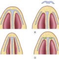
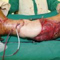
Operative techniques ( ; ; )
General concepts
- ▪
Reduction mammaplasty is performed by combining a skin incision/resection with a parenchymal resection.
- ▪
There have been many techniques described for incision placement, parenchymal resection, and dermoglandular pedicled position. And in general, any combination of these techniques can be utilized for a successful reduction.
- ▪
Preoperative markings and measurements are placed with the patient standing or seated in an upright position. This includes:
- •
The midline from sternal notch to xyphoid.
- •
The midclavicular and breast meridian line.
- •
The projection of the inframammary crease superimposed onto the midclavicular line.
- •
The appropriate skin incision pattern, i.e., vertical, Wise, etc.
- •
- ▪
The patients are placed in the supine position under general anesthesia.
- ▪
Make certain the patients hips are placed at the break in the bed to allow for the head to be raised during the surgery to assist with determination of symmetry.
- ▪
Lidocaine with epinephrine is infiltrated along the incisions and into the planned parenchymal resection.
- •
Infiltration of local anesthetic into the dermoglandular pedicle should be avoided.
- •
- ▪
Some authors have combined suction lipectomy with reduction mammaplasty techniques to assist with postoperative breast contour and reduction of axillary fullness or lateral chest rolls.
- •
Care must be taken to avoid infiltration of tumescent solution or passage of suction cannulas into the planned dermoglandular pedicle.
- •
- ▪
A nipple–areolar complex marker or cookie-cutter of desired size is used to mark the periareolar incisions.
- ▪
The periareolar incision is performed first, and the surrounding areas of the pedicle are de-epithelialized.
- •
Care must be taken to avoid getting too deep, as direct injury to the subdermal plexus could lead to vascular compromise of the nipple.
- •
- ▪
The remaining skin incisions are made and skin flaps created.
- ▪
After parenchymal resection and excess skin is removed, meticulous hemostasis should be obtained.
- ▪
Closure often involves a combination of deep shaping sutures within the remaining parenchyma, followed by layered closure of the skin.
- ▪
The new nipple areolar complex position is marked using the nipple areolar complex marker of desired size.
- ▪
The nipple areolar complex is inset into its new position and secured using a combination of dermal and subcuticular closure.
Related posts:


Stay updated, free articles. Join our Telegram channel

Full access? Get Clinical Tree


