Synopsis
- ■
The reconstructive surgery for the lower extremity has evolved from a staged approach to proving best solutions for functional and cosmetic outcome.
- ■
This chapter covers the classical approach with a gradual change of principle that advocates a one-stage elevator approach.
- ■
Special considerations should be given to overcome the complexity of lower extremity reconstruction, such as diabetes and chronic infection.
- ■
Finally, introduction of perforator flaps, the use of multiple flaps by combination, and supermicrosurgery will help you design and widen the reconstructive choice for the lower extremity.
Keywords
Lower, extremity, leg, foot, thigh, calf
Synopsis
- ■
The reconstructive surgery for the lower extremity has evolved from a staged approach to proving best solutions for functional and cosmetic outcome.
- ■
This chapter covers the classical approach with a gradual change of principle that advocates a one-stage elevator approach.
- ■
Special considerations should be given to overcome the complexity of lower extremity reconstruction, such as diabetes and chronic infection.
- ■
Finally, introduction of perforator flaps, the use of multiple flaps by combination, and supermicrosurgery will help you design and widen the reconstructive choice for the lower extremity.
Brief introduction
- ■
Lower extremity reconstruction following severe trauma, cancer ablation, and chronic infections remains challenging, as multiple structures including bone, muscle, vessels, nerves, and skin may be involved.
- ■
In the recent years, the management of lower extremity has evolved, with numerous new techniques and innovations leading to greater salvage opportunities.
- ■
If the extremity cannot be salvaged, the next goal is to maintain maximal functional length with good soft tissue coverage on the stump to bear the prosthesis for functional gait.
- ■
Extremity salvage is a long and complex process; thus, patient education, motivation, and compliance along with family support will be critical during physical and psychological recovery.
- ■
Although early amputation and prosthetic treatment was thought to offer the potential of faster recovery and lower cost, recent reports have provided different views.
- ■
The Lower Extremity Assessment Project, or LEAP, study showed no significant difference in outcome at 2 years.
Preoperative considerations
- ■
The primary goal of surgical reconstruction of the lower extremity wound is to restore or maintain function.
- ■
Although evaluations such as Mangled Extremity Severity Score, the Predictive Salvage Index, and the Limb Salvage Index can assist the team in making a decision for amputation, it must not be used as a sole criterion, and the decision to amputate must be individualized for each patient.
- ■
Whether acute or chronic, evaluation of lower extremity wounds and the eligibility for soft tissue reconstruction begins with vascular status evaluation.
- ■
If clinical and diagnostic examination reveals inadequate perfusion and the value of reconstruction is minimal, amputation should be individually decided.
- ■
An amputated or avulsed tissue should never be disregarded, especially in acute traumas, unless severely contaminated or lacking vascular structure.
- ■
The skin harvested from the degloved or amputated part can be utilized as biologic dressings to permanent skin grafts.
- ■
The leg length can be preserved using soft tissue distal from the zone of injury as fillet pedicled or free flaps.
- ■
Amputated bones can be banked or used as a flap to reconstruct the leg.
- ■
Once the wound is evaluated to have good vascular supply, stable skeletal structures, and a relatively clean wound, soft tissue coverage is then considered.
- ■
The concept of reconstructive ladder was proposed to achieve wounds with adequate closure using a stepladder approach from simple to complex procedures ( Fig. 13.1A ) .

Figure 13.1
The reconstructive elevator (B) requires creative thoughts and considerations of multiple variables to achieve the best form and function rather than a sequential climb up the ladder (A) . This paradigm of thought does not eliminate the concept of reconstructive ladder but replaces it as a ladder of wound closure and makes its mark in the field where variety of advanced reconstructive procedures and techniques are not readily available. Based on the reconstructive elevator, method of reconstruction should be chosen based on procedures that result in optimal function as well as appearance.
- ■
Although still valued and widely taught, in the era of modern reconstructive surgery, one must consider not only adequate closures but form and function.
- ■
Thus, a simpler reconstructive option may not necessarily produce optimal results, which is especially true for lower extremity coverage, where consequences of inadequate coverage will lead to complications such as additional soft tissue loss, osteomyelitis, functional loss, increased medical cost, and even amputation.
- ■
The reconstructive elevator requires creative thoughts and considerations of multiple variables to achieve the best form and function rather than a sequential climb up the ladder ( Fig. 13.1B ) .
- ■
Based on the reconstructive elevator, method of reconstruction should be chosen based on procedures that result in optimal function as well as appearance.
- ■
The initial evaluation of the lower extremity wound involves visual and manual examination. Neurological evaluation as well as vascular and skeletal evaluation is made to develop a plan for reconstruction.
- ■
After the decision is made to reconstruct the lower extremity, the first preoperative evaluation should start with vascular status.
- ■
Physical examination of palpable pulse, color, capillary refill, and turgor of the extremity allows assessment of initial status, and Doppler examination can provide additional information.
- ■
The use of preoperative arteriography for lower extremity reconstruction is considered when physical/Doppler exam reveals inconclusive vascular status or chronic vascular disease is suspected.
- ■
The use of computed tomographic angiography may obtain vascular information of the recipient region without the risk of complications from arterial puncture of the groin and also can provide vascular information of the donor flap, facilitating the planning and the surgical procedure.
- ■
Nerve injuries that are irreversible may require special considerations.
- ■
Peroneal nerve injuries result in foot drop and loss of sensation of the dorsum of the foot. Thus, lifelong splinting or tendon transfers may be required.
- ■
Complete loss of tibial nerve function results in loss of plantar flexion and is an absolute contraindication for reconstruction.
- ■
The loss of plantar sensation can be devastating and may hinder the need for reconstruction, although it is not an absolute contraindication.
- ■
An algorithm of approach is outlined in Fig. 13.2 .
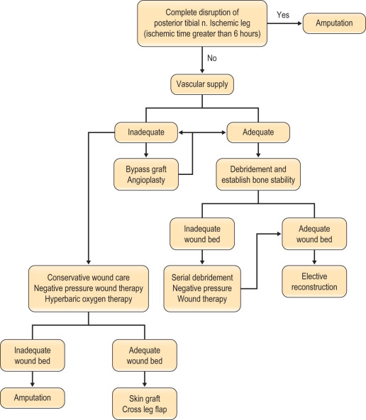
Figure 13.2
Algorithm of approach for soft tissue reconstruction of lower extremity.
Anatomical/technical pearls
Skin grafts and substitutes
- ■
Autologous skin grafts are used in a variety of clinical situations.
- ■
The split-thickness grafts are usually used as the first line of treatment where wounds cannot be closed primarily or undue tension is suspected.
- ■
In the extremity, often with complex wounds, bone exposure and/or avascular beds, infected wounds, and wounds with dead space and poorly coagulated beds, skin grafts should be avoided.
- ■
A skin substitute is defined as a naturally occurring or synthetic bioengineered product that is used to replace the skin in a temporary, semi-permanent, or permanent fashion.
- ■
Temporary epidermal replacements may be beneficial in superficial to middermal depth wounds.
- ■
Examples: EZ Derm and MediSkin (Brennen Medical-LLC, St Paul, MN), and Biobrane (UDL Laboratories Inc, Rockford, IL)
- ■
In deeper wounds, dermal replacements are of primary importance.
- ■
Examples: Allograft, AlloDerm (Life Cell Corporation, Woodlands, TX), Integra (Integra Life Sciences, Plainsboro, NJ), and Apligraf (Organogenesis Inc, Canton, MA).
Approach by location (local flaps)
Thigh
- ■
The thigh can be divided into three parts: the proximal thigh, midthigh, and the distal thigh (supracondylar knee) regions.
- ■
The medial portion of the proximal thigh can be especially challenging due to the location of vital structures and the likely formation of dead space.
- ■
Local lower extremity muscle or myocutaneous flap options include:
- •
Using the flaps based from the lateral circumflex femoral artery, such as tensor fascia lata, vastus lateralis, and rectus femoris flaps.
- •
Vertical rectus abdominis muscle or myocutaneous flap using the deep inferior epigastric artery.
- •
The gracilis muscle or myocutaneous flap based on the medial femoral circumflex artery may lack muscle bulk but is a good option when the dead space is not extensive.
- •
- ■
Now with increased knowledge of perforator and perforator based flaps, basically any perforator can be chosen as a source of vascular supply to the skin flap and be rotated to cover a defect.
- ■
When the use of local flaps is not feasible due to the complexity of the wound, free tissue transfer is indicated.
- ■
The midthigh wound, due to the anatomical character where femur is surrounded by a thick layer of soft tissue, rarely requires reconstruction using free tissue transfer and often is sufficiently reconstructed by skin graft or local flap.
- ■
Local muscle or musculocutaneous flaps based on the lateral or medial femoral circumflex artery can be used when available.
- ■
Any perforator can be chosen as a source of vascular supply to the skin flap and rotated to cover a defect.
- ■
If the patient has undergone massive resection or has special considerations such as postoperative radiation therapy, it may warrant free tissue coverage.
- ■
The wounds of the distal thigh (supracondylar knee) can be very difficult due to the limit of rotation from previously described local muscle or musculocutaneous flaps from the thigh.
- ■
Pedicled medial gastrocnemius muscle or musculocutaneous flap from the lower leg can be extended to cover this region.
- ■
Extensive or complex defects may require free tissue transfer or coverage using a perforator-based rotation/advancement skin flap.
Lower leg
- ■
The traditional planning for reconstruction of the lower extremity has been approached according to the location of the defect.
- ■
Divided into thirds, gastrocnemius muscle flap for proximal third, soleus muscle flap for middle third, and free flap transfer for the distal third of the leg.
- ■
Like the reconstructive ladder concept, this traditional approach can be useful, but the surgeon must individualize each wound and choose the initial procedure that can yield the best chance of success and avoid morbidity.
Microvascular free tissue transfer
- ■
Workhorse flaps for soft tissue coverage include muscle or musculocutaneous flaps such as latissimus dorsi, rectus abdominis, and gracilis.
- ■
The perforator flap, where a skin flap is based on a single or multiple perforators, such as the anterolateral thigh flap or thoracodorsal artery perforator flap, have been added on to the list.
- ■
Whichever flap you select, the guidelines for lower extremity reconstruction using free flaps remain the same:
- •
Anastomose the vessel outside the zone of injury.
- •
Make end-to-side arterial anastomosis and end-to-side or end-to-end venous anastomosis.
- •
Reconstruct the soft tissues first, and then restore the skeletal support.
- •
Primary limb amputation
- ■
Absolute indications include anatomically complete disruption of the posterior tibial nerve in adults and crush injuries with warm ischemia time greater than 6 h.
- ■
Relative indications include serious associated polytrauma, severe ipsilateral foot trauma, and anticipated protracted course to obtain soft tissue coverage and tibial reconstruction.
- ■
In these cases where limb salvage is not possible, attempts should be made to salvage as much limb length as possible.
- ■
Every effort should be made to save the functional knee joint, as below-knee amputation results in far superior ambulatory outcome and up to two- to threefold more full mobility compared to above-knee amputation.
- ■
The energy consumption is far less for below-knee amputation, and this allows these patients to walk significant daily distances, thus maintaining a good quality of life.
- ■
Though the ideal stump length below the knee is more than 6 cm, any length of tibia should be preserved.
- ■
If adequate soft tissue exists, the stump may be closed primarily, and where local tissue is inadequate, microsurgery allows preserving maximal length of the stump.
- ■
If the tissue distal to the amputation is usable, a fillet flap can be performed.
- ■
Other flaps, such as muscle, musculocutaneous, fasciocutaneous, and perforator flaps, can be used.
- ■
Muscle flaps may have a tendency to heal slowly and to shrink due to muscle atrophy, while skin flaps may provide better contour and sensibility.
Debridement
- ■
Bony stability is first established using external or internal fixation devices.
- ■
An external device is usually preferred if there is significant bone loss or bone devascularization and may facilitate coverage procedure.
- ■
Debridement must cover devitalized soft tissue and bone and be performed until fresh bleeding is noted.
- ■
Multiple stages of debridement may be needed to achieve adequate wound bed prior to soft tissue coverage.
- ■
The vacuum-assisted closure can be used to optimize the wound bed and minimize dressing changes until definitive reconstruction.
Timing of reconstruction
- ■
Regardless of the degree of contamination and extent of injury when indicated for salvage, there is no need to delay definitive coverage provided that the general condition of the patient and the status of the wound allow it.
- ■
General consensus favors early aggressive wound debridement and soft issue coverage.
- ■
Ideally, the wound is covered in the first 5–6 days after injury at the acute phase of the wound.
- ■
Godina further demonstrated that radical debridement and coverage within 72 h results in best outcome where only 0.75% of flaps fail, 1.5% are infected, and 6.8 months are needed for union of the bone.
- ■
The common idea behind early intervention is that it minimizes the risk for increasing bacterial colonization and inflammation leading to complications.
- ■
Acute coverage by day 5–7 is generally accepted as having a good prognosis in terms of decreased risk of infection, flap survival, and fracture healing.
Selection of recipient vessel
- ■
Many lower extremity wounds resulting from trauma are high-energy injuries with a substantial “zone of injury”.
- ■
This thrombogenic zone is known to extend beyond what is macroscopically evident, and failure to recognize the true extent of this zone is cited as a leading cause of microsurgical anastomotic failure.
- ■
Isenberg and Sherman demonstrated that clinical presentation of recipient vessel (vessel wall pliability and the quality of blood from transected end of vessel) was more important than the distance from the wound.
- ■
Based on these findings, one of the most important factors in selecting the recipient vessel may be the vascular quality itself.
Special considerations
Osteomyelitis
- ■
Osteomyelitis often follows severe open leg fractures with massive contamination or devascularized soft tissue and bone. Inadequate debridement or delayed coverage of the wound increases the chance for osteomyelitis, and early debridement remains to be the key to prevention.
- ■
To achieve the goal of infection control and the restoration of function, treatment principles for chronic osteomyelitis are debridement, including the complete resection of involved bone, flap coverage with vascularized tissue, and brief course of antibiotic treatment.
- ■
Although there has been controversy in selecting the type of flap for coverage, muscles have shown experimentally to have increased blood flow and antibiotics delivery, increased oxygen tension, increased phagocytic activity, and decreased bacterial counts in wounds reconstructed with muscle flaps rather than fasciocutaneous flaps.
- ■
Clinically, complete debridement and obliteration of dead space are the most important steps to treat osteomyelitis, and the type of flap seems less crucial.
- •
Bone defects can be managed with vascularized bone flap, secondary bone grafting, bone distraction lengthening, or a combination of these techniques.
- •
Not all chronic osteomyelitis can be salvaged. As with the indication for amputation, legs with nerves too damaged after osteomyelitis should not be salvaged.
- •
Diabetes
- ■
Patients with diabetes require additional concerns from chronic renal failure and nutrition to blood sugar control and are best approached by a multidisciplinary team.
- ■
Patients will frequently have chronic bacterial colonization, osteomyelitis, complex wounds, bone deformity, local wound ischemia, and vascular disease.
- ■
When patients with diabetes are required to undergo a reconstructive procedure of the extremity, vascular status must be evaluated to ensure success.
- ■
Any vascular problems must be addressed first and corrected. If not correctable, the surgeon may be faced with a high risk of failure.
- ■
One must consider the probability of successful reconstruction, based on eliminating the underlying problems of the diabetic wound and also take into account long-term ambulation after reconstruction ( Fig. 13.3 ) .
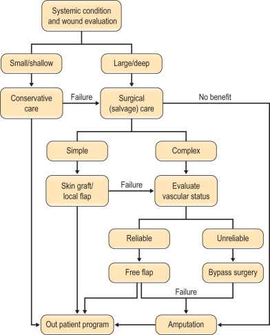
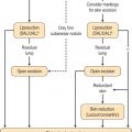
Figure 13.3
Algorithm for diabetic foot reconstruction.
- ■
Large and composite diabetic wounds must be aggressively debrided, including the necrotic bone, and covered with well-vascularized tissue.
Coverage after tumor ablation
- ■
As with any reconstructive procedure, the aim of reconstruction after tumor ablation is to maintain quality of life by preserving function and achieving acceptable appearance.
- ■
In addition, coverage must be able to withstand adjuvant therapy with radiation therapy and/or chemotherapy and play a role in achieving long-term local control of disease.
- ■
Skin grafts are always an option, especially for very extensive defects where flap coverage is not available.
- ■
For wounds scheduled for postoperative radiation therapy or located over joints and high-friction regions, skin graft should be avoided and be reconstructed with a durable flap.
- ■
Special consideration should be made to preoperative radiation therapy where skin would become fibrotic and ischemic around the cancer and thus will not allow local coverage.
- ■
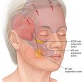
Various flaps from omentum, muscle with skin graft, musculocutaneous, and perforator flaps can be used for reconstruction depending on location, size, depth, adjuvant therapy, function, and cosmetic appearance ( Fig. 13.4 ) .
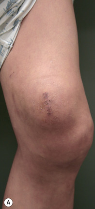
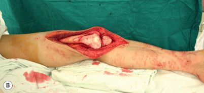
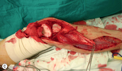
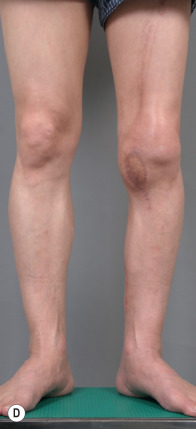
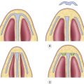
Figure 13.4
(A) A patient with soft tissue sarcoma of the knee region was noted. (B,C) After wide excision including the bone, a hemi-gastrocnemius muscle was elevated to resurface the knee joint. (D) Long-term results show good contour with acceptable function and appearance.
Exposed prosthesis
- ■
The traditional method to manage exposed hardware includes irrigation, debridement, antibiotics, and likely removal of hardware.
- ■
Factors such as location of the hardware, infection (type of bacteria and duration of infection), duration of exposure of hardware, and hardware loosening should be considered as important prognostic factors for successful management of exposed hardware.
- ■
If hardware is clinically stable, time of exposure is less than 2 weeks, infection is controlled, and the location of the hardware is for bony consolidation, then it may increase the likelihood of salvage of hardware using surgical soft tissue coverage.
- ■
Exposed vascular grafts present life- and limb-threatening complications. It should be managed with early debridement and muscle flap coverage to salvage the graft.
- ■
Local muscle flaps such as gracilis, sartorius, and tensor fascia lata are very useful in providing adequate coverage for exposed groin synthetic vascular prosthesis.
- ■
If the defect is extensive and inferiorly based, a vertical rectus abdominis musculocutaneous flap can be considered.
Soft tissue expansion
- ■
The use of tissue expansion in the lower extremity has not been successful as in other areas of the body, such as the breast and scalp.
- ■
The potential advantages of using expanded skin in the lower extremity include improved contour, coverage with like tissue, and improved aesthetic result.
- ■
However, use in the lower extremity has been associated with high rate of infection and extrusion of the implant.
- ■
The technique can be reserved for unstable soft tissues or scars of moderate size.
- ■
The implant is placed suprafascially in the subcutaneous pocket in the lower extremity, and application on the ankle and foot region must be avoided.
- ■
Transverse expansion has a lower failure rate compared to longitudinal advancement.
- ■
For avoidance of wound dehiscence, neurapraxia, and fat necrosis, expansion should proceed slowly, stopping before the onset of pain or, if it is measured, before intraexpander pressure exceeds 40 mmHg.
- ■
Flap prefabrication with tissue expansion may have a role in select reconstructions of the lower extremity.
Operative techniques
Muscle/musculocutaneous flaps
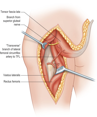
Tensor fascia lata
- ■
The tensor fascia lata is a small, thin, and short muscle with a long fascial extension from the iliotibial tract of the facia lata to the lateral aspect of the knee.
- ■
The muscle originates 5–8 cm anterior of the external lip of the anterior superior iliac crest immediately behind the sartorius and inserts to the iliotibial tract.
- ■
It abducts, medially rotates, and flexes the hip, acting to tighten the fascia lata and iliotibial tract but is an expendable muscle.
- ■
Its flat shape, excellent length, and reliable type I circulation pattern (dominant pedicle is the ascending branch of the lateral femoral circumflex artery and venae comitantes) make it useful in many reconstructive scenarios, both as a pedicled flap for local and regional coverage and as a free, composite unit that incorporates skin, muscle, and iliac bone.
- ■
Motor innervation is from the superior gluteal nerve entering the deep surface between the gluteus medius and gluteus maximus. Sensation is derived from T12, which innervates the upper skin territory, and the lateral femoral cutaneous nerve of the thigh (L2–3) innervates the lower skin.
- ■
When based on the dominant pedicle, located 8–10 cm below the anterior superior iliac spine, the anterior arc of location will reach the abdominal areas, groin, and perineum, while the posterior arc can reach the greater trochanter, ischium, perineum, and sacrum ( Fig. 13.5 ) .