Synopsis
Unilateral lip repair
Principles of unilateral cleft lip repair
- ▪
Presurgical nasoalveolar molding.
- ▪
Modification of surgical techniques.
- ▪
Mohler rotation incision.
- ▪
Mucosal flaps for nasal floor reconstruction, correction of mucosal deficiency in piriform area.
- ▪
Eliminate the perialar incision on advancement flap, limiting scars around the alar base and nostril floor.
- ▪
Mobilization of alar base.
- ▪
Nasal floor reconstruction with complete mucosal closure.
- ▪
Muscle release and reconstruction to simulate the philtral column.
- ▪
Anchoring of advancement flap to nasal septum for centralizing the Cupid’s bow.
- ▪
Correction of central vermillion deficiency with triangular vermillion flap from lateral lip.
- ▪
Semi-open rhinoplasty with a reverse-U incision on the cleft side and rim incision on the non-cleft side.
- ▪
Atraumatic dissection to release the fibrofatty tissue from lower lateral cartilages.
- ▪
Advancement and fixation of the cleft side lower lateral cartilage to the non-cleft side lower lateral cartilage and to the skin in an overcorrected position.
- ▪
Definition of the alar-facial groove with alar transfixion sutures.
- ▪
- ▪
Postoperative maintenance of overcorrection with silicone nasal conformer.
Bilateral lip repair
- ▪
A child born with bilateral cleft lip should not have to suffer because of an ill-conceived and poorly executed primary repair. The operative principles for synchronous nasolabial repair are established:
- ▪
Maintain symmetry.
- ▪
Secure primary muscular continuity.
- ▪
Design proper philtral size and shape.
- ▪
Construct the median tubercle from lateral labial elements.
- ▪
Position/secure the lower lateral cartilages and sculpt the nasal tip and columella.
- ▪
- ▪
The techniques based on these principles are within the repertoire of a well-trained surgeon whose practice is focused on children with cleft lip. Only the philtral columns and dimple seem just beyond the surgeon’s craft.
- ▪
Preoperative dentofacial orthopedic manipulation of the premaxilla is necessary to permit synchronous closure of the primary palate. The surgeon must repair the bilateral cleft lip and correct the nasal deformity in three dimensions based on knowledge of anticipated changes in the fourth dimension. Modifications of the techniques used in repair of the most common complete form are needed for the less common bilateral variants, such as binderoid, complete with intact secondary palate, symmetrical incomplete, and asymmetrical complete/incomplete.
- ▪
Outcomes can be assessed using preoperative and serial photography and documentation of revision rates. Direct anthropometry is the “gold standard” for quantification of the changing nasolabial features; however, it requires training and experience. Intraoperative anthropometry is used to record baseline dimensions and is repeated as the child grows. Two-dimensional photogrammetry is applicable for certain linear and angular measurements if properly scaled. Computerized three-dimensional photogrammetry is a new methodology for quantifying nasolabial appearance. It is both accurate and reliable, and someday could be employed in intra- and inter-institutional comparative studies.
Keywords
Cleft, Lip, Congenital, Hare lip, Complete, Fruste
Synopsis
Unilateral lip repair
Principles of unilateral cleft lip repair
- ▪
Presurgical nasoalveolar molding.
- ▪
Modification of surgical techniques.
- ▪
Mohler rotation incision.
- ▪
Mucosal flaps for nasal floor reconstruction, correction of mucosal deficiency in piriform area.
- ▪
Eliminate the perialar incision on advancement flap, limiting scars around the alar base and nostril floor.
- ▪
Mobilization of alar base.
- ▪
Nasal floor reconstruction with complete mucosal closure.
- ▪
Muscle release and reconstruction to simulate the philtral column.
- ▪
Anchoring of advancement flap to nasal septum for centralizing the Cupid’s bow.
- ▪
Correction of central vermillion deficiency with triangular vermillion flap from lateral lip.
- ▪
Semi-open rhinoplasty with a reverse-U incision on the cleft side and rim incision on the non-cleft side.
- ▪
Atraumatic dissection to release the fibrofatty tissue from lower lateral cartilages.
- ▪
Advancement and fixation of the cleft side lower lateral cartilage to the non-cleft side lower lateral cartilage and to the skin in an overcorrected position.
- ▪
Definition of the alar-facial groove with alar transfixion sutures.
- ▪
- ▪
Postoperative maintenance of overcorrection with silicone nasal conformer.
Bilateral lip repair
- ▪
A child born with bilateral cleft lip should not have to suffer because of an ill-conceived and poorly executed primary repair. The operative principles for synchronous nasolabial repair are established:
- ▪
Maintain symmetry.
- ▪
Secure primary muscular continuity.
- ▪
Design proper philtral size and shape.
- ▪
Construct the median tubercle from lateral labial elements.
- ▪
Position/secure the lower lateral cartilages and sculpt the nasal tip and columella.
- ▪
- ▪
The techniques based on these principles are within the repertoire of a well-trained surgeon whose practice is focused on children with cleft lip. Only the philtral columns and dimple seem just beyond the surgeon’s craft.
- ▪
Preoperative dentofacial orthopedic manipulation of the premaxilla is necessary to permit synchronous closure of the primary palate. The surgeon must repair the bilateral cleft lip and correct the nasal deformity in three dimensions based on knowledge of anticipated changes in the fourth dimension. Modifications of the techniques used in repair of the most common complete form are needed for the less common bilateral variants, such as binderoid, complete with intact secondary palate, symmetrical incomplete, and asymmetrical complete/incomplete.
- ▪
Outcomes can be assessed using preoperative and serial photography and documentation of revision rates. Direct anthropometry is the “gold standard” for quantification of the changing nasolabial features; however, it requires training and experience. Intraoperative anthropometry is used to record baseline dimensions and is repeated as the child grows. Two-dimensional photogrammetry is applicable for certain linear and angular measurements if properly scaled. Computerized three-dimensional photogrammetry is a new methodology for quantifying nasolabial appearance. It is both accurate and reliable, and someday could be employed in intra- and inter-institutional comparative studies.
Brief introduction
- ▪
The multidisciplinary approach is essential to the satisfactory treatment of the cleft patient including: surgeons, orthodontists, speech pathologists, pedodontists, prosthodontists, otolaryngologists, social workers, psychologists, as well as a photographer.
- ▪
The techniques presented for the unilateral cleft lip repair are based on the experience of the members of the Chang Gung Craniofacial Center over a period of 30 years in a Chinese population. They have also been tested in other racially diverse centers. The improved outcomes result from an integrated approach with presurgical management, surgical refinements, and postsurgical maintenance.
- ▪
It is generally believed that isolated cleft palate is a genetic entity distinct from unilateral cleft lip with or without cleft palate.
- ▪
This conclusion arises from both epidemiologic studies and the fact that embryologic events leading to cleft lip/palate and cleft palate occur at somewhat different times (3–7 weeks versus 5–12 weeks).
- ▪
It has long been assumed that both genetic and epigenetic factors play important roles in the etiology of clefts, and this is supported by the varying incidence of clefting with ethnicity, geographic location, and socioeconomic conditions.
- ▪
The incidence of cleft lip/palate in white newborns is approximately 1 in 1000 ( Table 11.1 ); isolated cleft palate occurs in about 0.5 in 1000.
Table 11.1
Incidence of cleft lip/palate in differing ethnic groups
Ethnicity
Incidence per 1000 births
American Indian
3.6
Japanese
2.1
Chinese
1.7
White
1.0
African American
0.3
Data from Wyszynski DF, Beaty TH, Maestri NE. Genetics of nonsyndromic oral clefts revisited. Cleft Palate Craniofac J. 1996;33:406–417. Vieira AR, Orioli IM. Candidate genes for nonsyndromic cleft lip and palate. ASDC J Dent Child. 2001;68:229, 272–279.
- ▪
While there are more than 250 syndromes associated with orofacial clefting, most cases occur as an isolated abnormality, so-called non-syndromic cleft lip/palate.
- ▪
In a large review of their center’s experience, Rollnick and Pruzansky identified other malformations in 35% of cleft lip/palate patients and 54% of cleft palate patients.
- ▪
Cleft lip/palate has an unequal gender distribution, favoring boys over girls, whereas this relationship is reversed in cleft palate only.
- ▪
Cleft lip/palate affects the left side more often.
- ▪
A common question from parents pertains to their risk of having another child with a cleft lip/palate:
- •
Unaffected (i.e., non-cleft) parents who have one child with cleft lip/palate have an estimated recurrence risk of 4%, increasing to 9% if two children are affected.
- •
If one parent is affected, the risk of having a child with cleft lip/palate is also 4%, increasing to 17% if there is already both an affected parent and an affected child.
- •
As the degree of familial relationship increases, recurrence risk decreases: first-, second-, and third-degree relatives have 4%, 0.7%, and 0.3% risk, respectively.
- •
Recurrence risk increases with the severity of the cleft.
- •
Preoperative considerations
- ▪
With the assistance of ultrasound, prenatal diagnosis of cleft lip is usually made after 16–20 weeks’ gestation.
- ▪
Three-dimensional ultrasonography has improved the accuracy of cleft lip diagnosis and is helpful for prenatal counseling because parents can visualize the face of the fetus clearly.
- ▪
A genetic diagnosis and evaluation for other systemic conditions should be done at the time when a prenatal diagnosis of the cleft is made.
- ▪
The newborn cleft baby should have a pediatric evaluation, and parents are counseled about feeding and given information for subsequent care and treatment.
- ▪
Depending on surgical preference, presurgical nasoalveolar molding (NAM) may be started at 2 weeks or even earlier.
- ▪
If utilized, it usually takes 3–4 months before the completion of the NAM.
- ▪
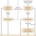
There are several different treatment plans for surgical correction of the cleft lip deformity ( Figs. 11.1 and 11.2 ) ; however, generally, timing is as follows:
- •
Alveolar/NAM:
- •
If utilized, typically begins by 2 weeks of age.
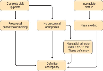
Figure 11.1
Overall cleft treatment plan in the Chang Gung Craniofacial Center.
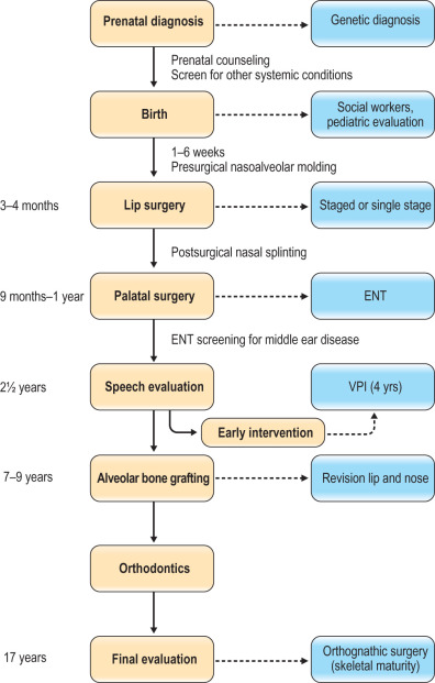
Figure 11.2
Surgical algorithm for unilateral cleft lip repair in the Chang Gung Craniofacial Center.
- •
- ▪
Lip repair typically occurs around 3 months of age:
- •
With presurgical NAM, a definitive cheiloplasty is done at the age of 3–5 months, when the alveolar gap is narrowed and nasal deformity is improved.
- •
When presurgical orthopedics is not available or if the child is older than 3 months, a definitive cheiloplasty with nasal correction is performed.
- •
If there is a wide cleft (>12–15 mm) and an associated tissue deficiency, a nasolabial adhesion cheiloplasty is done at 3 months, followed by a definitive cheiloplasty at about 9 months.
- •
- ▪
If the child has an associated cleft palate, a palatoplasty is typically performed at 9–12 months.
- ▪
Timing of alveolar bone grafting relates to the eruption of the central incisor and canine and is frequently determined by the orthodontist usually at the age of 7–11 years.
- ▪
Early intervention for velopharyngeal insufficiency is done as soon as possible on the basis of speech evaluation and nasopharyngoscopy.
- ▪
Secondary correction of nasal deformities and orthognathic surgery, when indicated, are delayed until facial growth is complete.
- ▪
Bilateral cleft lip presents in three major anatomic forms ( Fig. 11.3 ) :
- •
Bilateral symmetrical complete (50%).
- •
Bilateral symmetrical incomplete (25%).
- •
Bilateral asymmetrical (complete/incomplete) (25%).
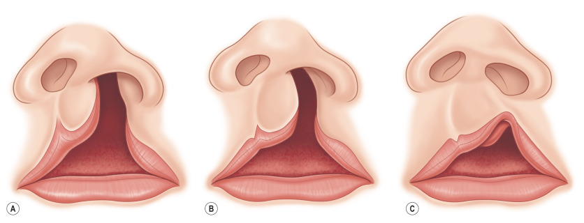
Figure 11.3
Examples of asymmetrical bilateral cleft lip. (A) Left, complete and right, minor form; (B) left, complete and right, microform; (C) left, incomplete and right, mini-microform.
- •
- ▪
The extent of the palatal cleft usually corresponds to the severity of the labial clefts.
- ▪
Minor-form cleft lip extends 3–5 mm above the normal Cupid’s bow peak, i.e., 50% or less of the normal cutaneous labial height.
- ▪
Other features are deficient vermillion on medial side of the cleft, cutaneous groove and muscular depression, hypoplastic median tubercle, and minor nasal deformity.
- ▪
Microform cleft lip is characterized by a notched vermillion–cutaneous junction in which the Cupid’s bow peak is elevated less than 3 mm above normal.
- ▪
Other features are the same as in a minor form, but they are less obvious.
- ▪
Nasal deformities include small depression of the sill, slightly slumped alar genu, and 1–2 mm lateral displacement (and often under-rotation) of the alar base.
- ▪
Mini-microform cleft lip is distinguished by a disruption of the white roll (vermillion–cutaneous junction) without elevation of the Cupid’s bow peak.
- ▪
Usually there is a notch of the free mucosal margin. Muscular depression (particularly noticeable below the nostril sill) is variable as is the cleft nasal deformity.
- ▪
This detailed subcategorization of the contralateral side in an asymmetrical bilateral cleft lip is important because the extent of vermillion–cutaneous disjunction determines the operative strategy.
- ▪
Synchronous bilateral nasolabial repair is indicated for a contralateral incomplete cleft lip, including a minor form.
- ▪
Correction of a contralateral microform or mini-microform is usually deferred until closure on the greater side.
- ▪
Alignment of the three maxillary elements sets the skeletal stage for synchronous bilateral nasolabial repair and minimizes the nasolabial distortions that occur during rapid growth of early childhood.
- ▪
After retrusion and centralization of the premaxilla, the philtral flap can be designed in proper proportions, the nasal tip cartilages can be anatomically positioned, and the alveolar clefts can be closed, which stabilizes the maxillary arch and usually eliminates oronasal fistulas.
- ▪
There are two dentofacial orthopedic strategies: passive and active.
- ▪
Passive strategies include NAM devices.
- ▪
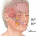
Active strategies include the Latham appliance ( Fig. 11.4 ) .
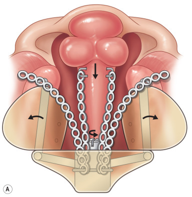
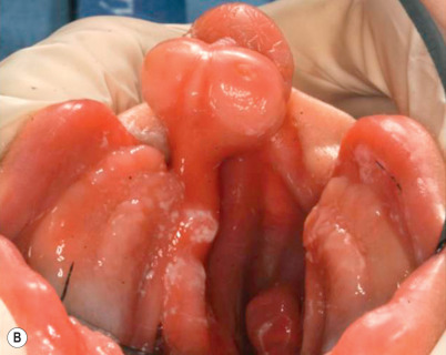
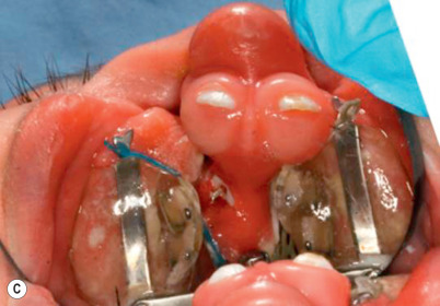
Figure 11.4
(A) Latham appliance; (B) prior to insertion of device; (C) 6 weeks following dentofacial orthopedic manipulation.
Anatomical/technical pearls
- ▪
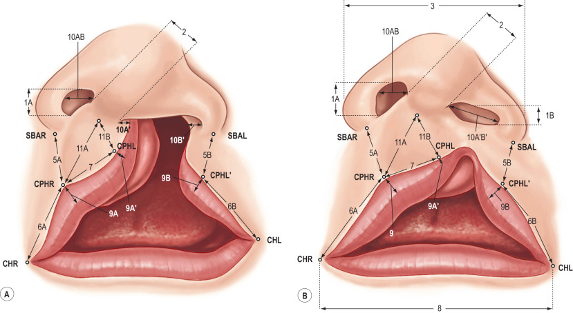
Areas of vital concern to the surgeon: the amount of tissue medial to the base of the ala, the vertical height of the lateral lip, the horizontal length of the lateral lip, and the epidermal extension from the columella onto the premaxilla.
- ▪
The discrepancy between the height from the central point of the base of columella to the two peaks of the Cupid’s bow is critical for leveling of the Cupid’s bow.
- ▪
Vertical length of the lip is more important aesthetically compared with the horizontal length.
- ▪
Therefore, vertical length is seldom sacrificed for horizontal length columella and nasal floor skin.
- ▪
Inadequate reconstruction of this deficient vermillion will result in free border deformities as seen in a straight-line vermillion closure.
- ▪
The vermillion medial to the base of the philtral column fits into the deficient vermillion beneath the Cupid’s bow.
- ▪
The key concepts of the rotation advancement surgical technique in the unilateral cheiloplasty are as follows:
- •
Mohler rotation incision.
- •
Mucosal flaps for nasal floor reconstruction, correction of mucosal deficiency in piriform area.
- •
Eliminate the perialar incision on advancement flap, limiting scars around the ala base and nostril floor.
- •
Mobilization of alar base.
- •
Nasal floor reconstruction with complete mucosal closure.
- •
Muscle release and reconstruction to simulate the philtral column.
- •
Anchoring of advancement flap to nasal septum for centralizing the Cupid’s bow.
- •
Correction of central vermillion deficiency with triangular vermillion flap from lateral lip.
- •
Semi-open rhinoplasty with a reverse-U incision on the cleft side and rim incision on the non-cleft side.
- •
Atraumatic dissection to release the fibrofatty tissue from lower lateral cartilages (LLCs).
- •
Advancement and fixation of the cleft side LLC to the non-cleft side LLC and to the skin in an overcorrected position.
- •
Definition of the alar-facial groove with alar transfixion sutures.
- •
- ▪
The following principles for repair of bilateral cleft lip were induced based on study of the literature and observations of residual deformities:
- •
Maintain nasolabial symmetry . Even the slightest differences between the two sides of the lip and nose will become more obvious with growth. Symmetry is the one advantage a bilateral cleft lip has over its unilateral counterpart. It must be maintained.
- •
Secure muscular continuity . Construction of a complete oral ring permits normal labial function, eliminates the lateral bulges, and minimizes later distortion of the philtrum and interalar widening.
- •
Design the philtral flap of proper size and shape. The philtrum rapidly elongates and widens, particularly at the columellar–labial junction.
- •
Construct the median tubercle using lateral vermillion-mucosal elements. There is no white roll in the prolabium. Retained vermillion lacks normal coloration and fails to grow to full height.
- •
Position the slumped/splayed LLCs and sculpt excess soft tissue in nasal tip and columella . These maneuvers are necessary to establish normal nasal tip projection and columellar length/width.
- •
Operative techniques
Unilateral cleft lip ( ; )
Alveolar molding: external taping
- ▪
The external taping (non-surgical lip adhesion) is the simplest technique for both presurgical molding of the maxillary halves and approximation of the alveolus.
- ▪
A strip of Micropore tape is placed across the cleft to approximate the upper lips. The objective of the tape is to simulate effects of an adhesion cheiloplasty and reposition the maxillary segments into proper alignment.
Nasoalveolar molding
- ▪
Liou’s method utilizes a molding bulb attached to a dental plate as an outrigger to mold the nose along with external taping of the lip.
- ▪
The device is held to the palate with dental adhesives.
- ▪
The force from taping and counterforce from the molding bulb provide the combined force necessary to bring the alveolus into proper position.
- ▪
The nasal molding and alveolar molding are done at the same time, taking approximately 3 months.
- ▪
Newer devices contain an internal spring.
- ▪
Grayson’s method utilizes nasal molding after alveolar approximation to avoid overstretching the nasal cartilage.
- ▪
The appliance consists of an acrylic or resin plate, which fits over the maxillary dental arch (alveolar ridges), an acrylic retention arm or button, and a nasal stent.
- ▪
This technique should be started within the first 2 weeks after birth; careful monitoring is required every 1–2 weeks for a period of 3–6 months to complete it.
Rotation advancement cheiloplasty for complete clefts
- ▪
The following points and measurements are made, with a caliper, at the time of surgery:
- •
The points of the Cupid’s bow on the epidermis–vermillion junction line (the white skin roll).
- •
The vermillion–mucosa junction line (the red line).
- •
This clearly defines the intervening vermillion and also helps identify the deficient vermillion beneath the cleft-side Cupid’s bow.
- •
The base of the ala and the commissure (see Fig. 11.5 ).
- •
The base of the cleft-side philtral column is difficult to identify.
- •
It is located where the white skin roll changes direction and where the vermillion first becomes widest, usually 3–4 mm lateral to the converging point of red line and white skin roll.
- •
Medial incisions
- ▪
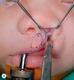
After the above markings are made, a Mohler rotation incision line is marked as a curving line from cleft side Cupid’s bow (CPHL) going upward into the base of columella and then turning back to the nasolabial junction of the non-cleft side philtral column ( Fig. 11.6 ) .
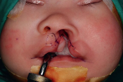
Figure 11.6
Preoperative marking: CPHR, IS, CPHL, CPHL′ and CHL, as described in Fig. 11.5 . The C-flap (C) and C-flap mucosa (CM) are marked. The dotted line on the lip is the red line, which is the junction between vermillion and mucosa. Incision lines are shown on the lip extending from point CPHL lateral to the columella on the skin edge overlying the premaxilla extending superiorly along the junction line of columella skin and septal cartilage mucosa. The cleft-side base of the philtral column is also marked (CPHL′). The proposed incision lines are marked with the rotation incision in a Mohler fashion. A small triangular white skin roll flap is designed above the CPHL′.
- ▪
The height of this rotation incision should be the same as the height of the non-cleft side philtral column.
- ▪
The angle of the back cut is dependent on the width of columella.
- ▪
If the columella is wide, a wider angle can be made.
- ▪
The incision across the free border of the lip at CPHL should be at right angles to the axis of the white skin roll to facilitate subsequent lip closure.
- ▪
After the incision is made, the muscle is freed from the skin in the subdermal plane for a distance of 2–3 mm ( Fig. 11.7 ).
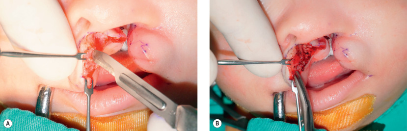
Figure 11.7
(A) Muscle layer is dissected along the skin edge initially with a blade. (B) The muscle dissection is then continued with tenotomy scissors. The abnormal muscle insertion beneath the columellar base and nasal floor is released.
- ▪
The muscle dissection on the non-cleft side should reach the nasal floor of the non-cleft side for adequate releasing of the abnormal muscle insertion to the columellar base.
- ▪
Traction on the free border of the lip will determine if the rotation is adequate, that is, both sides of the Cupid’s bow at the same level ( Fig. 11.8 ) .
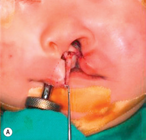
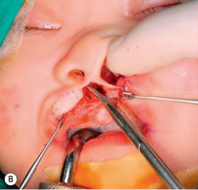
Figure 11.8
(A) Traction on the free border of the lip helps to determine if the rotation is adequate. (B) The muscle dissection on the rotation flap should reach the nasal floor on non-cleft side.
- ▪
Even if the rotation is inadequate, extending the incision across the non-cleft side philtral column should be avoided, as it will result in a vertically long lip. If the rotation fails to level the Cupid’s bow, nothing further is done until after muscle repositioning.
- ▪
The C-flap incisions are made on a line that extends from point CPHL along the junction of skin and mucosa to the most lateral point of the skin overlying the premaxilla ( Fig. 11.9 ) .