Keywords
Ptosis, Blepharoplasty, Levator, Müllerectomy, Müller’s, Eyelid
History
The evolution of ptosis surgery can be studied and categorized from an anatomical point of view. These categories include skin excision, frontalis suspension, resection of tarsus and/or Müller’s muscle, and levator resection. Each technique has undergone periods of transformation, falling in and out of favor over time .
The origin of ptosis surgery focusing on advancing the levator muscle complex is credited to Bowman in 1857 when he described resection of the levator aponeurosis and a portion of the tarsus via an internal incision . The first to target the levator from an external incision was Everbusch in 1883 . Over the course of the following 75 years both the internal and external approach to levator and tarsal resection underwent refinement, but fell out of favor because of unpredictable results. It was not until the 1960s that new concepts in ptosis surgery surfaced. In 1961 Fasanella and Servat described the combined conjunctival-tarsal-Müller’s muscle resection, known as the Fasanella-Servat procedure . It was more predictable and reproducible than previous such tissue resections, making it a popular choice among surgeons. Putterman and Urist modified the Fasanella-Servat procedure by resecting only conjunctiva and Müller’s muscle and leaving the tarsus intact. This procedure, published in 1975, has become a standard among ptosis surgeons . Improvements in the levator-based approach to ptosis surgery also developed during this era. Although initially described and performed in the 1800s, it was not until the published works of pioneers such as Beard, Jones, Fox, and Anderson that levator-based surgery came to the forefront .
Personal Philosophy
The numerous descriptions of ptosis surgery over the years speak to the challenging nature of the procedure. With experience, the technical aspects of ptosis repair have become less cumbersome; however, the difficulty in attaining consistent and predictably symmetric height and contour of the eyelid continues. The nuances of anatomical variability, differences in levator function (LF), and asymmetrical involutional changes all prevent the surgeon from applying a universal method for correcting all clinical presentations of ptosis.
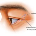
There are four general surgical approaches that elevate the eyelid margin. Firstly, standard blepharoplasty, in the setting of skin overhanging the eyelid margin (dermatochalasis), may result in an improved lid position by alleviating mechanical ptosis. The remaining three procedures are true ptosis corrections, two of which are commonly used in conjunction with aesthetic blepharoplasty. These include external levator advancement (ELA) and a posterior approach, Müller’s muscle conjunctival resection (MMCR). In ELA the levator aponeurosis/muscle is isolated and advanced through an external eyelid crease incision to elevate the upper eyelid margin. This technique is used for variable degrees of ptosis with adequate levator muscle function. The procedure requires patient cooperation to set and adjust eyelid height and contour, and sometimes requires adjustment of the aponeurotic position in the early postoperative period. Conversely, in the MMCR procedure, Müller’s muscle is resected and advanced from an internal (transconjunctival) approach. This technique works well for small amounts of ptosis with good LF and is particularly helpful if avoidance of an eyelid crease incision is desired (such as in unilateral ptosis repair). As opposed to ELA surgery, patient cooperation is not needed, and the predictability of the postoperative eyelid height and maintenance of existing eyelid contour are both easier to achieve. This approach is less technically demanding than the ELA procedure, but is limited by the degree of ptosis that can be corrected, typically 2 mm or less.
A final approach to ptosis correction involves linking the eyelid proper to the frontalis muscle via a variety of autologous or alloplastic materials (frontalis suspension procedure). This technique is used in cases of ptosis associated with poor LF. As the procedure is rarely employed in conjunction with aesthetic blepharoplasty, it will not be covered in this chapter.
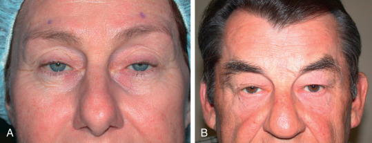
It is not unusual for a patient to present with both dermatochalasis and true eyelid ptosis ( Fig. 11.2 ). Performing isolated blepharoplasty in this situation may lead to unhappy patients. For this reason it is incumbent upon the aesthetic eyelid surgeon to master the procedures needed to address these combined eyelid deficits or work closely with an oculofacial plastic surgeon who can assist with such cases. In this chapter the critical elements of evaluation and surgery needed to address patients with ptosis who desire blepharoplasty will be reviewed.
Anatomy
The upper eyelid margin normally sits 4 mm above the central corneal light reflex and peaks just nasal to the center of the pupil. The eyelid crease is the natural indentation of upper eyelid skin that results from levator aponeurotic attachments to the dermis. This is best seen when the patient looks downward. The crease typically lies 7 to 10 mm above the lid margin and tapers nasally and temporally to the respective canthi. When aponeurotic ptosis is present the crease often lies higher than this level. The lid fold (if present) is the overhang of skin that can obscure the crease. The strip of pretarsal skin that sits between the eyelid margin and the lid fold is called the tarsal platform ( Fig. 11.1 ). Symmetry of the tarsal platform is an important aesthetic goal in blepharoplasty/ptosis surgery. In addition to overhanging upper eyelid skin, changes in eyebrow and upper eyelid position can alter the eyelid crease and fold, with resultant changes in tarsal platform show. As an example, the patient with asymmetrical eyebrow position may have less tarsal platform show on the side of the more ptotic eyebrow, even if all other features of the eyelids are identical.
Levator Muscle
The levator palpebrae superioris is the main retractor of the upper eyelid. It is innervated by the third (oculomotor) cranial nerve, and is composed of a muscular and tendinous (aponeurotic) portion. The muscular portion originates in the orbital apex from the lesser wing of the sphenoid bone. It is approximately 40 mm in length. It courses directly above the superior rectus muscle and transitions to aponeurosis 15 to 20 mm superior to the tarsal plate near a horizontal condensation of connective tissue, visible as a distinct white band called the superior transverse orbital ligament, or Whitnall’s ligament ( Fig. 11.3 ). This ligament has strong attachments at the medial and lateral orbital bones. The levator muscle complex redirects from a horizontal to vertical direction as it reaches Whitnall’s ligament, and continues as the aponeurosis for 14 to 20 mm until it attaches on the anterior tarsal surface. It is thought that Whitnall’s ligament acts as a pulley to reorient the force generated by the levator muscle from horizontal to vertical.
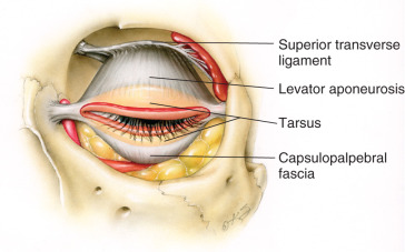
Beyond Whitnall’s ligament, the levator aponeurosis fans out to form both medial and lateral horns, the latter of which is better defined. The lateral horn separates the orbital and palpebral lobes of the lacrimal gland and inserts on the lateral orbit at Whitnall’s tubercle. Some of its fibers contribute to the lateral canthal tendon. The medial horn has loose attachments to the medial canthal tendon as well as the posterior lacrimal crest (see Fig. 11.3 ).
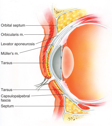
As discussed in the chapter on upper lid blepharoplasty ( Chapter 10 ), the levator aponeurosis lies immediately deep to the preaponeurotic (central) eyelid fat pad and fuses with the orbital septum prior to inserting on the anterior aspect of the tarsal plate and pretarsal fascia. It attaches most firmly to the tarsal plate about 3 mm above the eyelid margin. Proximal to its tarsal attachments, the aponeurosis sends numerous small fibers anteriorly through the orbicularis and subcutaneous tissue to the skin. These fibrous attachments form the natural eyelid crease.
Müller’s Muscle
Müller’s muscle, a secondary upper eyelid retractor, is smaller than the levator muscle but plays an important role in the upper eyelid position. The muscle is sympathetically innervated, and interruption of this autonomic function results in ipsilateral ptosis (Horner’s syndrome). Müller’s muscle originates from the underside of the levator muscle just distal to Whitnall’s ligament and inserts on the superior edge of the tarsal plate (8–12 mm distance) . The contraction of Müller’s muscle (epinephrine dependent) elevates the eyelid margin, but it may also contribute to eyelid position by transmitting the force of the levator complex to the superior tarsus ( Fig. 11.4 ). Just deep to Müller’s muscle is the conjunctiva. Frequently, the peripheral arterial arcade can be seen running horizontally on the anterior surface of Müller’s muscle. This is an important landmark in ELA ptosis surgery to help define the plane between the levator aponeurosis and Müller’s muscle. When the surgeon sees the arcade vessel, it means that the aponeurosis is either thinned, dehiscent, or that it has been surgically elevated.
Tarsal Plate
The upper tarsal plate, a rigid structure comprised of dense fibrous connective tissue, provides support and a uniform contour to the upper eyelid. As stated, it serves as a point of attachment for the eyelid retractors. Its thickness varies, but on average is 1 mm. The central vertical height is 10 to 12 mm and then gradually tapers to 2 to 3 mm medially and laterally as it reaches the canthal tendons . There is no tarsal plate medial to the punctum. The Meibomian glands, holocrine glands that secrete the lipid portion of the tear film, originate within the tarsus, and their small openings can be seen at the posterior aspect of the lid margin. Loss of the tarsal tissue or loss of tarsal rigidity makes ptosis repair more challenging.
Preoperative Evaluation
Patient Selection
A complete history and focused physical examination is essential when evaluating patients with ptosis and dermatochalasis. Some patients will state specific goals for how they want their eyelids to appear, whereas others will simply want their eyes “lifted.” It is up to the surgeon to interpret the patient’s desires and then to determine what is in the patient’s best interest. The authors have found that having a mirror and drawing pad available to show the patient the features of the eyelid that are being discussed, and how surgery will address the issues, is a useful tool to provide clarity to any surgical evaluation and discussion.
History
A general medical, surgical, and ocular history is identified. The discontinuation of any medications, prescription or herbal, which predispose to bleeding must be discussed with the patient and their primary care physician or cardiologist. A history of previous blepharoplasty or ptosis repair should be sought, as this may influence the surgical plan. Multiple prior failed attempts at ptosis repair suggest a more complicated procedure and referral may be warranted. A history of dry eye or frequent use of topical lubricants is important. Worsening or inducing dry eye syndrome is a significant concern of blepharoplasty and ptosis surgery. Appropriate preoperative education regarding this risk is essential.
The vast majority of patients with ptosis and dermatochalasis seeking surgical intervention will give a vague history of a gradually worsening appearance and drooping of the eyelids, and/or an awareness of blockage of peripheral vision by the overhanging eyelid. This generally suggests an involutional etiology. Some patients will have a history of contact lens use. In this instance ophthalmic care may be needed prior to surgical intervention. On rare occasions a neurological cause of ptosis may be present (i.e., Horner’s syndrome, third nerve paresis, myasthenia gravis). A discussion of such entities is beyond the scope of this chapter, but a basic understanding of these conditions should be sought by the aesthetic eyelid surgeon who performs ptosis surgery.
Examination
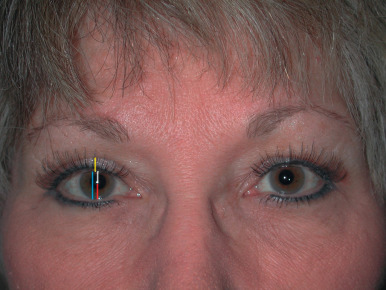
Working from the top to the bottom, the examination begins by assessing the position of the brow. The interplay of brow and eyelid position, and eyelid fullness, is a complex and constantly evolving subject. Aesthetic eyelid surgeons should familiarize themselves with this literature. Any procedure that elevates the eyelid, true or apparent, can reduce frontalis compensatory drive, and thereby lower the brow. Thus in the presence of ptosis or dermatochalasis, latent eyebrow ptosis may be present but not manifest until after surgery. The preoperative assessment then moves to the eyelids. The degree of upper eyelid skin redundancy, the presence and location of orbital fat prolapse, and upper eyelid sulcus is then assessed. Next, the position of the eyelid margin in relation to the central corneal light reflex is evaluated. This measurement is known as the margin reflex distance 1 (MRD1). It bears repeating that patients presenting for droopy eyelid skin may not realize they have concomitant ptosis. When deemed clinically appropriate, the surgeon will manually elevate the eyelid skin to get an adequate view of the eyelid margin. As previously discussed, a normal MRD1 varies from person to person, but on average is 4 mm. The margin reflex distance 2 (MRD2) is the measurement from the central corneal light reflex to the lower eyelid margin. A normal MRD2 is about 5 mm ( Fig. 11.1 ). An abnormally large MRD2 indicates possible lower lid retraction. When lower eyelid retraction is present, performing blepharoplasty or ptosis repair may exacerbate corneal exposure.
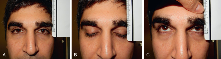
The most critical factor in planning ptosis repair surgery is quantifying lid excursion (LF). To measure this, the patient looks down while the examiner applies pressure to the patient’s eyebrow with their thumb to restrict the action of the frontalis muscle. The patient then looks up and the amount of LF is measured in millimeters (mm) with a ruler. A normal LF is greater than 10 mm, an intermediate level falls between 6 and 10 mm, and poor LF is 6 mm or less ( Fig. 11.5 ). With poor LF (<6 mm) the likelihood of successful ptosis repair with either an ELA or MMCR is poor. As a general rule, for the surgeon who is not an eyelid specialist, when LF is at this level it is best to refer the patient for further evaluation of the cause of ptosis. Often these patients have a congenital ptosis or an acquired neuromyopathic etiology of their lid malposition. In this setting, frontalis suspension surgery (not discussed) may be appropriate ( Fig. 11.6 ). Patients with LF greater than 10 mm have an excellent chance of responding appropriately to either ELA or MMCR surgery. Most adult patients presenting with ptosis fall into this category and have what is classified as “involutional” ptosis. These patients typically have excellent LF and no lagophthalmos. The term involutional can be a misnomer, as patients who are not elderly may also present this way