Abstract
Protozoan organisms continue to represent a major disease burden worldwide. Diseases such as toxoplasmosis affect much of the population, and other diseases like leishmaniasis have undergone a resurgence in many parts of the world. This chapter reviews the cutaneous manifestations of protozoan diseases.
Keywords
Amebiasis, Infection, Leshmaniasis, Protozoan, Toxoplasmosis, Trypanosomiasis
- •
Trichomoniasis typically presents with vaginal pruritus and a frothy discharge, and responds well to treatment with metronidazole.
- •
Systemic therapy for leishmaniasis is recommended for patients who are immunosuppressed or who acquire infection in areas where mucocutaneous disease has been reported.
- •
Suramin remains useful to treat early Rhodesian trypanosomiasis, while melarsoprol is the drug of choice for central nervous system (CNS) disease. Pentamidine is still used for Gambian disease.
- •
For American trypanosomiasis, nifurtimox and benznidazole are used to reduce the severity of the acute illness.
- •
Acanthamoeba affects immunocompromised individuals, while Balamuthia mandrillaris causes erythema and induration of the central face and CNS invasion in previously healthy patients.
Trichomoniasis
Clinical Manifestations
Trichomoniasis is caused by Trichomonas vaginalis , a flagellate protozoan. Trichomoniasis typically presents with vaginal pruritus, a burning sensation, and a frothy discharge. Male sexual partners may present with balanoposthitis.
Diagnosis
A wet mount typically demonstrates the motile organism, but direct fluorescent antibody and polymerase chain reaction (PCR) assays are also available.
Treatment
Treatment includes metronidazole, 2 g in a single oral dose or 500 mg twice a day for 7 days. A topical form is also available. Patients should be warned about disulfiram-type effects if alcohol is consumed. In pregnant women, topical clotrimazole may be used instead.
Leishmaniasis
Clinical Manifestations
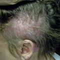
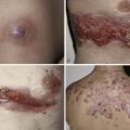
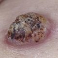
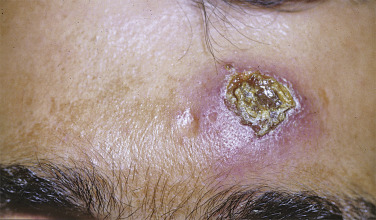
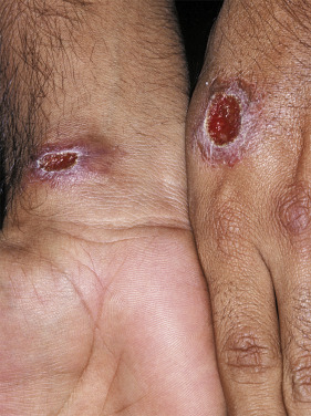
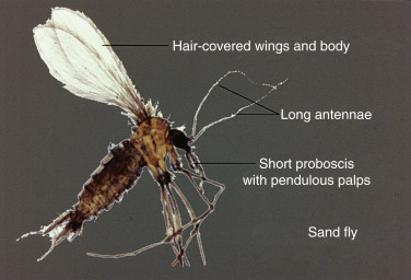
The rural type of Old World leishmaniasis is characterized by moist chronic ulcers that heal within 6 months. Rodent reservoirs carry the organism, which is transmitted via a sand fly vector. Dry lesions and recurrent lesions (leishmaniasis recidivans) are associated with the urban type of disease, caused by Leishmania tropica . New World disease may consist of only cutaneous lesions, especially with the mexicana variety. The primary lesion begins as a papule that becomes crusted, verrucous, or ulcerated, with an infiltrated red border ( Figs 34-1 and 34-2 ). Subcutaneous peripheral nodules represent a lymphangitic pattern of spread and lymphadenopathy may be present. On the Yucatan Peninsula and in Guatemala, the workers who harvest chicle for chewing gum develop chiclero ulcers on the ear. The etiologic agent is Leishmania mexicana and the sand fly vector Lutzomyia flaviscutellata ( Fig. 34-3 ). Uta occurs in the Peruvian highlands. As with the chiclero ulcer, lesions are found on exposed sites and mucosal lesions do not occur.
Disseminated cutaneous leishmaniasis occurs with both New and Old World diseases, but the most destructive manifestations occur with mucocutaneous New World disease. Diffuse anergic leishmaniasis may resemble lepromatous leprosy or Lobo’s disease.
Old World L. tropica , Leishmania major , Leishmania aethiopica , and Leishmania infantum cause cutaneous leishmaniasis. The latter also produced the Mediterranean form of visceral leishmaniasis. New World L. mexicana does not induce mucosal disease. Leishmania braziliensis guyanensis , Leishmania braziliensis braziliensis , and Leishmania braziliensis panamensis produce cutaneous lesions and the latter two are also associated with mucocutaneous disease. Leishmania donovani spp. donovani , infantum , and chagasi cause visceral leishmaniasis.
Cutaneous leishmaniasis is endemic in Southwest Asia, the Mediterranean and Latin America. In the United States, cutaneous leishmaniasis is most common in South Texas, but rare reports of cutaneous disease have occurred as far north as Pennsylvania and the Midwest.
Dogs and rodents are the natural reservoir hosts. The Old World vector is the Phlebotomus sand fly, whereas Phlebotomus perniciosus and Lutzomyia sand flies are the vectors for New World leishmaniasis. In humans, the aflagellare form (amastigote) is found in tissue.
Diagnosis
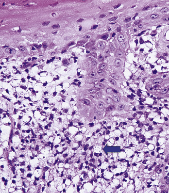
Leishmania are nonencapsulated and are seen as intracellular organisms within histiocytes, containing a nucleus and a paranucleus. Within the histiocytes the organisms often line up at the periphery of a vacuole like the bulbs surrounding an old-fashioned movie marquee ( Fig. 34-4 ).