Abstract
Erythroderma is defined as erythema covering more than 90% of the body and can present with or without scale. Although many cases are idiopathic, investigation of underlying causes is important. Frequently, exacerbation of an underlying skin disorder including psoriasis, cutaneous T-cell lymphoma, atopic dermatitis, or a medication reaction is responsible for erythroderma. Workup is targeted on identifying the underlying driving process if possible, and includes a biopsy, although in some cases this can be nonspecific. Hospitalization also should be considered for monitoring of fluid–electrolyte balance, adequate nutrition, and skin care. Treatment is based upon the underlying skin disorder and includes both skin-directed therapy with topical steroids and systemic medications. Caution should be used in prescribing systemic immunosuppressants if a definitive diagnosis is unclear to avoid exacerbation of an underlying malignancy or cutaneous lymphoma. Prognosis is also dependent on the inciting cause. Development of sepsis, cardiac failure, or erythroderma related to a malignancy all tend to be poor prognostic factors.
Keywords
Cutaneous lymphoma, Exfoliative erythroderma, Medication/drug, Pustular psoriasis
- •
Erythroderma is erythema, with or without scaling, involving more than 90% of the cutaneous surface.
- •
Many cases of erythroderma are idiopathic, but the most common causes are exacerbation of an underlying skin disease, a drug hypersensitivity reaction, and cutaneous T-cell lymphoma.
- •
Although pathology can be nonspecific, a biopsy should typically be performed because it may give clues to the underlying diagnosis.
- •
Hospitalization may be required for appropriate monitoring of fluid–electrolyte balance, nutrition, and thermoregulation, as well as for diligent skin care.
- •
Treatment should be focused on the underlying disease, if known, in addition to skin-directed therapy with topical corticosteroids applied twice daily under moist occlusion.
- •
Prognosis depends on the underlying condition but tends to be worse for those with malignancy-related erythroderma and for those who develop high-output cardiac failure or sepsis.
Introduction
Erythroderma is the clinical finding of erythema, with or without scaling, on more than 90% of an individual’s body surface area. The term “exfoliative dermatitis” is used in some contexts and is often considered to be the same entity, but erythroderma is a more encompassing and often favored term. Numerous conditions can result in erythroderma, and identifying the exact inciting cause or underlying pathology may be challenging. Extreme disruption in the barrier function of the skin can lead to systemic manifestations such as fluid and electrolyte abnormalities, tachycardia, and problems with thermoregulation. Management of erythroderma should be focused on treatment of the underlying cause, if possible, in addition to supportive care for the skin and systemic manifestations.
The incidence of erythroderma is difficult to determine given lack of standardized reporting, and the incidence likely varies depending on location. In the United States and Europe, the incidence has been estimated to be between 1 and 2 cases per 100,000 individuals/year, but some reports from India have reported an incidence as high as 35 cases per 100,000 dermatologic outpatients. Erythroderma is more commonly seen in patients older than 45 years, with an average age of 55 years, which may be related to an increase in conditions that result in erythroderma in older individuals. There also appears to be a male predominance, with males being affected 2 to 3 times more often than females.
Cause and Pathogenesis
Erythroderma represents a final clinical endpoint for many dermatological and systemic disease states ( Table 14-1 ). The most common causes are exacerbation of an underlying skin disease such as psoriasis or atopic dermatitis, a drug hypersensitivity reaction, and cutaneous T-cell lymphoma; however, many cases are considered idiopathic, as an inciting cause is not identified. Erythroderma is likely secondary to a complex interaction of cytokines and cellular adhesion molecules affecting T-cells within the skin. Keratinocytes and Langerhans cells both produce IL-1, which increases vascular permeability, acts as a chemotactic factor for T-cells, and upregulates ICAM-1. ICAM-1 is found on venous endothelial cells and plays an important role in T-cell binding to both endothelial cells and keratinocytes. This complex interaction of T-cells and cytokines leads to the erythema and desquamation seen clinically. Increased cell turnover in the epidermis occurs secondary to an increase in both the number of proliferative cells and the mitotic rate. These rapidly shed epidermal cells contain amino acids and proteins normally retained by the skin, leading to significantly higher than normal levels of protein loss.
| Primary Skin Diseases | Malignancy | Other |
|---|---|---|
| Psoriasis Atopic dermatitis Pityriasis rubra pilaris Contact dermatitis Chronic actinic dermatitis Bullous pemphigoid Pemphigus foliaceus Ichthyoses Seborrheic dermatitis (rare) Stasis dermatitis | Cutaneous T-cell lymphoma Systemic lymphoma Leukemia Myelodysplastic syndrome Solid organ malignancy Paraneoplastic reaction | Drug hypersensitivity reaction Dermatomyositis Hepatitis HIV Graft-versus-host disease Omenn syndrome Dermatophyte infection Crusted scabies |
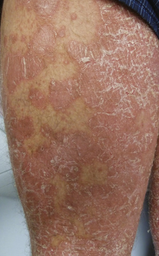
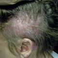
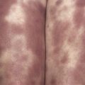
Exacerbation of an underlying skin disease is a common cause of erythroderma. Psoriasis, atopic dermatitis, contact dermatitis, stasis dermatitis, and seborrheic dermatitis can all lead to erythroderma. Psoriatic erythroderma can be triggered by abrupt withdrawal of a systemic medication, including oral corticosteroids, addition of a new medication, topical irritants such as tar or phototherapy burns, pregnancy, or systemic illness. Erythrodermic psoriasis can present with extensive involvement of characteristic psoriatic plaques ( Fig. 14-1 ) or with diffuse erythroderma, often with small pustules or coalescing “lakes” of pus. Erythroderma from atopic dermatitis tends to present as part of a chronic course with extensive lichenification following an uncontrolled flare, but may also present with a more acute erythroderma. New-onset pityriasis rubra pilaris (PRP) is another important consideration in the differential diagnosis of erythroderma. Lesions in PRP tend to have an orange-red hue and are classically separated by islands of sparing ( Fig. 14-2 ). The eruption typically starts on the face or scalp and spreads downwards, with an associated palmoplantar keratoderma. Photosensitivity is a common feature. Follicularly based, hyperkeratotic papules on the fingers, wrists, and elbows may be another clue to the diagnosis.
Related posts:
Stay updated, free articles. Join our Telegram channel

Full access? Get Clinical Tree


