Clinical Presentation
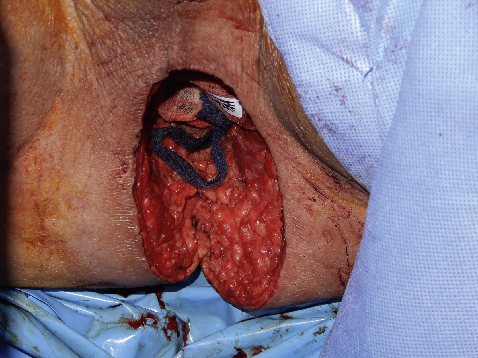
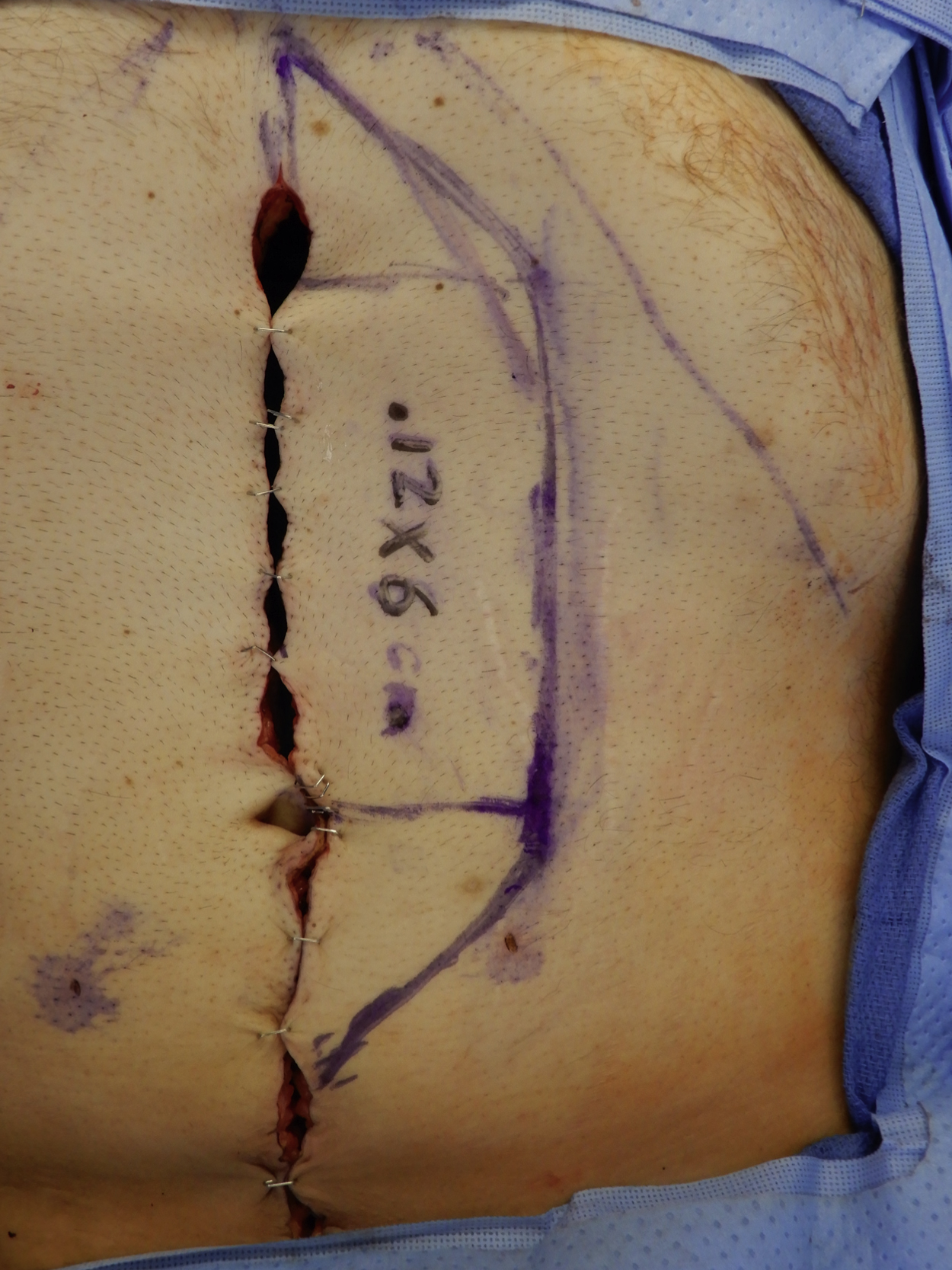
A 34-year-old White male was diagnosed with advanced rectal cancer and had been scheduled for an abdominoperineal resection (APR) by the colorectal service. Because his perineal resection could be extensive with a potential dead space within the pelvic cavity, the plastic surgery service was asked to perform a perineal reconstruction with a filling of the flap tissue into the pelvic cavity after an APR. In the operating room, an APR was completed by the colorectal service, which left a 10 × 6 cm perineal defect ( Fig. 35.1 ).
Operative Plan and Special Considerations
A vertical rectus abdominis myocutaneous (VRAM) flap can be a classic option for a patient’s perineal soft tissue reconstruction with the soft tissue filling of the dead space within the pelvic cavity. With proper design, the flap can carry a large skin paddle but only sacrifice a small amount of the anterior rectus sheath if perforators can be identified and incorporated within the skin paddle. The flap can be completely elevated and tunneled through the pelvis to fill the dead space within the pelvic cavity and be brought out for perineal soft tissue reconstruction. If only a small amount of the anterior rectus sheath is harvested with the flap, the actual fascial defect can be closed primarily and no mesh is needed for the abdominal donor site closure.
Operative Procedures
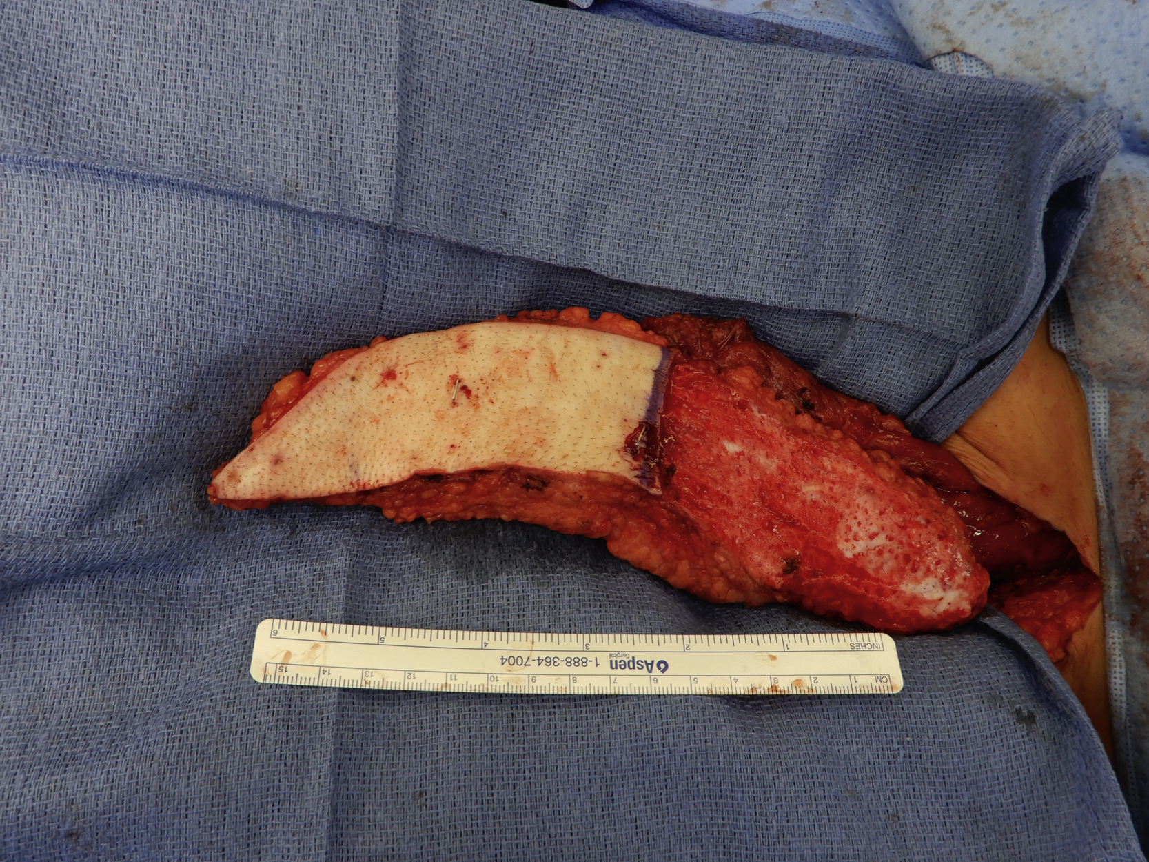
Under general anesthesia with the patient in the lithotomy position, the perineal wound was assessed after an APR procedure was completed by the colorectal service. It measured 10 × 6 cm and communicated with the pelvic cavity. A 12 × 6 cm skin paddle was designed and two perforators within the skin paddle were confirmed by a handheld Doppler ( Fig. 35.2 ). The maximum width of the skin paddle could be estimated by a skin pinch test. The proposed incision was infiltrated with 1% lidocaine with 1:100,000 epinephrine.
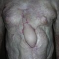
The procedure was started by making a skin incision of the skin paddle down to the anterior rectus sheath. By further dissection toward the midline, the anterior rectus sheath was incised. The rectus abdominus muscle was identified. The dissection was performed to free the lateral boarder of the left rectus abdominus muscle and each tendinous intersection was also dissected free. Several tacking sutures were used to attach the muscle to the skin paddle. At this point, the muscle close to the xiphoid was divided with electrocautery and care was taken to control the superior epigastric vessels completely. The muscle and its skin paddle was elevated from the posterior rectus sheath and the dissection was done toward the pedicle. During dissection, the inferior epigastric vessels were identified and dissected free toward to the inguinal area. The inferior insertion of the muscle was completely divided while the pedicle vessels were well protected. The entire flap was tunneled through the pelvis and brought out to the perineal area. The proximal portion of the flap was de-epithelialized after it was placed back on the abdomen ( Fig. 35.3 ). The flap was again tunneled through the pelvis and inset into the perineal area without any tension.