Other Dermatologic Conditions
Jennifer L. Greenman
This chapter will review six common dermatologic conditions that do not neatly fit into the previous chapters. All of the conditions discussed are benign, but it is important to distinguish them from malignancy and infection, as well as offering appropriate treatment when indicated.
Erythema Nodosum
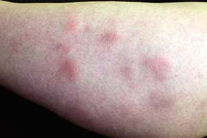 |
A 26-year-old woman presents with a 5-day history of painful, red plaques and nodules distributed over her shins (Fig. 27-1). The lesions are moderately well demarcated with dusky erythema. She complains of mild generalized aching that began at the onset of the eruption. Her only routine medication is an oral contraceptive, which she has used for the past 8 years. She denies any history of fever, fatigue, cough, gastrointestinal (GI) symptoms, malaise, mucosal ulcerations, foreign travel, or illicit drug use. She has never developed similar lesions in the past.
Background/Epidemiology
Panniculitis refers to a group of disorders involving inflammation of the subcutaneous fat. Erythema nodosum (EN) is the most common form of panniculitis and in most cases represents a hypersensitivity response to a remote focus of infection or inflammation. Overall, the prevalence of EN in the United States is approximately 1 to 5 per 100,000 persons, with peak incidence occurring between the ages of 20 and 30 years. In adults, it is more common among women, with a female to male ratio of 6:1. In children, the sex ratio is 1:1.
Key Features
Erythema nodosum (EN) is the most common form of panniculitis.
Reactions are mostly in young women, often from oral contraceptive medications.
Other associations with EN include bacterial or deep fungal infections, tuberculosis, sarcoidosis, inflammatory bowel disease, and cancer.
Erythematous nodules on shins are the most common presentation.
First-line treatment is with nonsteroidal anti-inflammatory agents (ibuprofen).
The course is usually self limiting, typically lasting 3 to 6 weeks.
Pathogenesis
Although most often idiopathic, EN has been associated with a wide variety of disease processes, including bacterial or deep fungal infections, tuberculosis, sarcoidosis, inflammatory bowel disease, and cancer. Medications including oral contraceptives and some antibiotics have also been implicated as triggers (Table 27-1). When a patient presents with EN, it is imperative to search for its etiology, because management of underlying disease is often the most definitive means of alleviating symptoms.
Clinical Presentation
Clinically, EN appears as symmetrically distributed, erythematous, deep nodules located on the shins, ankles, and knees. The skin nodules are usually painful or tender. Patients may describe nonspecific flu-like symptoms occurring
1 to 2 weeks before the eruption. Fever with generalized aching sometimes accompanies the onset of skin lesions and arthralgias are reported by a majority of patients. EN may also appear as traumatic bruises, but the patient’s history should discriminate between these two conditions. Superficial thrombophlebitis produces tender lesions on the lower legs, but these lesions are usually located on the lateral aspect of the lower legs. In addition, they consist of hard, irregular, and fibrotic cords or plaques, rather than the erythematous nodules seen in EN.
1 to 2 weeks before the eruption. Fever with generalized aching sometimes accompanies the onset of skin lesions and arthralgias are reported by a majority of patients. EN may also appear as traumatic bruises, but the patient’s history should discriminate between these two conditions. Superficial thrombophlebitis produces tender lesions on the lower legs, but these lesions are usually located on the lateral aspect of the lower legs. In addition, they consist of hard, irregular, and fibrotic cords or plaques, rather than the erythematous nodules seen in EN.
 Figure 27-1 Erythema nodosum on the shins of a young woman. |
Usually, EN is diagnosed clinically, but the physician should consider alternative diagnoses (Table 27-2). Lesions of EN appear as erythematous, well localized, deep nodules that are 1 to 5 cm in diameter (Fig. 27-1). Nodules may coalesce to produce larger plaques. Typically, multiple lesions are present and are distributed symmetrically. Pretibial involvement is most common, although the extensor surfaces of the forearms, thighs, and trunk may also be affected. Lesions begin bright red in color, but become deeper red or purple within several days. Ultimately, they evolve into brownish-yellow, bruise-like discolorations with indistinct borders. Lesions typically regress spontaneously without ulceration, scarring, or atrophy.
Diagnosis
Because EN has an extensive list of etiologies (Table 27-3), a rational, cost-effective diagnostic approach is desirable. A complete clinical history should be
elicited in all patients, with reference to previous diseases, recent infection, current medications, foreign travel, pets, as well as familial cases. Suggestions for initial evaluation are summarized in Table 27-3.
elicited in all patients, with reference to previous diseases, recent infection, current medications, foreign travel, pets, as well as familial cases. Suggestions for initial evaluation are summarized in Table 27-3.
Table 27-1 Causes of EN | |
|---|---|
|
Table 27-2 Differential Diagnosis of EN | ||||||||||||
|---|---|---|---|---|---|---|---|---|---|---|---|---|
|
Beta-hemolytic streptococcal infection is the most common identifiable cause of EN, accounting for up to 44% of cases in adults and 48% of cases in children. Nodules tend to develop within 3 weeks of streptococcal pharyngitis. Patients should undergo throat culture for group A streptococci and have antistreptolysin-O (ASO) titers drawn or a polymerase chain reaction (PCR) analysis performed. Recent streptococcal infection is likely when ASO titers change by at least 30% in two consecutive determinations performed in a 2- to 4-week interval. PCR assays more rapidly evaluate for viral infection and have been considered an effective stand-alone alternative to rapid antigen immunoassays for the identification of streptococcal infection.
A chest radiograph should be performed in all patients with EN to rule out pulmonary diseases as the cause of the cutaneous eruption. EN in combination with radiographically demonstrable bilateral hilar lymphadenopathy, arthritis, and no evidence of tuberculosis characterize Lofgren syndrome. In most cases, this represents an acute variant of pulmonary sarcoidosis with a benign course. This condition is more frequent in females, specifically during pregnancy and puerperium. All patients with EN should also be stratified by risk for tuberculosis exposure.
The patient’s geographic location and recent travel history may also be relevant when considering disease etiology. In western and south-western areas of the United States, approximately 5% of patients infected with
coccidioidomycosis, also known as San Joaquin Valley fever, develop EN. Prevalence of EN increases to 15% to 40% in patients with acute respiratory symptoms. Because resolution of fungal infection often coincides with the resolution of EN, patients who present with relevant physical findings and travel history should be evaluated for coccidioidomycosis and treated, if applicable. Diagnosing coccidioidomycosis involves either the recovery of Coccidioides species from clinical specimens or the detection of specific anticoccidioidal antibodies in serum or other body fluids. The diagnosis is most often made by serologic testing for antibodies. A commercial immunodiffusion kit is currently available.
coccidioidomycosis, also known as San Joaquin Valley fever, develop EN. Prevalence of EN increases to 15% to 40% in patients with acute respiratory symptoms. Because resolution of fungal infection often coincides with the resolution of EN, patients who present with relevant physical findings and travel history should be evaluated for coccidioidomycosis and treated, if applicable. Diagnosing coccidioidomycosis involves either the recovery of Coccidioides species from clinical specimens or the detection of specific anticoccidioidal antibodies in serum or other body fluids. The diagnosis is most often made by serologic testing for antibodies. A commercial immunodiffusion kit is currently available.
Table 27-3 Key Diagnostic Studies for EN | |
|---|---|
|
In patients who present with diarrhea or gastrointestinal (GI) symptoms, practitioners should obtain a stool culture, evaluate for ova and parasites, and consider a workup for inflammatory bowel disease. In adults, EN is more often associated with ulcerative colitis than Crohn disease. EN often parallels disease course: cutaneous eruptions often coincide with GI disease flares, while lesion resolution often occurs with bowel disease calming.
Medications are often implicated as the cause of EN. Antibiotics, especially sulfonamides, have been cited as one the most common drugs responsible for outbreaks. If patients develop EN while taking antibiotics; however, it is often difficult to determine if the medication or the initial infection is responsible for the cutaneous lesions. Oral contraceptives (OCPs) may also cause EN, although these drugs are becoming less frequently implicated. Presumably, the much lower amounts of hormones in newer OCP preparations account for this decline.
EN is often diagnosed clinically, but a tissue biopsy can be useful when a patient presents atypically. If lesions persist beyond 6 to 8 weeks, become ulcerated, or are not located on the shins, a deep incisional or excisional biopsy should be performed for histologic evaluation. Small punch biopsies should be avoided as they often produce inadequate samples.
Therapy
Initial therapy should be aimed at treating any underlying disease, if one has been identified. Resolution of an underlying process often leads to resolution of EN.
For exquisitely tender lesions, pain can be managed conservatively with nonsteroidal anti-inflammatory drugs (aspirin 325 to 650 mg PO every 4 to 6 hours, ibuprofen 400 to 800 mg PO every 6 to 8 hours, naproxen 275 mg PO every 6 to 8 hours, or indomethacin 25 to 50 mg PO three times daily).
Bed rest and avoidance of contact irritation of affected areas is also helpful.
Oral potassium iodide prepared as a supersaturated solution in a dosage of 400 to 900 mg per day for 1 month is another therapeutic option. This treatment is most effective in providing symptomatic relief when initiated at the onset of lesion eruption:
Prolonged use should be avoided to minimize the risk of hyperthyroidism, and this treatment is contraindicated in pregnancy as it may produce goiter in the fetus.
Intralesional, topical, or systemic corticosteroids can be therapeutic as well. Injection of triamcinolone acetonide (Kenalog), in a dosage of 5 mg/mL, into the center of the nodules may cause them to resolve. Applying topical corticosteroids with kitchen-grade plastic wrap occlusion at night may also help to reduce inflammation.
Systemic steroids should be reserved for cases that become recurrent or prolonged, or in patients with significant discomfort:
Prednisone 40 to 60 mg per day in a tapering dose has yielded nodule resolution within a few days.
Although a short course of systemic corticosteroids can provide dramatic relief, steroids should only be offered as a therapeutic option if underlying infection, risk of bacterial dissemination, or sepsis and malignancy has been excluded through thorough evaluation.
“At a Glance” Treatment
Identify underlying cause
First-line: nonsteroidal anti-inflammatory drugs:
Aspirin 325 to 650 mg PO every 4 to 6 hours, ibuprofen 400 to 800 mg PO every 6 to 8 hours, naproxen 275 mg PO every 6 to 8 hours, or indomethacin 25 to 50 mg PO three times daily
Bed rest and avoidance of contact irritation:
Oral potassium iodide supersaturated 400 to 900 mg per day for 1 month
Intralesional triamcinolone acetonide (Kenalog) 5 mg/mL, into the center of the nodules may cause them to resolve
Systemic steroids should be reserved for cases that become recurrent or prolonged, or in patients with significant discomfort:
Prednisone 40 to 60 mg per day in a tapering dose
Patients with persistent or extremely painful lesions, or those that appear in atypical locations should be referred to a dermatologist for evaluation.
Consider systemic fungal or mycobacterial infection in appropriate cases.
The most common cause is strep or medications, exclude those first.
Course and Complications
EN is usually self-limiting, typically lasting 3 to 6 weeks. More severe cases require about 6 weeks to resolve. Lesions do not scar. Relapses are exceptional, but they are more common in patients with idiopathic EN and EN associated with upper respiratory tract infections. When associated with inflammatory bowel disease, EN may parallel GI disease course, recurring with bowel flares and regressing between episodes. Finally, in elderly patients, especially those with severe venous insufficiency and gravitational edema of the lower extremities, an acute episode of EN may be followed by a persistent erythematous swelling of the ankles.
ICD9 Code
| 695.2 | Erythema nodosum |
Granuloma Annulare
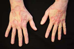 |
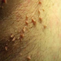
A 32-year-old woman presents with an annular, pinkish lesion on the dorsum of her right foot, lateral ankle region, and dorsal hands (Fig. 27-2). The lesion was first noticed approximately 1 month ago. Although it has not bothered the patient, she is concerned because this pinkish ring had slowly expanded in size. On examination, the lesion has slightly elevated peripheral borders with palpable and mobile subcutaneous granules. What is the most likely diagnosis?
Background/Epidemiology
Granuloma annulare (GA) is a benign, self-limited, papular eruption most often distributed on the dorsal aspect of the hands and feet (Fig. 27-2). Usually, it is asymptomatic and comes to the physician’s attention because of cosmetic concerns. Its name derives from its histological and clinical appearances. Clinically, lesions present as ringlike or annular groups of papules. GA may affect patients at any age, but most cases are diagnosed during the first three decades of life. Incidence is highest in women, with a ratio of 2:1. It is also seen more frequently in atopic individuals.
Key Features
Granuloma annulare (GA) is a benign, self-limited, papular eruption most often distributed on the dorsal aspect of the hands and feet.
It may affect patients at any age, but most cases are diagnosed during the first three decades of life.
Etiology is unknown and diagnosis is clinical. Rigorous studies are unnecessary.
GA resolves between 2 months and 2 years after initial presentation in 75% of patients.
Pathogenesis
Although the etiology of GA remains unclear, some studies have shown it to follow viral infections, insect bites, tuberculin skin tests, cell-mediated hypersensitivity reactions, and trauma. In children, GA has not been associated with any systemic illness. In adults, several reports have also documented cases of
GA occurring in patients with acquired immunodeficiency syndrome, specifically at the site of a previous herpes zoster infection.
GA occurring in patients with acquired immunodeficiency syndrome, specifically at the site of a previous herpes zoster infection.
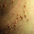
 Figure 27-2 Granuloma annulare on the dorsal hands. |
Clinical Presentation
GA can take many clinical forms. The four main variants are localized (Fig. 27-3), disseminated (Fig. 27-4), subcutaneous, and perforating (Table 27-4). Seventy-five percent of patients present with localized GA. Each subtype is discussed below.
Localized Type
Localized GA typically begins as a small, expanding ring or arc of 2- to 4-mm papules that are usually flesh-colored but sometimes erythematous, violaceous, or brown (Fig. 27-4). These papules are closely set and often give the ring’s border a “beaded” appearance. Although overlying epidermal markings may be distorted, they are intact. Scaling, vesicles, erosions, and pustules are absent. As the condition progresses, ring centers may become slightly depressed and hyperpigmented. Lesion borders remain distinct and feel slightly indurated on palpation. Several lesions may become confluent and merge to form larger annular plaques. In general, lesions are asymptomatic, nontender, and nonpruritic. Most often, localized GA is found on the dorsal aspects of the hands and feet. More than 50% of patients will have spontaneous resolution within 2 years.
Disseminated Type
Disseminated or generalized GA is similar in appearance to the localized variant, but is more widespread. Multiple flesh-colored or erythematous papules may fuse
to form annular rings on the extremities, trunk, and neck. These lesions occur almost exclusively in adults and tend to persist longer than localized lesions.
to form annular rings on the extremities, trunk, and neck. These lesions occur almost exclusively in adults and tend to persist longer than localized lesions.
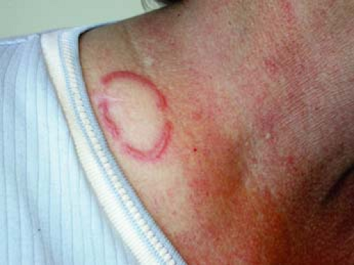 Figure 27-3 Localized granuloma annulare. |
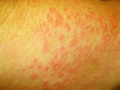 Figure 27-4 Disseminated granuloma annulare. |
Table 27-4 Types of GA | |
|---|---|
|
Subcutaneous Type
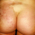
Subcutaneous GA is rare and mostly diagnosed in children between the ages of 2 to 5 years. Lesions are asymptomatic, rapidly growing soft-tissue nodules on the extremities, hands, scalp, buttocks, and pretibial and perioral areas. On palpation, the lumps are firm, nontender, and nonmobile and vary in size from 1 to 5 cm. Subcutaneous nodules are often solitary, but they may also occur in clusters. Lesions may resolve spontaneously or recur after excision.
Perforating Type
Perforating GA is another rare variant most commonly seen in children and young adults. It is characterized by 1- to 4-mm umbilicated, crusted, or scaly papules on the extremities. Most patients are asymptomatic, although 25% reported pruritus, and 25% complained of pain in at least one study.
Although GA is a common dermatologic condition in the pediatric and adult populations, it is frequently misdiagnosed. The ability to distinguish GA from other lesions is important, because it allows the clinician to counsel patients and parents appropriately and to avoid unnecessary and expensive medical evaluations. A number of common annular skin conditions, including tinea corporis, nummular eczema, psoriasis, erythema migrans, and pityriasis rosea can appear similar to GA (Table 27-5). The key feature that distinguishes GA from these other conditions is its lack of skin surface changes.
Diagnosis
Often, the diagnosis of GA is clinical and rigorous studies are unnecessary. Potassium hydroxide preparations are simple and efficiently help rule out fungal infections, if scaling at lesion borders is subtle. In addition, a punch biopsy may be helpful in clinically confusing cases, especially with the subcutaneous variant of GA.
Imaging studies may also be helpful when evaluating for the subcutaneous variant of GA. Radiographs show a nonspecific soft-tissue mass without calcification or bone involvement. Ultrasonographic examination reveals a hypoechoic area in the subcutaneous tissues. Magnetic resonance imaging shows a mass with indistinct margins, isointense or slightly hyperintense to muscle with T1-weighted images.
Therapy
In most cases, treatment is not necessary, and physicians should reassure their patient and his or her family about the good prognosis of this poorly understood entity. GA resolves between 2 months and 2 years after initial presentation in 75% of patients. However, 40% of children may have recurrent lesions,
often at the original eruption site. Above all, the physician should educate patients and family members on the natural course and benign nature of GA. Individuals should be reassured that GA is neither infectious nor contagious and usually resolves in several months to years.
often at the original eruption site. Above all, the physician should educate patients and family members on the natural course and benign nature of GA. Individuals should be reassured that GA is neither infectious nor contagious and usually resolves in several months to years.
Table 27-5 Differential Diagnosis for GA | ||||||||||||||||||||||||
|---|---|---|---|---|---|---|---|---|---|---|---|---|---|---|---|---|---|---|---|---|---|---|---|---|
|
Related posts:

Stay updated, free articles. Join our Telegram channel

Full access? Get Clinical Tree


