Pigmentary Disorders
Vinod E. Nambudiri
Much like the height and weight of individuals, the spectrum of skin pigmentation is a continuous, fluid distribution of hues. The pigment-containing cells of the skin, the melanocytes, are thought to be present in roughly equal number across all skin types. However, the distribution of melanosomes, subcellular organelles rich in the primary skin pigment melanin contained within melanocytes, varies in concentration. Greater melanosome density results in darker baseline pigmentation. This chapter examines disorders in pigmentation, with consideration given to the common specific pigmentary aberrations of melasma and vitiligo, as well as generalized disease processes such as postinflammatory hyperpigmentation.
Melasma
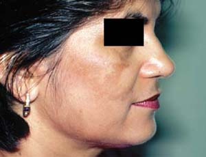 |
A 39-year-old primigravid Indian woman presents to her physician at 30 weeks gestation concerned about the appearance of several new dark-colored patches of skin on her cheeks and forehead. These have appeared over the last few weeks. On physical exam, you note that she has a baseline brown complexion. There are multiple darker brown, nonelevated, blotchy patches of skin on her face, primarily involving the forehead, cheeks, and nose (Fig. 14-1). On further history taking, she reveals that she spends a considerable time outdoors enjoying the sunny weather. She also recalls her mother and sister mentioning similar episodes during their respective pregnancies. What is the most likely diagnosis? What if any treatments are you able to offer her?
Background/Epidemiology
The term “melasma” derives from the Greek words describing a “black spot” and was originally used in medical literature to refer to the increased skin pigmentation arising in the lower extremities due to venous stasis. In modern clinical dermatology, the term has been used to refer to conditions resulting in patchy or generalized hyperpigmentation, most often of the face. When occurring in pregnant women, the term “chloasma” is used to describe the hyperpigmented appearance of the face—often referred to as the “mask of pregnancy.”
Pathophysiology
While the disease may appear in any individual, females and those of Asian, Hispanic, and Middle Eastern descent are more commonly affected by melasma than others. The condition is one that typically affects individuals in the second and third decades of life. The hyperpigmentation in melasma is due to the deposition of melanin in the epidermis, dermis, or both. Interestingly, despite the
association with several putative etiologic factors that contribute to the development of melasma, the exact pathogenic mechanism remains unknown.
association with several putative etiologic factors that contribute to the development of melasma, the exact pathogenic mechanism remains unknown.
Key Features
Melasma is a hormonally related skin hyperpigmentation, most often on the cheeks and upper lip.
It is often seen during and after pregnancy or with use of oral contraceptives.
Treatment includes daily sunscreen use and bleaching agents such as hydroquinone.
Clinical Presentation
The presence of hyperpigmentation—an increase in pigment deposition resulting in darkening—is most easily apparent against the background of lighter skin tones. However, melasma, a clinical diagnosis based on the presence of acquired hyperpigmentation, may arise in individuals of virtually any baseline skin type. Melasma most commonly is found on sun-exposed areas of skin. There is often a symmetric appearance of the hyperpigmented, which range in color from ashy gray to light- to dark-brown and are nonelevated (Fig. 14-1). The three classic distributions for facial melasma hyperpigmentation are (a) centrofacial, involving the forehead, cheeks, chin and upper lip; (b) malar, involving primarily the cheeks; and (c) mandibular, classically involving the chin and jaw line. Melasma is not limited to the face, and is often seen in the dorsal extremities and sun-exposed areas.
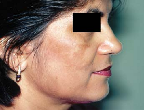 Figure 14-1 Facial melasma. From Goodheart HP. Goodheart’s Photoguide of Common Skin Disorders, 3rd ed. Philadelphia: Lippincott Williams & Wilkins, 2009. |
Diagnosis
The diagnosis of melasma is often made clinically based on the physical appearance and patient history. The use of a Wood’s lamp (an ultraviolet light source) can aid in determining the level of pigment deposition in melasma. Classically, pigment deposited in the epidermis will result in light brown pigmentation of the affected skin that exhibits enhanced contrast under Wood’s lamp examination. Pigment deposition in the dermis often produces ashen or bluish discoloration of the skin that does not change or enhance under Wood’s lamp examination. In individuals with pigment deposition in both layers of the skin, the hyperpigmented regions appear an intermediate darker brown in color and will show variable enhancement under Wood’s lamp examination. Diagnosis of any of the aforementioned variants may be more complex in individuals with darker background skin complexion. A skin biopsy is not required to make the diagnosis of melasma.
The differential diagnosis for melasma includes other disorders of hyperpigmentation described in this chapter, such as postinflammatory hyperpigmentation, or other inflammatory-based conditions such as contact dermatitis, or drug-related hypersensitivities and photosensitivity. A thorough history is important when melasma is the suspected clinical diagnosis, as several potential underlying causes may be considered (Table 14-1).
Treatment
Varying courses of melasma have been reported, from self-resolving episodes to recurrent or chronic hyperpigmentation. Cessation of an obvious external
trigger—such as particular contraceptives or topical cosmetics—is indicated when such factors are believed to be part of the causal etiology. Thyroid hormone levels should be checked if there is suspicion of an underlying thyroid disorder, as these states are more common in individuals with melasma. First and foremost, assiduous daily application of a high-number UVA/UVB blocking sunscreen is mandatory if medical therapy or cosmetic procedures are to be undertaken. Months of treatment can be quickly undone by several hours of unprotected sun exposure.
trigger—such as particular contraceptives or topical cosmetics—is indicated when such factors are believed to be part of the causal etiology. Thyroid hormone levels should be checked if there is suspicion of an underlying thyroid disorder, as these states are more common in individuals with melasma. First and foremost, assiduous daily application of a high-number UVA/UVB blocking sunscreen is mandatory if medical therapy or cosmetic procedures are to be undertaken. Months of treatment can be quickly undone by several hours of unprotected sun exposure.
Table 14-1 Factors Associated with Melasma Development | ||||||||
|---|---|---|---|---|---|---|---|---|
|
A variety of medical treatments are available to diminish existing hyperpigmentation. Available in 2% or 4% topical cream or topical gel preparation, hydroquinone is the topical medications most commonly prescribed for the treatment of melasma. By inhibiting the action of the enzyme tyrosinase, hydroquinone is effective at blocking a key step in melanin synthesis. For melasma treatment, the 4% hydroquinone is applied twice daily on the affected areas for 2 to 3 months or until results are noted. Preparations of hydroquinone with sunblock are also available by prescription and would be useful for individuals with large sun-exposed areas of hyperpigmentation due to melasma.
Tretinoin 0.05% cream can also be used for the treatment of melasma. Tretinoin acts by increasing the speed of turnover of the stratum corneum (the outermost, keratin-rich layer of the skin) and may be used daily at bedtime for 3 to 6 months for melasma. A more potent preparation of hydroquinone 4%, tretinoin 0.05%, and topical steroid fluocinolone acetonide 0.01% is also available by prescription (Tri-Luma) for the treatment of melasma and can be applied nightly for up to 8 weeks. Another topical hypopigmentation agent that may be used as an alternative to hydroquinone-based preparations is 20% azelaic acid.
Risks of topical hypopigmenting agents include drying of the skin, hypersensitivity reactions, and bleaching of surrounding skin. Hypopigmenting topical agents are relatively contraindicated in the treatment of melasma in pregnant individuals.
Further therapies available for the treatment of refractory melasma include chemical peels. Salicylic acid peels, glycolic acid peels, and trichloroacetic acid peels have been reported to decrease pigmentation in conjunction with the aforementioned depigmenting agents. Other reported modalities include dermabrasion and laser therapy.
“At a Glance” Treatment
All patients with melasma: daily application of a high SPF sunscreen/block with UVA protection
Primary medical therapy:
Apply 4% hydroquinone BID on the affected areas for 2 to 3 months
Tretinoin 0.05% cream QHS for 3 to 6 months
A mix of hydroquinone 4%, tretinoin 0.05%, and fluocinolone acetonide 0.01% (Tri-Luma) QHS for up to 8 weeks
Secondary treatments:
Facial chemical peels
Dermabrasion
Laser therapy
Melasma that is refractory to treatment with topical hypopigmentation agents should be referred to a dermatologist for possible evaluation of alternative treatment modalities.
Referral to a dermatologist interested in cosmetic procedures is recommended for those desiring chemical peels or laser treatments.
Course and Complications
Melasma is not a life-threatening disease but may cause the sufferer significant concern given its visibility on the face. Despite the incomplete understanding of underlying pathogenesis contributing to melasma, several treatments have evolved to combat the increased pigmentation. Limiting
sun exposure and daily use of opaque sunblock regardless of sun exposure should be stressed to the patient to prevent further progression of melasma.
sun exposure and daily use of opaque sunblock regardless of sun exposure should be stressed to the patient to prevent further progression of melasma.
All patients with melasma should apply a high-number sunscreen to affected areas daily.
ICD9 Code
| 709.09 | Other dyschromia |
Vitiligo
 |
A healthy 28-year-old man presents to your clinic with questions about his skin. Over the past few months he has begun to notice patchy areas of his skin that have become lighter than what he describes as his “normal” skin tone. The areas that he identifies are distributed on his face, trunk, both arms, and fingers (Fig. 14-2). They appear chalk-white in color, and have sharp borders demarcating them from his type IV (olive-brown) colored skin. The patches do not itch and are not painful, but he has become increasingly self-conscious about wearing short sleeves that reveal the discolored patches on his skin. He does note that the light-colored areas appear to be growing in size, and also notes that his sister has some similar patches on her arms as well. He is concerned about the possibility of the patches spreading and affecting his face or other parts of his body. What is his most likely diagnosis? What is the pathologic process at work? What preventative strategies and therapeutic treatments are available for his condition?
Background/Epidemiology
At the opposite end of the spectrum from the hyperpigmented, dark-colored patches of melasma, the tell-tale skin findings in vitiligo are patches of pale skin with a partial or total lack of pigmentation. While not limited to one specific skin type, the lesions of vitiligo are more readily apparent in individuals with darker baseline pigmentation due to the contrast between normal and affected skin. Also known as leukoderma, vitiligo characteristically presents during childhood or early adulthood and may persist throughout an individual’s lifetime. The time course for the initial presentation and the chronicity of disease is variable; the depigmentation may be progressive over weeks to months and may continue to expand, remain stable, or even self-resolve in individual cases. Both men and women may be affected by vitiligo, and the disease has been described around the world with a similar prevalence (0.5% to 2.0% of the population). Onset is usually early in life, averaging around age 20 years. Given the easier detection in individuals with darker baseline pigmentation, vitiligo may be more frequently reported—or treatment may be more frequently sought—by individuals with skin of color.
Key Features
Vitiligo presents as hypo- or depigmented patches, characteristically symmetric.
It is caused by death of normal epidermal melanocytes and is associated with thyroid disease and other autoimmune conditions.
Treatment is often at best partially successful. Topical steroids and tacrolimus may be useful. Narrowband UVB for widespread disease may offer slow improvement.
Pathophysiology
The underlying etiology of vitiligo is poorly understood. Putative hypotheses have been put forward to explain the pathophysiologic mechanism of the disease, but this remains an area of active research. An understanding of the disease process emerges from the histologic findings seen on microscopic examination of biopsies from vitiligo patches: normal skin but for the total absence of melanocytes.
Prevailing theories about the evolution of vitiligo include a cellular immune-mediated phenomenon (aberrant cytotoxic T-lymphocytes activated against melanocytes), a humoral immune-mediated phenomenon (circulating autoantibodies targeted against melanocytes), a melanin biosynthetic pathway mutation (increased generation of free radicals within melanocytes), as well as combinations of the above postulates. While there is not a direct Mendelian
inheritance pattern associated with vitiligo, up to 30% of individuals with the disease will report at first-degree family member with vitiligo as well.
inheritance pattern associated with vitiligo, up to 30% of individuals with the disease will report at first-degree family member with vitiligo as well.
Clinical Presentation
An individual with vitiligo often notices the development of focal patches of off-white to chalk-white skin. Often seen in the face (Fig. 14-2), trunk, extensor surfaces, extremities, groin, and axillae, vitiligo is a condition that develops after birth, most commonly presenting in the first three decades of life. The white patches of skin may start as small, millimeter sized areas and expand to involve several square-centimeters of skin. Patients may report a history of trauma, sunburn, or viral illness preceding the onset of depigmentation in vitiligo, though none of these is a necessary prerequisite.
In addition to the dermatologic manifestations of vitiligo, it is important to question about the psychological impact of the disease as well. Vitiligo affects areas of the skin that are easily visible to both the patient and other individuals—such as the face, arms, and hands—the condition may become a source of concern not only for the patient but for those in their immediate surroundings. In addition to the uncertainty of cause underlying the depigmentation that the patient must face, in some cultures there is social stigma to having areas of nonpigmented skin and this generates a psychosocial burden of embarrassment and stress that must also be dealt with.
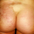
The physical findings in vitiligo of hypopigmented and depigmented macules may be found distributed throughout the body. The pattern of depigmentation with surrounding hypopigmented patches and normal skin is termed trichrome vitiligo (Fig. 14-3), with a common predilection for the face, trunk, extensor surfaces, extremities, groin, and axillae, as noted above; there is often involvement of peri-orificial tissue such as perioral and perianal regions. The patches of vitiligo retain their normal sensation and do not illicit pain or other discomfort such as itching or burning. Hair growing in the area of depigmentation may turn white (poliosis) due to the loss of melanocytes in these skin appendages as well.
The most common distribution of vitiligo is generalized patches of depigmentation spread throughout the body with relative midline symmetry,
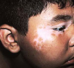
termed vitiligo vulgaris. Another variant involves primarily the face and distal extremities, so-called acrofacial vitiligo. When confined to a single dermatome, the patchy depigmentation is referred to as segmental vitiligo (Fig. 14-4), often presenting when the patient is in the first or second decades of life. Finally, an isolated patch of chalk-white depigmentation may be termed focal vitiligo (Fig. 14-5). Given the unpredictable nature of the disease, however, it is possible for conversion between distribution subtypes; for example, progression from focal to segmental may occur with time. When the majority of the body surface is involved, the condition is termed vitiligo universalis. These categorizations are summarized in Table 14-2.
termed vitiligo vulgaris. Another variant involves primarily the face and distal extremities, so-called acrofacial vitiligo. When confined to a single dermatome, the patchy depigmentation is referred to as segmental vitiligo (Fig. 14-4), often presenting when the patient is in the first or second decades of life. Finally, an isolated patch of chalk-white depigmentation may be termed focal vitiligo (Fig. 14-5). Given the unpredictable nature of the disease, however, it is possible for conversion between distribution subtypes; for example, progression from focal to segmental may occur with time. When the majority of the body surface is involved, the condition is termed vitiligo universalis. These categorizations are summarized in Table 14-2.
Related posts:

Stay updated, free articles. Join our Telegram channel

Full access? Get Clinical Tree


