Ultraviolet Light Reactions
My-Linh T. Nguyen
Sunburn is acute inflammation of the skin in response to ultraviolet (UV) damage. Its characteristic erythema and pain are well known to many patients. Sunburns present a problem not only because of their immediate symptomatology, but also because they are a marker of excessive sun exposure and thus cancer risk. Patients with a history of sunburn carry a twofold relative risk of developing melanoma versus patients with no sunburn history. Treatment for sunburns involves symptomatic relief, fluid replacement, prophylaxis for possible infection, and preventative counseling.
There are many other reactions to the sun that may mimic sunburn or have more of an eczematous nature. Commonly encountered conditions would include polymorphous light reaction, photoallergic contact dermatitis, and phytophotodermatitis. These will be discussed in further detail.
Sunburn
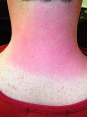 |
A 23-year-old white man presents with sharply demarcated, uniform erythema with pain on his neck (Fig. 22-1). History reveals that he had been skiing earlier in the day. What is the most likely diagnosis? How should he be treated? What counseling should he be given?
Background/Epidemiology
For lighter-skinned individuals, most have experienced sunburn as a result of prolonged sunlight exposure. In the United States, 30% to 40% of adults and 83% of children report at least one sunburn in the preceding year. Individuals with darker skin types are less likely to burn, but occasionally may experience sunburn while on a photosensitizing medication or in some cases as a result of a systemic illness that includes photosensitivity. In general, sunburns are classified as a superficial or first-degree burn. Sunburns are more common in men and, in general, children are burned more frequently than adults.
Key Features
Sunburn is caused by excessive UV radiation causing delayed, sharply demarcated, uniform edema in exposed skin with possible pain, pruritus, edema, and vesicles/bullae.
Predisposing factors include: light skin, light hair, and photosensitizing medications.
Treatment:
Symptomatic therapy (cold compress, moisturizing lotion, nonsteroidal anti-inflammatory drugs [NSAIDs], corticosteroids).
Fluid replacement.
Infection prophylaxis (topical antibiotic if vesicles/bullae present).
Sun avoidance during healing.
Preventative counseling.
Pathogenesis
The exact mechanisms leading to the sunburn reaction are not fully known. Current evidence suggests that UV irradiation leads to cutaneous vasodilation, causing erythema, edema, and neutrophilic infiltration. UVB radiation causes apoptosis through a variety of pathways, including DNA mutagenesis, direct activation of membrane-bound death receptors, and generation of intracellular reactive oxygen species.
Table 22-1 Fitzpatrick’s Skin Phototypes | |||||||||||||||||||||
|---|---|---|---|---|---|---|---|---|---|---|---|---|---|---|---|---|---|---|---|---|---|
|
Clinical Presentation
Manifestation
The presentation of sunburn can range from mild (sharply demarcated, uniform erythema on sun-exposed areas with sparing of covered skin [Fig. 22-1]) to severe (erythema accompanied by edema, vesicles, bullae, pain, and/or pruritus). Erythema typically arises 6 hours after sun exposure, peaks at 12 to 24 hours, and resolves after 72 hours. In severe cases, fever, fatigue, headache, and tachycardia may be present.
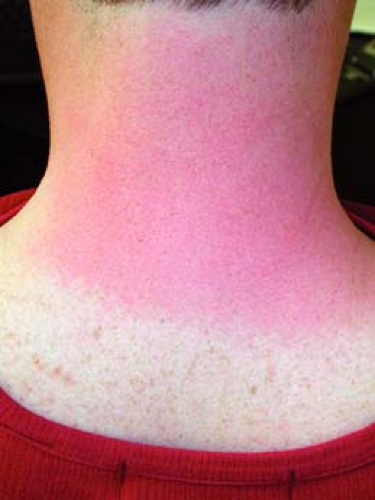 Figure 22-1 Acute sunburn on the neck. Note the sharp cutoff where the shirt protected the skin. |
History and Risk Factors
UV exposure: the amount of UV exposure correlates with severity of sunburn. Low latitude, high altitude, and midday exposure (11 AM to 4 PM) represent the greatest risk for skin damage. Sunburns can occur on cloudy days, as UV radiation penetrates cloud cover, and in the shade, because UV can reflect off of snow, water, and concrete to reach skin.
Skin type: the Fitzpatrick skin phototypes scale (Table 22-1) classifies skin based on its baseline color and reaction to sun exposure. Skin types I and II are at greatest risk for sunburns.
Hair color: lighter hair such as blonde, red, or light brown is associated with increased risk of sunburns.
Photosensitizing medications: summarized in Table 22-2.
Table 22-2 Medications Associated With Photosensitivity | |
|---|---|
|
Table 22-3 Differential Diagnosis of Sunburn Reactions | ||||||||||||||||||
|---|---|---|---|---|---|---|---|---|---|---|---|---|---|---|---|---|---|---|
|
Diagnosis
Diagnosis is relatively straightforward given a history of recent sun exposure and physical exam showing characteristic skin reaction in sun-exposed areas.
History: UV exposure 3 to 6 hours before onset of erythema
Physical exam:
Skin: uniform erythema in sun-exposed areas with sharp demarcation versus nonexposed areas. In severe sunburn, erythema may be accompanied by edema, vesicles, and bullae.
General: in severe cases, fever, malaise, and rapid pulse may be present.
Mucous membranes: rarely, tongue may be sunburned if mouth is open during physical activity
The differential diagnosis of sunburn reactions is broad. Causes range simply from prolonged UV exposure to systemic illnesses, which cause increased sensitivity to the sun. This differential is summarized in Table 22-3.
Treatment
Sunburn is managed through symptomatic treatment, fluid replacement, prophylaxis for infection if needed, and avoidance of sun exposure during recovery. Currently, there are no recommended therapies that significantly shorten recovery time or reduce epithelial injury.
For hyperpigmentation following phytophotodermatitis, hydroquinone 4% cream may be useful in lightening the dark pigment.
Symptomatic Treatment
The patient may find moisturizing lotion, aloe vera gel, cool water soaks, and/or cold compresses helpful in relieving discomfort.
Topical or oral NSAIDs decrease erythema if used before or immediately after UV exposure, but their benefits drop off 24 hours after UV exposure. One exception is topical diclofenac 0.1% gel (5 mg/cm2), which, when applied 6 and 10 hours after UV exposure, can relieve pain and erythema for up to 48 hours.
Topical corticosteroids may boost the benefits of NSAIDs if applied shortly after UV exposure. They do not provide clinically significant benefit after 30 hours post-UV exposure or when used without NSAIDs.
Antihistamines are not recommended, as they have not been shown to produce clinically significant benefits.
Although there is little published evidence documenting the efficacy of topical anesthetics in sunburn, some patients may find relief with agents such as benzocaine. Benzocaine may cause allergic contact dermatitis.
Fluid replacement: oral rehydration is recommended to compensate for increased fluid loss through the injured skin.
Infection prophylaxis: if vesicles/bullae are present, a topical antibiotic such as silver sulfadiazine or bacitracin may be used to decrease the risk of infection.
Sun avoidance during healing: further sun exposure should be avoided for 1 to 2 weeks, as the injured skin is more susceptible to sunburns during this time. See “Prevention” below.
“At a Glance” Therapy
Symptomatic Treatment
Moisturizing lotion, aloe vera gel, cool water soaks, and/or cold compresses
Topical (diclofenac 0.1% gel applied 6 and 10 hours after UV exposure) or oral NSAIDs
Topical corticosteroids should be used in conjunction with topical NSAIDs
Antihistamines produce clinically significant benefits.
Although there is little published evidence documenting the efficacy of topical anesthetics in sunburn, some patients may find relief with agents such as benzocaine. Benzocaine use may cause allergic contact dermatitis.
Prevention
To avoid further sunburns as well as to reduce chronic sun damage and cancer risk, UV exposure should be limited using the following techniques:
Avoid sun exposure between 10 AM to 4 PM, and especially between 11 AM to 2 PM. This should be done even on cloudy days, as UV radiation penetrates cloud cover.
Wear sunglasses, a wide-brimmed hat, and dark, loose-fitting clothing.
Wear sunscreen with at least an SPF (sun protection factor) of 30. SPF is the factor by which a substance prolongs the minimal duration of UV exposure required to produce erythema. If a patient normally develops erythema after 10 minutes of exposure, for instance, then with SPF-15 sunscreen, he/she would theoretically develop erythema after 150 minutes of UV exposure. However, this is an overestimate, as most people significantly under-apply sunscreen (recommended application is 2 mg/cm2 or 30 to 35 mL/body application) and realistically achieve only 20% to 50% of the expected SPF. Sunscreen should be applied 15 to 30 minutes before sun exposure and re-applied every 2 to 3 hours.
Oral and topical antioxidants such as vitamins A, C, and E help protect against sunburns. However, antioxidants should be used in conjunction with sunscreen, not as a replacement.
Although tanning through gradual sun exposure or tanning beds can decrease the risk of sunburn, it is not recommended as a preventative technique, because tanning may not prevent chronic sun damage and skin cancer and may carry its own negative risks. Artificial “sunless” tanning uses dyes to give the appearance of tanned skin and does not confer sunburn protection.
Course and Complications
Rupturing of vesicles and bullae can lead to infection.
Severe sunburns can damage melanocytes, leading to mottled depigmentation, or induce melanocyte proliferation, causing solar lentigines.
Sunburns are a marker of excessive UV exposure and indicate increased risk of dermatoheliosis, actinic keratoses, and skin cancer. A history of sunburn confers a 2.0 relative risk of developing melanoma versus no sunburn history.
If symptoms do not resolve after 2 weeks, consider consulting a dermatologist.Related posts:

Stay updated, free articles. Join our Telegram channel

Full access? Get Clinical Tree