Geriatric Dermatology
Gregory L. Wells
Peter C. Schalock
With advances in medicine and nutrition, the aging population continues to grow. Common causes of geriatric dermatologic visits include a multitude of dermatoses that are related to the aging of the skin and human body. Some common causes of geriatric skin problems are benign keratoses such as seborrheic keratoses and cherry angiomas, as well as neoplastic problems such as basal cell/squamous cell carcinomas and melanomas. These are reviewed in their respective chapters. Other conditions that are seen more commonly in the older individual will be reviewed here and include angular cheilitis, generalized pruritus, Grover disease, xerosis, and neoplasm-related dermatoses. Stasis dermatitis and skin ulcerations are also frequently encountered in older individuals. These topics are covered in Chapter 20.
Angular Cheilitis
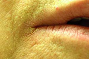 |
A 72-year-old man describes a painful rash at the corners of the mouth (Fig. 17-1). During examination, it is noted that he has fissured pink patches affecting the bilateral angles of the mouth. He describes poor-fitting dentures. What is the most likely diagnosis?
Background
Angular cheilitis (AC) is a common dermatosis affecting the corners of the lips and adjacent skin. The causes of this condition are multifactorial. AC is most commonly seen in older individuals, though any age may be affected. The risk of developing AC is three times higher for those wearing dentures, and it is estimated that 30% to 50% of those with upper and lower dentures will develop AC. Another group at higher risk for developing AC is acquired immunodeficiency syndrome (AIDS) patients with lower CD4+ counts, thus allowing more frequent candidal infections. Patients with anemia, malignancy, or diabetes mellitus are also at higher risk for developing this condition.
Key Features
Angular cheilitis (AC) is a reaction due to excessive wetness or dryness.
Presentation is with fissured pink-red to gray-white papules and plaques at the angles of the mouth.
Perform a potassium hydroxide (KOH) exam to rule out Candida or culture to rule out Staphylococcus aureus infection.
Treat with antifungals or antibacterials and consider low- to mid-potency topical steroids or emollients if necessary.
Pathogenesis
Angular cheilitis results from chronic inflammation most often caused by saliva contacting the angles of the mouth (maceration/irritant dermatitis also known as perlèche). In elderly patients, ill-fitting dentures or resorption and atrophy of the alveolar bone may lead to poor occlusion of the lips, allowing chronic saliva leakage. In many, a fine crease at the corners of the mouth allows saliva to move out of the mouth by capillary action, thus creating a moist
environment that is conducive for fungal or bacterial growth. The most commonly encountered organisms in AC are S. aureus and Candida albicans.
environment that is conducive for fungal or bacterial growth. The most commonly encountered organisms in AC are S. aureus and Candida albicans.
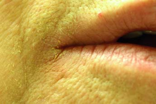 Figure 17-1 Angular cheilitis in an older man. |
Another consideration is allergic contact dermatitis, often secondary to products being used to treat the primary AC. An example would be the patient who uses a neomycin and bacitracin-containing ointment to treat her AC, and who then develops pruritic dermatitis in addition to the primary AC (Fig. 17-2). Causes of AC are summarized in Table 17-1.
 Figure 17-2 Angular cheilitis secondary to allergic contact dermatitis to applied neomycin. |
Clinical Presentation
Angular cheilitis initially presents as bilateral fissuring, erythematous plaques, and scaling at the corners of the mouth (Figs. 17-1, 17-2). These changes sometimes extend laterally and inferiorly. In many cases, the cutaneous lip and cheek skin are fissured. Depending on the process causing this condition, there are often other findings. Honeycomb crusting would suggest bacterial impetigo, while satellite pustules and a deep red appearance and fine scale suggest a fungal infection. Symptoms include pain, pruritus, or burning.
Table 17-1 Causes of Angular Cheilitis | |
|---|---|
|
Diagnosis
Angular cheilitis is a clinical diagnosis. To evaluate for fungal infection, one should consider a KOH preparation or send a culture for fungus/yeast. Bacterial infection is appropriately evaluated by taking a swab for culture. In general, a skin biopsy or further invasive testing is not indicated.
In patients in whom an underlying systemic cause is suspected, appropriate further workup is recommended. Consider checking a complete blood count, blood glucose, vitamin levels, and in patients with risk factors, a human immunodeficiency virus (HIV) test. Nutritional status should be evaluated. If allergic contact dermatitis is suspected, simply discontinuing all topical medications other than white petrolatum for at least 3 weeks should show considerable improvement. If definite identification of an allergen is necessary, the patient should be referred to a dermatologist for patch testing.
Therapy
Therapy is based on the underlying cause of AC. In the patient with dentures, refitting problematic/ill-fitting prostheses should be helpful. Treat infections appropriately with agents such as topical antifungals (i.e., ketoconazole, clotrimazole, miconazole); topical antibiotics (i.e., mupirocin); or topical anti-inflammatory agents such as calcineurin inhibitors (i.e., tacrolimus and pimecrolimus). If there is a component of atopic or allergic dermatitis, the mainstay of therapy is low- to mid-potency topical steroids (i.e., desonide or triamcinolone acetonide). In cases of recalcitrant yeast infection, consider oral fluconazole therapy.
Once the acute dermatitis is controlled, one may consider zinc oxide or white petrolatum as a barrier for maintenance. In difficult cases, one may consider treating with injectable intradermal fillers such as hyaluronic acid to reshape the oral commissures.
“At a Glance” Treatment
Refit ill-fitting dentures.
Treat based on etiology of infection or irritation:
Bacterial infection: mupirocin ointment BID × 1 to 2 weeks then PRN
Atopic or allergic dermatitis: desonide 0.05% cream BID for up to 3 weeks
Fungal: ketoconazole 2% cream BID × 1 to 2 weeks then PRN
Recalcitrant yeast: fluconazole PO
Refer treatment-resistant AC.
Cases where ACD is suspected should be referred to dermatology if the allergen cannot be identified by avoidance regimens.
Ill-fitting dentures should be refit by the prosthodontist.
Secondary infection with S. aureus or yeasts.
Poor fitting dentures need to be refit.
Consider secondary allergic contact dermatitis to topical medicaments.
Course and Complications
Angular cheilitis generally follows a benign course without significant complication. Identification of the underlying cause of AC is essential to prevent acute cases from becoming chronic and also treating/curing chronic cases of AC.
ICD9 Codes
| 528.5 | Diseases of lips |
| 692.0 | Contact dermatitis and other eczema due to detergents |
| 692.1 | Contact dermatitis and other eczema due to oils and greases |
| 692.2 | Contact dermatitis and other eczema due to solvents |
| 692.3 | Contact dermatitis and other eczema due to drugs and medicines in contact with skin |
| 692.4 | Contact dermatitis and other eczema due to other chemical products |
| 692.5 | Contact dermatitis and other eczema due to food in contact with skin |
| 692.6 | Contact dermatitis and other eczema due to plants (except food) |
| 692.70 | Unspecified dermatitis due to sun |
| 692.71 | Sunburn |
| 692.72 | Acute dermatitis due to solar radiation |
| 692.73 | Actinic reticuloid and actinic granuloma |
| 692.74 | Other chronic dermatitis due to solar radiation |
| 692.75 | Disseminated superficial actinic porokeratosis (dsap) |
| 692.76 | Sunburn of second degree |
| 692.77 | Sunburn of third degree |
| 692.79 | Other dermatitis due to solar radiation |
| 692.81 | Dermatitis due to cosmetics |
| 692.82 | Dermatitis due to other radiation |
| 692.83 | Dermatitis due to metals |
| 692.84 | Contact dermatitis and other eczema due to animal (cat) (dog) dander |
| 692.89 | Contact dermatitis and other eczema due to other specified agents |
Generalized Pruritus
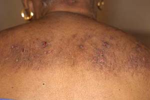 |
A 52-year-old woman with chronic kidney disease secondary to diabetic nephropathy presents with a several-month history of diffuse pruritus. She has been on hemodialysis for the last 5 years. She denies any rash but has been scratching her skin uncontrollably (Fig. 17-3). What is the most likely diagnosis? What tests can support the diagnosis?
Background
Pruritus or itch is the most common symptom of the skin. It can be caused by both cutaneous and systemic disorders. Systemic generalized pruritus is defined by the underlying disease that causes the symptom of cutaneous itch. Fifty percent of dialysis patients report cutaneous pruritus, as do 25% of those with jaundice. A thorough workup of primary pruritus patients is essential, considering an underlying systemic illness is found in 10% to 50% of primary pruritus patients. General categories of systemic pruritus include renal pruritus, cholestatic pruritus, hematologic pruritus, endocrine pruritus, pruritus related to malignancy, and idiopathic generalized pruritus.
Key Features
Pruritus is the most common skin symptom.
It can be caused by cutaneous and systemic disease or xerosis.
Generalized pruritus without primary skin lesion merits laboratory investigation.
Treatment includes topical and systemic antipruritics.
Pathogenesis
Unmyelinated C and A-delta neurons in the epidermis respond to pruritogenic and thermal stimuli. These neurons synapse with central neurons in the dorsal ganglia and travel through the spinothalamic tracts to the thalamus and cerebral cortex. These signals are processed in various areas of the brain. Multiple mediators of pruritus are responsible for the cutaneous sensation of itch. These mediators include histamine, tryptase, prostaglandin E, substance P, opioid peptides, nerve growth factor, and interleukin-2. The pathogenesis of systemic pruritus is related to the underlying cause (Table 17-2).
Clinical Presentation
Patients with pruritus may have a primary dermatologic disease or a systemic disease leading to itch. In patients with generalized pruritus, or pruritus affecting the entire body, the practitioner must rule out a systemic source
for the sensation. Clinical findings may include skin without a primary lesion, or may demonstrate cutaneous changes due to scratching or rubbing, as shown in Figure 17-3, or prurigo nodularis/lichen simplex chronicus, as shown in Figure 17-4. These skin findings may include multiple crusted papules, plaques, and ulcers with surrounding or overlying erosions and excoriations. Affected areas may include the arms, legs, abdomen, chest, and buttocks with sparing of the back or other unreachable body sites.
for the sensation. Clinical findings may include skin without a primary lesion, or may demonstrate cutaneous changes due to scratching or rubbing, as shown in Figure 17-3, or prurigo nodularis/lichen simplex chronicus, as shown in Figure 17-4. These skin findings may include multiple crusted papules, plaques, and ulcers with surrounding or overlying erosions and excoriations. Affected areas may include the arms, legs, abdomen, chest, and buttocks with sparing of the back or other unreachable body sites.
Table 17-2 Pathogenesis of Systemic Pruritus | ||||||||||||||
|---|---|---|---|---|---|---|---|---|---|---|---|---|---|---|
|
Diagnosis
The differential diagnosis of generalized pruritus is large and covers a wide range of systemic illness (Table 17-3).
 Figure 17-3 Excoriations and postinflammatory hyperpigmentation on the upper back of an elderly woman. |
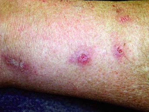 Figure 17-4 Prurigo nodularis. |
Table 17-3 Differential Diagnosis of Generalized Pruritus | ||||||||||||||||||||||||||
|---|---|---|---|---|---|---|---|---|---|---|---|---|---|---|---|---|---|---|---|---|---|---|---|---|---|---|
|
Related posts:

Stay updated, free articles. Join our Telegram channel

Full access? Get Clinical Tree


