Neurobiology of Scars: Managing Pain and Itch


Neurobiology of Scars: Managing Pain and Itch


Kendra Grim

Michael E. Nemergut


The history of procedural sedation and anesthesia for children has been a story of extremes.1 Although experience in adult patients dictated the importance of providing analgesia, hypnosis, and amnesia to all such patients undergoing invasive diagnostic and therapeutic procedures, the importance of providing these goals for infants and children has historically been less clear. Indeed, in the past many have questioned the ability of neonates to sense pain, develop memories (explicit and/or implicit), as well as possess the higher order cognition necessary for consciousness.2,3 These abilities were more than just academic—they had profound implications on therapeutic decision making. Indeed, because modulating nociception, memory formation, and/or consciousness confers overt risk (e.g., hypotension, apnea, airway obstruction, etc.), one might argue it illogical to provide analgesia to a person insensate, amnesia to a person without memory formation, and/or hypnosis to an unconscious person. In its extreme, such a point of view led to the practice of allowing infants to undergo major surgical procedures (such as an exploratory laparotomy) in the presence of paralysis but absence of sedative or analgesic medications.4,5

Today, it would be at variance with care standards to have a pediatric patient undergo a significant invasive procedure in the absence of analgesia as there are incontrovertible data that newborns not only feel pain, but that failure to provide adequate analgesia may result in both short- and long-term adverse consequences.6,7,8,9 In this chapter, we will review the basic pathways involved in the transmission of pain and itch, as well as review pharmacologic methods for their assessment and for controlling these unpleasant sensations. It should be noted that although much is known about the nuances of these pathways, comparatively little is known with regard to how these pathways interact specifically with scars; furthermore, data with regard to stimulus mitigation are limited. When able, we will expound upon extant data with regard to scars and we will indicate when inferring from more generalized data. Surgical methods are discussed in a separate chapter (see Chapter 12).


Physiology


Pain


Pain Transmission

The sensation, delivery, and interpretation of pain sensation are complex. The discussion below will highlight the mechanisms involved, which have clinical implications of how patients sense/describe pain as well as provide a basis for pharmacologic treatment.

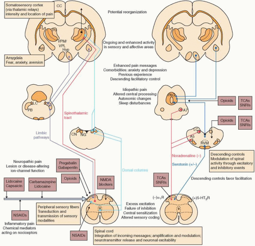
In its most simplified form, nociception signals begin in primary afferent neurons, which are subsequently transmitted through the dorsal horn of the spinal cord and along ascending pathways to the thalamus and somatosensory cortex. Primary afferents are the principal sensory neurons for nociception. The cell bodies of these sensory neurons are located in the dorsal root ganglia (DRG) or in the corresponding ganglion of specific cranial nerves (Fig. 11-1).10 As sensory neurons can be very diverse, they are categorized in the Erlanger-Gasser classification according to the size of the axon and presence or absence of myelin. The three major categories are, in order from largest diameter to
smallest, A, B, and C (Table 11-1).10 Neurons of Groups A and B are coated with myelin, which enhances nerve conduction speed. Group A is further subdivided into four subgroups, of which Aβ and Aδ are important to pain sensation and signaling. Aβ provide cutaneous touch and pressure afferents, whereas Aδ are mechanoreceptors, nociceptors, thermoreceptors, and sympathetic postganglionic fibers.10 Group B are sympathetic preganglionic fibers.10 Group C are small, unmyelinated mechanoreceptors, nociceptors, thermoreceptors, and sympathetic postganglionic fibers.10


FIGURE 11-1 Pain pathway transmission and modulation. The ascending sensory and affective components are shown on the left. Processes by which higher centers can alter spinal function are shown on the right. The major functional roles of different neural components in these pathways are summarized in boxes. Changes that occur after tissue or nerve damage are listed, and pharmacologic agents are shown at their sites of action. Peripheral inputs are indicated by arrows. (-)α2R=inhibition of neuronal activity. (+) 5-HT3R, stimulation of neuronal activity; Am, amygdala; A5 and A7, brainstem nuclei containing noradrenergic neurons; CC, cerebral cortex; CN, cuneate nucleus; Hyp, hypothalamus; LC, locus coeruleus; NG, nucleus gracilis; NMDA, N-methyl-D-aspartate; NSAID, nonsteroidal anti-inflammatory drug; PAG, periaqueductal gray matter; PB, parabrachial nucleus; Po, posterior nuclei of the thalamus; RVM, rostroventral medial medulla; SNRI, serotonin-norepinephrine reuptake inhibitor; TCA, tricyclic antidepressant; VPM and VPL, ventrobasal thalamus, medial and lateral components. (Gilron I, Baron R, Jensen T. Neuropathic pain: principles of diagnosis and treatment. Mayo Clin Proc. 2015(Apr);90(4):532-545.)

The primary afferents that contribute to pain sensation are Aδ and C fibers, and each transmits different types of pain sensation. As Aδ are myelinated, they transmit acute pain rapidly, typically sensed as “sharp” by a patient. C fibers are small and unmyelinated, and thereby transmit pain in a more delayed fashion, often described as “burning” in
quality. Free nerve endings of both Aδ and C fibers respond to varied stimuli intensity including tissue injury, possible tissue injury, and even threatened injury.10 This graded response may be intended to prevent injury, although it may also contribute a mechanism to development of chronic pain states.


Table 11-1 Peripheral Nerve Fiber Classifications


Fiber


Innervation


Conduction Velocity (mm/s)


Primary muscle spindle, motor to skeletal muscle


100


Cutaneous touch and pressure afferents


50


Motor to muscle spindles


20


Mechanoreceptors and nociceptors, sympathetic postganglionic


15


B


Sympathetic preganglionic


7


C


Mechanoreceptors, nociceptors, thermoreceptors, and sympathetic postganglionic fibers


1


Adapted from Davis PJ, Cladis FP, Motoyama EK. Smith’s Anesthesia for Infants and Children. New York, NY: Elsevier Health Sciences; 2011. Chapter 15, Table 15-4.


Cell bodies for both Aδ and C fibers are located in the DRG of the spinal cord or respective cranial nerves they serve. Most of the sensory neurons terminate in the ipsilateral dorsal horn. However, the spinal neurons bifurcate into ascending and descending branches to innervate several spinal segments. The dorsal horn is divided anatomically into locations known as Rexed laminae. Aδ and C fibers converge on lamina I (marginal zone), lamina II (substantia gelatinosa), and lamina X (central canal). Lamina V comprises wide dynamic range neurons. In lamina V, Aβ, Aδ, and C fibers converge, which leads to the phenomenon of “referred pain.” An example of referred pain is how visceral pain from myocardial infarction can be felt as pain in the left arm.

In the dorsal horn, the sensory nerves connect with the second-order neurons of the spinothalamic and spinoreticulothalamic tracts. Sensory nerves to the spinothalamic tract cross the contralateral ventrolateral tract and ascend the ventral horn to the thalamus. Sensory nerves to the spinoreticulothalamic tract ascend ipsilaterally in the ventrolateral tract, have medullary projections, and finally ascend to the thalamus.

From the thalamus, a nociceptive signal is transmitted to the somatosensory cortex for discriminative sensation, or to the anterior cingulate gyrus or inferior insula for sensation of the affective-motivational sensation of pain. These pathways are responsible for the affective phenomenon of how pain is experienced physically and emotionally.

Anatomically, the vascular supply of the spinal cord has important clinical implications. Although the posterior spinal cord is supplied by two posterior spinal arteries, the anterior spinal cord is supplied by a solitary anterior spinal artery. The largest anterior spinal artery, the artery of Adamkiewicz, has variable anatomic locations and is vulnerable to injury or spasm. Infarction of the anterior spinal cord is a devastating complication that leads to paraplegia with intact proprioception.


Table 11-2 Neuropathic Pain Taxonomy


Type


Definition


Allodynia


Pain generated by a typically non-noxious stimulus (light touch)


Hyperalgesia


Increased pain from a typically noxious stimulus


Hyperpathia


Abnormally painful reaction to a stimulus, especially repetitive stimulus (raised threshold, painful response)


Dysesthesia


Unpleasant abnormal sensation, spontaneous or evoked


Paresthesia


Abnormal sensation, spontaneous or evoked (with or without pain)


Hyperesthesia


Increased sensitivity to stimulation (with or without pain)


Hypoalgesia


Diminished response to normally noxious stimulus (raised threshold, decreased response)


Hypoesthesia


Decreased sensitivity to stimulation, excluding special senses


From International Society of Pain Taxonomy 2012.


http://www.iasp-pain.org/Taxonomy. Accessed November 12, 2016.


Mechanisms of Neuropathic Pain

Understanding the mechanisms of pain is important, as the sensation described by the patient can identify the type of injury/aberrancy that governs treatment. Mechanisms involved in neuropathic pain are multiple and overlapping. As such, a single patient may report multiple mechanisms of pain with one nidus or injury. This understanding supports the clinical importance of multimodal analgesia for treatment of pain.

As the description of pain is important to understanding its mechanism, the International Society of Pain developed a taxonomy of pain descriptors (Table 11-2; http://www.iasp-pain.org/Taxonomy). These descriptors can be divided into positive and negative symptoms, based on the response of the patient to stimuli. Positive painful symptoms include allodynia (pain generated by a nonpainful stimulus, such as light touch), hyperalgesia (increased pain from a painful stimulus), and hyperpathia (raised threshold or heightened pain response). Positive symptoms that may or may not be associated with pain include dysesthesia (unpleasant abnormal sensation, spontaneous, or evoked), paresthesia (abnormal sensation, may not be unpleasant, spontaneous, or evoked), and hyperesthesia (increased sensitivity to stimulation, but may not be painful). Negative symptoms include hypoalgesia (decreased sensitivity to painful stimulus) and hypoesthesia (decreased sensitivity to stimulus, not necessarily painful stimulus).

There are at least five accepted mechanisms for neuropathic pain that explain the sensations described above. These mechanisms include ectopic activity, peripheral sensitization, central sensitization, impaired inhibitory modulation, and activation of microglia (Table 11-3).11 Ectopic activity is thought to be secondary to hyperexcitability
and spontaneous firing of neurons occurring after an insult, thereby explaining spontaneous symptoms such as paresthesia and dysesthesia; such neuropathic pain can be constant, intermittent, or paroxysmal. Ectopic activity has been attributed to changes in voltage-gated sodium and potassium channels, and hyperpolarization of cyclic nucleotide-gated channels.11


Table 11-3 Neuropathic Pain Mechanisms


Type


Mechanism


Associated Symptomatology


Ectopic


Spontaneous, sodium and potassium channel changes


Neuroma, demyelination, trigeminal neuralgia


Peripheral sensitization


Hyperexcitability of sensory neurons, TRPV1 channel changes


Hyperalgesia, allodynia


Central sensitization


Change in Aβ fibers. Increase CGRP, substance P, and NMDA


Fibromyalgia, IBS, chronic fatigue syndrome


Impaired sensory modulation (inhibition)


Apoptosis of GABAergic spinal inhibitory neurons


Postinjury pain, postherpetic neuralgia, entrapment neuropathy


Activation of microglia


Upregulation of chemokine receptors, release of glial cytokines


Central sensitization, hyperalgesia, allodynia, anticipation


Peripheral sensitization results from hyperexcitability and reduced activation threshold of primary afferent neurons, described by the patient as hyperalgesia or allodynia.11 Peripheral sensitization may be thought of as inflammatory pain, and may be due to changes in the transient receptor potential cation channel subfamily V member 1 (TRPV1) ion channel.12

Central sensitization is classified as central neuroplastic changes involving spinal or supraspinal nerves. The patient may describe exaggerated response to painful stimulus, hyperalgesia, allodynia, and/or anticipation. Aβ touch fibers develop phenotypic changes including increased neuropeptide expression of substance P and increased excitatory amino acid transmission via N-methyl-D-aspartate (NMDA) receptors.11 These changes may explain some of the purported analgesic benefit of NMDA modulating agents such as ketamine (see below). Microglial activation occurs following injury by upregulation of chemokine receptors and release of glial cytokines and growth factors.11 These changes are believed to contribute to central sensitization and increased pain sensation reported by the patient.

Inhibitory neurons project from the central nervous system (CNS) and serve to modify painful stimuli. Apoptosis of spinal inhibitory interneurons has been reported as a consequence of some injuries, which may explain abnormalities in inhibitory neuron signaling.13 Without modulation from inhibitory neurons, a peripheral painful stimulus may be interpreted by the CNS as pain of significantly increased severity.


Itch

Centuries ago, Samuel Haffenreffer defined itch as an unpleasant sensation that causes the urge to scratch, the act of which is subsequently rewarded by a pleasant sensation and/or itch relief. Itch is primarily carried by C-fibers, then transmitted by the spinothalamic tract to the brain.14 In research, itch is induced by histamine, extracts from cowhage (a tropical legume), or electrical stimulation. Importantly, cowhage-induced itch cannot be inhibited by antihistamines and suggests different mechanisms are involved in pruritus. Indeed, the transmission between histamine and cowhage appears to be different in the peripheral nervous system as well as the spinal cord and brain15; nevertheless, both involve regions that are also involved in pain transmission. The mechanism for electrically evoked itch, while preliminary, does appear to involve C-fibers.15 Centrally, multiple areas of the brain respond to both histamine and cowhage-induced pruritus, including the prefrontal cortex, supplementary motor area, premotor cortex, primary motor cortex, primary somatosensory cortex, parietal cortex, cingulate cortex, precuneus, opercular cortex (secondary somatosensory cortex and insular cortex), claustrum, and basal ganglia.15 Of particular interest, the precuneus is thought to participate in pain modulation and has been shown to activate on functional magnetic resonance imaging to itch stimuli.16 The primary somatosensory cortex and secondary somatosensory cortex are thought to be the primary regions for sensory interpretation, in addition to their roles in the perception of pain intensity.15 The cingulate cortex has also been shown to activate in response to itch.

Once transmitted, the sensation of itch is modulated centrally. Similar to nociception, both spinal cord and supraspinal areas participate in stimulus modulation. Inhibitory neurons attenuate signals from ascending the spinal cord. Supraspinal inhibition has been shown to contribute to itch suppression; however, the exact mechanism is not known.15 Periaqueductal gray area and rostral medulla are well documented to be involved with descending inhibitory control for pain transmission, and modulation of itch is hypothesized to involve the same or a similar pathway.15

The pleasure induced by scratching an itch is hypothesized to involve the reward system of the CNS, including the midbrain, striatum, medial prefrontal cortex, and anterior cingulate cortex.15 Dopamine release has been purported to be central to the pleasure sensation associated with scratching, and the midbrain has a particularly high density of dopaminergic receptors.15

Chronic pruritus can be agonizing for patients, as compulsive scratching may lead to a cycle of further tissue damage, further histamine release, and further pruritus (i.e., the so-called “itch-scratch-itch” cycle). Scratching can also become a maladaptive habit. A central mechanism for perpetuation of itch has also been hypothesized, as patients with atopic dermatitis have shown higher activity in the basal ganglia,17 and patients with pruritus secondary to
end-stage renal disease have higher density of gray matter with enhanced baseline activity in the basal ganglia.18 Further research is required to understand the central mechanisms that contribute to chronic itch.


Assessment


Clinical

Pain is defined as an unpleasant sensory and emotional experience caused by a noxious or perceived noxious stimulus. No two individuals experience the same pain in an identical manner, and the ability to communicate pain does not correlate with the pain experienced.19 Nociception describes how neurons respond to noxious stimuli including autonomic and behavioral changes, such as elevated heart rate and withdrawal reflex, respectively. Nociception, however, does not always induce pain. Neuropathic pain includes spontaneous positive and negative symptoms as described above, and it is imperative to understand the patient’s particular pain experience to formulate an appropriate treatment plan.19,20

Itch is also an unpleasant sensory experience, and patients “self-treat” by scratching or applying a painful stimulus. Scratching causes a sensation of pleasure and relief. However, prolonged scratching can lead to excoriation, skin disruption, secondary infection, and poor wound healing and/or scarring.


Psychosocial Impact

It is difficult to overstate the impact of pain on psychosocial function. Pain is often associated with significant negative emotional changes and maladaptive behavior problems; in response, emotion and mental health can change how pain signals are interpreted centrally.21 It is recommended that patients with severe acute or chronic pain be evaluated for concurrent mental health illness as comorbid conditions such as anxiety and depression are common (see Chapter 24).22 In addition, preexisting anxiety and depression are commonly worsened by pain, even if previously well controlled. Mental health changes associated with chronic pain or itch can have impact on the patients’ overall health, quality of life, and productivity.


Oct 15, 2018 | Posted by in Dermatology | Comments Off on Neurobiology of Scars: Managing Pain and Itch

Full access? Get Clinical Tree

Get Clinical Tree app for offline access