Scar Histopathology and Morphologic Classification


Scar Histopathology and Morphologic Classification


Molly Powers

David Ozog

Marsha Chaffins


The assessment and classification of scars in various conditions is essential for guiding therapy and advancing research. However, to our knowledge no consensus has been drawn on the best method to evaluate scar morphology. This chapter will help to review the basic histologic and morphologic characteristics of the various scar types and to discuss the classification of these lesions.


Scar Histology

There is limited research relating to scar histology. Hypertrophic and keloidal scars are better described, though to our knowledge there is no conclusive body of literature that examines the histology of each of the varying types of scars. The standard histologic description of a mature scar notes thicker collagen bundles with spindle cells that are arranged parallel to the epidermis, as compared with the basket weave appearance of the surrounding normal dermis as demonstrated in Figure 5-1.1 The underlying vessels are typically oriented vertical (perpendicular) to the epidermis and are equal in number to the uninjured dermis after approximately 12 months of scar maturation.1,2 In a mature scar there is often loss of elastic tissue with the effacement of the epidermis (Fig. 5-2).3

From the moment of injury to the skin and through the lifetime of the scar, there is a consistent change and remodeling, which is reflected in the corresponding histology. For example, in an early scar there are more inflammatory cells, but this usually normalizes after approximately 1 month absent any derangement in the scar maturation process.1 Dermal vasculature also undergoes dynamic changes. One to three months after tissue injury, scars have a higher density of blood vessels in the dermis. However, as the scar continues to mature, the density decreases but blood vessel size increases in comparison to earlier scars.1 Bond et al.1 evaluated “poor” and “excellent” scar outliers based on their appearance in comparison to the representative group. Scars deemed “poor” were found to have higher density of blood vessels. “Excellent” scars had a lower blood vessel density and approached that of normal skin after a shorter duration of injury. These findings support the use of vascular lasers in younger scars (see Chapter 13). As a scar continues to mature, the vasculature of nonpathologic scars (excluding hypertrophic scars and keloids) approaches that of normal skin.4 Clinically, this is associated with diminishing erythema. A maturing scar, however, does not fully recover its preinjury histology. There is complete loss of skin appendages, incomplete reformation of the rete ridges and papillary dermis, as well as alterations in the collagen fiber bundles.1 This maturation process is further outlined in Table 5-1.


Hypertrophic (Fibroproliferative) Scars

There are important histopathologic distinctions among different scarring processes that may guide prognosis and management. However, distinguishing a hypertrophic scar from a keloid histopathologically is sometimes very difficult.4 Keloids and hypertrophic scars both differ from normal skin as they have increased deposition of connective tissue as well as rich vasculature.5 In the literature there is conflicting information regarding the vasculature of keloids and hypertrophic scars. Many theories suggest the role of hypoxia in the formation of pathologic scars. Kischer et al.6 found an increased number of occluded or partially occluded

microvessels in hypertrophic and keloidal scars in comparison to normal scars and normal dermis because of endothelial cell swelling and increased endothelial cell density. Additionally the degree of vascular occlusion was higher in keloids with fewer and more flattened vessels, especially centrally within the lesion, suggesting less vascular supply in a keloid as compared with a hypertrophic scar. The growth of fibroblasts and increased collagen deposition may result in this occlusion.7,8 On the contrary, other studies have demonstrated increased vasculature of keloids and hypertrophic scars in comparison with normal scars and normal skin.1 Amadeu et al.4 reported that the volume occupied by vessels in the papillary dermis was 79.7% higher in hypertrophic scars (P < 0.01) and 62.5% higher in keloids (P < 0.05) as compared with normal skin. Similar findings were noted in the reticular dermis, with hypertrophic scars having 62.9% higher vessel volume (P < 0.025) and keloids having 68.5% higher vessel volume than normal skin (P < 0.001). The benefit of pressure dressings and vascular lasers for the treatment of these pathologic scars contradicts our understating of the role of hypoxia in their pathogenesis. At this point, further research needs to be conducted to fully understand this interplay.


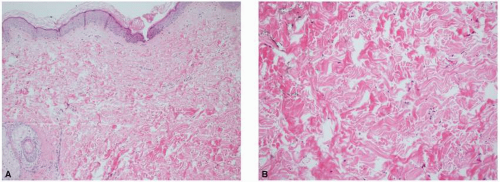
FIGURE 5-1 Histology of normal skin. A: Low-power image of normal skin with fine collagen and elastic fibers noted in the papillary dermis. B: Higher power image of normal reticular dermis with more compact collagen and thicker elastic fibers.


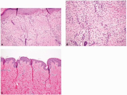
FIGURE 5-2 The histologic comparison of an early and a late scar. A: Low-power image of a newly forming scar demonstrating the cellular reticular dermis, increased vasculature, and absence of rete ridges and papillary dermis. B: Higher power image of a newly forming scar with fine, horizontally oriented collagen bundles and increased vasculature with vessels oriented vertically. C: An established scar with horizontally oriented thicker and more densely packed collagen bundles and loss of all skin appendages.


Table 5-1 Flowchart Describing the Histologic Maturation of a Scar Over Time


Months 1-3


Months 4-6


Months 7-9


Months 10-12


  • Flat DEJ


  • Cellular reticular dermis


  • ECM immature with fine collagen bundles


  • Increased blood vessels and fibroblasts


  • No increased number of inflammatory cells seen after 1 mo


image


  • Rete ridge and papillary dermis reformation minimal


  • Collagen fibers thicker and denser around month 4


  • Scars highly vascular with larger vessels but still reduced from months 1 to 3


image


  • Rete ridge reformation has begun


  • Collagen fibers becoming thicker and denser


  • Small reduction in blood vessel density


image


  • Some scars with rete ridge and papillary dermal formation


  • Collagen fiber bundle maturity equal to the surrounding normal dermis


  • Fibers were dense and arranged horizontally


  • Blood vessel density higher than normal skin at 10 mo, but equaled that of normal skin at 12 mo


DEJ, dermoepidermal junction; ECM, extracellular matrix.


Aside from their vasculature, keloids and hypertrophic scars each have a high mesenchymal cell density and inflammatory cell infiltration, with an absence of subepidermal appendages including sebaceous glands and rete ridges.1 Both lesions have a predominant active fibroblast cell type, but keloids tend to have more quiescent forms.9 The dermis of a keloid comprises characteristic large, thick collagen bundles that are brightly eosinophilic and hyalinized with hematoxylin and eosin staining as demonstrated in Figure 5-3. These bundles are closely packed with thin fibrils and form the characteristic “keloidal collagen.” Lee et al.10 reported distinct keloidal collagen in approximately 55% of all keloids included in their study. They concluded that the histologic absence of keloidal collagen does not rule out the diagnosis of a keloid, but its presence is of high diagnostic value as it is only discovered in keloidal scars. Keloidal collagen is arranged in a nodular fashion in contrast to the parallel architecture of collagen in ordinary scars. In comparison, hypertrophic scars contain distinct smaller nodules present in the dermis (demonstrated in Fig. 5-4), aiding in the histologic distinction from keloids and normal scars, which often lack these nodules. The compaction of the collagen bundles in keloids has no effect on the overlying epidermis.7,11 Additionally, hypertrophic scars have an accumulation of myofibroblasts expressing α-smooth muscle actin, whereas keloidal myofibroblasts often lack the expression of α-smooth muscle actin.5


Atrophic Scars

In contrast to keloids and hypertrophic scars, the histologic appearance of striae can be very subtle and may be difficult to distinguish from normal skin (Fig. 5-5). As with other scar types, the histologic appearance of striae depends on the stage of evolution. In earlier (younger) striae, there is often dermal edema with perivascular infiltrates. The epidermis can appear normal in earlier stages, but later may flatten and have blunting of the rete ridges (atrophy). Earlier striae may also demonstrate elastolysis and a relative lack of mast cells. There are structural changes seen in collagen bundles with prominent fibroblasts and reduced microfibrils. Earlier striae have been referred to as striae rubra, as they often appear erythematous in color.12

Well-established striae may be termed striae alba, owing to their hypopigmented (white) appearance over time. Striae alba demonstrate epidermal atrophy and decreased dermal thickness. Collagen fibers are generally found running parallel to the skin and transverse to the direction of the individual stria. There are variable alterations in the elastic fibers with reduced and fragmented dermal elastin on special stains. There is also loss of skin appendages including hair follicles and adnexal structures, as with all scars.9


Dyspigmentation

There are very few studies that examine the histologic findings associated with clinical dyspigmentation in scars. However, Travis et al.13 evaluated the optical and histologic properties of wounds and their relationship with dyspigmentation in a porcine model. Hyperpigmented scars, hypopigmented scars, and uninjured tissue were evaluated by fixing and embedding these samples for histologic examination using Azure B stain and primary antibodies to
S100B, HMB45, and α-melanocyte-stimulating hormone (α-MSH) for comparison. They discovered no statistically significant difference in melanocyte number between hyperpigmented and hypopigmented scars and uninjured skin samples. There was, however, a statistically significant difference in the amount of melanin and α-MSH, and immunohistochemical evidence of stimulated melanocytes in hyperpigmented versus hypopigmented scars.


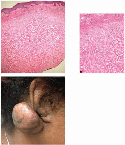
FIGURE 5-3 Histology and clinical appearance of a keloid. A: Low-power image demonstrating the large, thick collagen bundles that are closely packed with thin fibrils. B: Higher power image illustrating the unique thick nodular “keloidal collagen.” C: Clinical picture of a keloid. Keloid on the right earlobe of this young African-American woman. The earlobe is a common site for keloid formation after ear piercing.


Histologic Effects of Fractional Laser Treatment

Whereas there has been extensive research conducted on certain scar types, elucidation of the relationship between clinical scar appearance and the corresponding histology may help guide treatment and evaluation of the response. For example, Ozog et al.14 delineated the histologic changes
in collagen typing between laser-treated and -untreated burn scars. It is well described that normal skin contains a combination of type I and type III collagen; fetal skin contains a higher proportion of type III collagen, and burn scars contain a higher proportion of type I to type III collagen. A course of three ablative fractionated CO2 laser treatments to burn scars eventuated in a collagen profile approaching that of normal skin, with a posttreatment increase in type III collagen as demonstrated by the Herovici stain (Fig. 5-6). Additionally, Taudorf et al.15 found statistically significant clinical improvement in various scar types (normal, hypertrophic, and atrophic) after three monthly nonablative fractional laser treatments (P < 0.0001 vs. untreated), with corresponding histology indicative of collagen remodeling. There is a predominance of thickened collagen within a scar as compared with the normal surrounding dermis. The healing process that follows fractionated photothermal injury ultimately leads to the remodeling and reorganization of collagen that begins to approach that of normal skin. A later study completed by Connolly et al.16 discovered that, counterintuitively, treatment of erythematous burn scars with a fractionated CO2 laser led to a statistically significant increase in vascular density as determined by anti-CD31 immunostaining, despite a decrease in clinically apparent erythema during the treatment course (Fig. 5-7). These histologic findings as illustrated above have both supported and challenged our understanding of the mechanisms leading to clinical improvement in scars. Perhaps we may use these predicted and unforeseen findings to further propel scientific discovery and improved management.


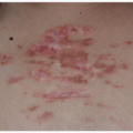
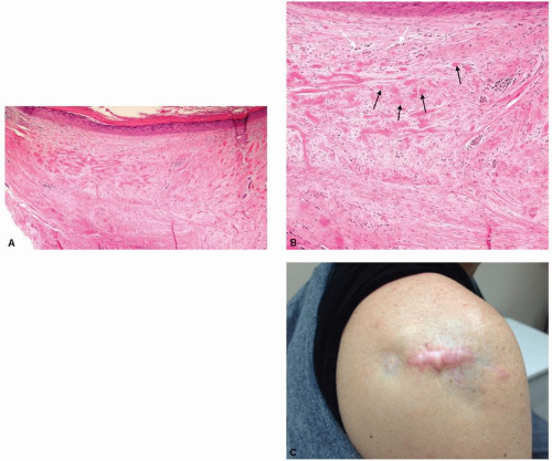
FIGURE 5-4 Histology and clinical appearance of a hypertrophic scar. A: Low-power view demonstrating the smaller nodules in the dermis. B: Higher power view illustrating again the smaller nodules as demonstrated by the black arrows and occluded microvessels superficially (white arrows) with increased deposition of connective tissue. C: Clinical picture of a scar with areas of atrophy and hypertrophy. Shoulder of a woman who underwent multiple intralesional steroid injections for a hypertrophic scar, resulting in steroid-induced atrophy.


Reflectance Confocal Microscopy

To date, light microscopy of tissue has been the principal technique to evaluate the histology and microstructure of the skin. In vivo reflectance confocal microscopy (RCM) is a relatively new, noninvasive technology that has recently been utilized for the evaluation of various types of scars.17,18,19,20 RCM provides real-time viewing with similar
resolution to classical histology, without tissue damage (Fig. 5-8). Additionally, it may be used to detect the dynamic microscopic changes of a particular skin lesion over time. RCM may provide promising in vivo evaluation and comparison of many scar types, and may also be used for the pre- and posttreatment analyses of scars, among other dermatologic conditions.


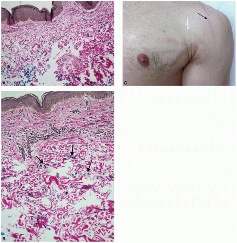
FIGURE 5-5 Histology of striae with elastin stain demonstrating reduced elastin fibers, and a representative clinical photo. A: Lower power; B: Higher power with arrows demonstrating the elastic fibers. C: Clinical picture of striae with the white arrow indicating striae alba, and the black arrow indicating striae rubra. Multiple large striae on the chest and shoulder of a young man because of a sudden increase in muscle mass.

Figure 5-8 provides a clinical comparison of a hypertrophic scar with the associated confocal microscopic imaging. At a depth of 152.4 µm below stratum corneum, collagen fibers and bundles (red arrow) are present inside the lesion since it is raised compared with adjacent normal skin.

Only gold members can continue reading. Log In or Register to continue

Stay updated, free articles. Join our Telegram channel

Oct 15, 2018 | Posted by in Dermatology | Comments Off on Scar Histopathology and Morphologic Classification

Full access? Get Clinical Tree

Get Clinical Tree app for offline access