Medical Management of Scars


Medical Management of Scars


Julian Poetschke

Markus Reinholz

Gerd G. Gauglitz


Scars are common and can be cosmetically, physically, and psychologically debilitating. As recently described by our group,1 keloids, atrophic acne scars, self-harm scars, and burn scars may significantly affect patients in their daily well-being. Thus, adequate assessment of associated symptoms and appropriate treatment remains crucial to improve the overall quality of life of these patients. Although many studies have been published on the pathophysiology of excessive scarring, the complex alterations underlying hypertrophic and keloid scar formation remain relatively poorly understood (see Chapter 6). The lack of appropriate animal models in this area may significantly contribute to this fact.


Pathophysiology Underlying Physiological and Excessive Scar Formation

In order to understand the altered wound healing response underlying excessive scarring, it may be best compared to physiological wound healing.2

Scars regularly occur after trauma of the deeper dermis following a complex physiological wound healing cascade. This cascade can be subdivided into three phases: inflammation, proliferation, and remodeling. Traumatic injuries are immediately followed by the onset of hemostasis. As a fibrin-rich blood clot develops, it serves as a scaffold for the ensuing wound repair and the degranulation of activated thrombocytes sets free a variety of chemotactic and proinflammatory agents. Macrophages and neutrophil granulocytes are attracted and dissolve necrotic tissue. Released cytokines like epidermal growth factor, platelet-derived growth factor, insulin-like growth factor I, and transforming growth factor (TGF)-β mediate the stimulation of fibroblasts and keratinocytes and mark the transition into the proliferative phase.2,3 During this phase, a scaffold of extracellular matrix (ECM), consisting of immature type III collagen, elastin, proteoglycans, and hyaluronic acid, is constructed by the activated fibroblasts. Mediated by vascular endothelial growth factor, vascular ingrowth is induced. As myofibroblasts facilitate wound contraction and closure, the remodeling phase begins. During this final phase, abundant ECM is degraded and immature matrix components are converted into their mature form. This is most notably achieved through the transformation of procollagen type III into mature type I collagen.2,3,4,5

Typically, immature scars (Fig. 10-1) have a reddish appearance. Sometimes they cause itching and are slightly elevated. Within months, they regularly turn into a flat, frequently depigmented scar without any further symptoms (Fig. 10-2).6 Based on a published study observing the natural history of scar redness and maturation after incisional and excisional wounds, scars commonly fade within approximately 7 months.7

The successful conversion of a wound clot into granulation tissue necessitates a balance between degradation and deposition of ECM proteins.2 Alteration of this process can lead to abnormalities in scarring. Several risk factors have shown to increase abnormal (pathological) scarring such as specific anatomic locations, infection, genetic susceptibility, and delayed epithelialization (Table 10-1). The occurrence of keloids has been reported after negligible surgical or laser procedures, and even without apparent trauma—primarily in predisposed individuals.2

Both keloids (Fig. 10-3) and hypertrophic scars (Fig. 10-4) reveal fundamental aberrations in the wound healing cascade, which may be best characterized by an imbalance between the anabolic and catabolic phases.8 A scar is densely infiltrated with inflammatory cells that secrete fibrogenic factors, such as TGF-β1 and TGF-β2. Increased
levels of TGF-β1 and TGF-β2 promote an accumulation of ECM, whereas their degradation through decreased levels of TGF-β3 and matrix metalloproteinases (MMPs) is impaired. Both the severity of inflammation and the type of immune response predispose to excess scar formation.9 An increased TH2 response stimulates fibrogenesis, whereas a TH1 preponderance reduces tissue fibrosis.10,11


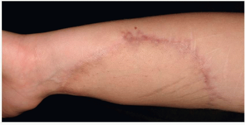
FIGURE 10-1 Immature scar with raised edges and erythema.


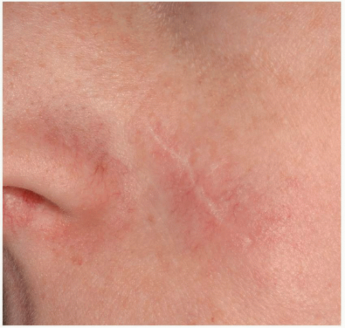
FIGURE 10-2 Physiological scar healing after surgery. (From Reinholz M, Poetschke J, Schwaiger H, et al. The dermatology life quality index as a means to assess life quality in patients with different scar types. J Eur Acad Dermatol Venereol. 2015;29(11):2112-2119.)


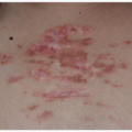
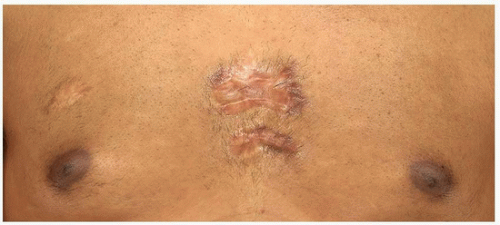
FIGURE 10-3 Spontaneous presternal keloid.


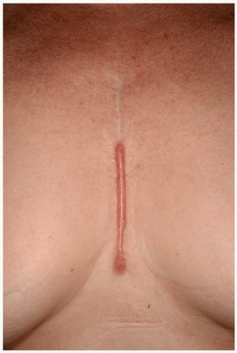
FIGURE 10-4 Hypertrophic scar after open heart surgery; the scar remains within the confines of the original injury. (From Reinholz M, Poetschke J, Schwaiger H, et al. The dermatology life quality index as a means to assess life quality in patients with different scar types. J Eur Acad Dermatol Venereol. 2015;29(11):2112-2119.)


Table 10-1 Main Risk Factors for Hypertrophic Scars and Keloids


Hormones (higher incidence during puberty and pregnancy)


Genetic predisposition


Topography


Chronic inflammation (e.g., acne)


Tension on wound margins


Delayed epithelialization (>21 d)


There is evidence that a more prolonged inflammatory period with increased infiltrates of immune cells present in the scar tissue of keloids may contribute to increases in fibroblast activity with greater deposition of ECM proteins.9 This may help explain why keloids continue expanding beyond the margins of the initial injury, whereas hypertrophic scars (in which the immune cell infiltrate decreases over time) remain restricted to the initial wound bed and commonly show involution over time.9


Therapeutic Approaches for the Treatment of Hypertrophic Scars and Keloids

Various studies elaborating on the formation of excessive scarring have resulted in a multitude of therapeutic strategies (Table 10-2). A vast number of articles on the prevention or improvement of keloid and hypertrophic scars have been published. Despite the rapidly increasing options in this area, some considerations are worth being highlighted in this context:


  • Many of the therapeutic approaches available to date are being used for both prevention and treatment of excessive scarring.


  • Only a limited number of these strategies have been supported by controlled and well-designed prospective studies.


  • Differentiating between keloids and hypertrophic scars is crucial prior to any therapeutic manipulation because of augmented recurrence rates with keloids (Table 10-3).


  • Lasers continue to play an increasingly important role and are being marketed accordingly.

But:


  • By utilizing basic options such as silicone sheeting/gel, intralesional triamcinolone acetonide (TAC), cryotherapy, or pressure, improvement can frequently be observed without any extensive cost for the respective patients.

The following paragraph will provide a summary of the currently available options for the prevention and treatment of scarring, starting with the most common.


Surgical Approaches for the Prevention and Therapy of Hypertrophic Scars and Keloids

Five main principles should be taken into consideration when considering any surgical approach for the prevention or treatment of excessive scarring2:


  • Common prophylactic approaches in order to reduce the possibility of postoperative pathological scarring:


    • Delayed epithelialization beyond 10 to 14 days is well known to increase the incidence of hypertrophic scarring12; fostering rapid epithelialization is crucial to avoid excessive scar growth.


    • Wounds subjected to high tension due to motion, body location, or tissue loss are at increased risk of excessive scarring and spreading of scars (see Chapter 7). Patients undergoing surgery in these body areas should be aware of this significant risk prior to any surgery.13


    • The modern understanding of wound healing is based on the knowledge of restoring skin anatomy and function, and a thoughtful selection of suture materials and closure techniques. Surgical approaches should precisely coapt the tissue layers and margins. This is necessary to minimize new tissue formation within the wound. Suitable surgical closing techniques will also eliminate a potential cavity by approximating the subcutaneous tissue. If misalignment and dead space is minimized adjacent to the opposed wound edges, new tissue has limited room to grow. Atraumatic handling of tissue in combination with avoiding tight closures result in better aesthetic outcomes. Additionally, reduced tension on the wound margins facilitated by cautious undermining and loosening of the surrounding tissue contributes to better results. As an example, a combination of polydioxanone (PDS II, Ethicon Inc.) monofilament synthetic absorbable subcutaneous sutures (which provide extended wound support for up to 6 months) with absorbable sutures or Steri-Strip (3M) for optimal epidermal wound closure might be used. It has been shown that subcutaneous fascial tensile reduction sutures in a predisposed patient population, where the tension is placed on the layer of deep fascia and superficial fascia, revealed good clinical results. Here 2-0 or 3-0 PDS II sutures were preferred for subcutaneous/fascial sutures, and 4-0 or 5-0 PDS II sutures for dermal sutures.14


  • If hypertrophic scarring occurs, determining the optimal timing for procedural intervention (including surgical excision/revision) is key. Hypertrophic scars have the potential to mature over a year or more after injury and significantly flatten and soften without any medical intervention, potentially obviating the need for surgical excision.15 Nevertheless postexcisional relapse rates of the original hypertrophic scar are commonly negligible.16,17 On the other hand, if scar (joint) contractures occur, then invasive approaches that release contractures should be performed earlier (see Chapter 12).14


  • The development of hypertrophy is facilitated when there is an increased tension on wound margins. Consequently, employing surgical techniques such as Z- or W-plasty, grafts, or local skin flaps to interrupt the vicious circle between scar tension and scar thickening due to permanently stimulated ECM production may help mitigate pathological scar formation.18


  • Delayed would healing (e.g., after deep dermal burn or wound infection) may increase the risk of hypertrophic scars and keloids. Transforming selected lesions by surgery (excision with suture or graft) into a wound with appropriate healing time may reduce the risk of new excessive scar formation.18


  • If excessive scar tissue is surgically removed, a situation similar to a fresh wound is created. In this new setting disproportionate scarring might be reduced by adjuvant conservative therapy straight from the beginning.18 Nonetheless, the excision of keloids without any further conservative therapy (e.g., corticosteroid or 5-Fluorouracil [5-FU] injections, intrainterventional cryotherapy, pressure, or radiation) should be rigorously avoided because of a strongly elevated risk of recurrence (45% to 100%). The postexcision scar may even grow larger than the initial one in this new area of trauma.19,20 Remarkably good cosmetic results were obtained after a surgical repair approach (core excision with low-tension wound closure or shave excision) of earlobe keloids with postinterventional

    corticosteroid or 5-FU injections, postoperative pressure (e.g., pressure earrings), application of imiquimod 5% cream or intraoperative cryotherapy on the incision site.21


Table 10-2 Hypertrophic Scars and Keloids: Current Therapeutic Strategies


Treatment


Use


Mechanisms of Action


Indications, Efficiency, and Comments


Prophylaxis


Pressure therapy


Continuous pressure (15-40 mm Hg) for at least 23 h/d ≥6 mo of scar healing


Collagen synthesis↓, Apoptosis↑ via limiting the supply of blood and oxygen to scar tissue


  • Prophylaxis of hypertrophic burn scars, ear keloids (postexcision)


  • Controversial success


  • Reduced compliance due to frequent patient discomfort


Silicone gel sheeting


≥12 h/d for ≥2 mo beginning 2 wk after wound healing


Pressure↑; hydration of the stratum corneum; temperature ↑→ collagenase activity↑


  • Prophylaxis of hypertrophic scars and keloids


  • Controversial benefit


  • No effects on mature keloids and hypertrophic scars


Flavonoids


e.g., Contractubex gel (Merz Pharma, Frankfurt, Germany), Mederma Skin Care Gel (Merz, Pharmaceuticals, Greensboro, NC, USA). Twice daily for 4 to 6 mo


Antiproliferative, anti-inflammatory, reduction in TGF-β expression


  • Limited to management and prophylaxis of hypertrophic scars and keloids


Current Therapies


Corticosteroids


Intralesional injections of triamcinolone acetonide (10-40 mg/mL), several treatments once or twice a month


Anti-inflammatory effects.


Vasoconstriction↑→ oxygen and nutrients↓


Anti-mitotic effects on keratinocytes and fibroblasts


α2-macroglobulin↓→ collagenase↑


Production of TGF-β1 and 2↓


  • First-line therapy for the treatment of keloids and second-line therapy for the treatment of hypertrophic scars


  • Combination with surgery, PDL, and cryotherapy


  • Beware: skin and subcutaneous fat atrophy, telangiectasias


Cryotherapy


Contact/spray freezing with liquid nitrogen using 10-20 s freeze-thaw cycles


Induction of vascular damage → anoxia and tissue necrosis. Collagen synthesis↓


  • Overall effective for hypertrophic scars, for keloids in combination with triamcinolone acetonide injections recommended


  • Limited to management of smaller scars


  • Beware: blistering and pain


Scar revision


Excision with linear, tension-free closure, split- or full-thickness skin grafting, Z-plasty, W-plasty


Surgical removal of excessive scarring


  • Efficacious for treatment of hypertrophic scarring


  • Recurrence rates of 45%-100% after keloid excision


Radiotherapy


Superficial X-rays, dosages15-20 Gy, overall limit 40 Gy. Over 5-6 sessions in the early postoperative period


Inhibition and apoptosis of proliferating fibroblasts


  • Overall good efficiency rates of adjuvant radiotherapy after keloid excision


  • Beware: potential risk of malignant change/carcinogenesis


Laser therapy


Pulsed dye laser (585-nm PDL) with doses ranging from 6.0 to 7.5 J/cm2 (7-mm spot) or from 4.5 to 5.5 J/cm2 (10-mm spot), 2-6 treatments every 2-6 wk


Local ischemia via destroying blood vessels → tissue hypoxia↑


  • Excellent therapeutic option for the treatment of younger keloids and hypertrophic scars


  • High recurrence rates with other (ablative) laser techniques for the treatment of keloids


Emerging Therapies


Interferon


Intralesional injection of INF-α2b (1.5-2 million International Units) twice daily over 4 d


Antiproliferative properties


  • Clinical studies report overall effectiveness


  • Beware: flu-like symptoms on injection


5-FU


Intralesional injection of 5-FU 50 mg/mL


Increased fibroblast apoptosis via inhibiting DNA synthesis


  • Overall effective for the treatment of keloids and hypertrophic scars


  • Beware: blood count monitoring (anemia, leukopenia, thrombocytopenia). No therapy in pregnant women or patients with bone marrow suppression


From Gauglitz GG, Korting HC, Pavicic T, et al. Hypertrophic scarring and keloids: pathomechanisms and current and emerging treatment strategies. Mol Med. 2011;17(1/2):113-125.


Table 10-3 Hypertrophic Scars and Keloids: Epidemiological, Clinical, and Histological Differences


Hypertrophic Scarring


Keloids


Incidence


40%-70% following surgery, up to 91% following burn injury.150,151,152


6%-16% in African populations.153,154


Equal in sex distribution with highest incidence in the second to third decade.155,156


Predilection sites


Shoulders, neck, presternum, knees, and ankles.17,157,158


Anterior chest, shoulders, earlobes, upper arms, and cheeks.154,159,160


Less affected: Eyelids, cornea, palms, mucous membranes, genitalia, and soles.154


Time course


Within 4-8 wk following wounding,161 rapid growth phase for up to 6 mo, then regression over a period of a few years.158,162,163


Within years after minor injuries or spontaneous formation on the mid-chest in the absence of any known injury. Persistence for long periods of time. No spontaneous regression.41


Low recurrence rates after excision of the original hypertrophic scar.16,17


High recurrence rates following excision.


Appearance


Do not extend beyond the initial site of injury.164


Project beyond the original wound margins.164,165


Both lesions are commonly pruritic, but keloids may even be the source of significant pain or hyperesthesia.158,161


Histological characteristics


Primarily fine, well-organized, wavy type III collagen bundles oriented parallel to the epidermis surface with abundant nodules containing myofibroblasts and plentiful acidic mucopolysaccharide.3


PCNA/p53-level/ATP expression low.168


Disorganized, large, thick, type I and III hypocellular collagen bundles with no nodules or excess myofibroblasts.3,166 Poor vascularization with widely scattered dilated blood vessels.167


PCNA/p53-level/ATP expression high.168


From Gauglitz GG, Korting HC, Pavicic T, et al. Hypertrophic scarring and keloids: pathomechanisms and current and emerging treatment strategies. Mol Med. 2011;17(1/2):113-125.


Silicone-Based Products

Silicone-based products are currently mostly available as gels, patches, and sheets. Silicone is also frequently being combined with pressure, and a multitude of ready-to-use products are available to date. Silicone gel sheeting has been incorporated frequently into scar management paradigms since its introduction in the early 1980s.22,23,24,25,26 The mechanism of action appears to involve a normalization of transepidermal water loss, among other possible pathways, rather than any inherent antiscarring property of silicone.15,27 Silicone-based products should usually be employed for 12 to 24 hours per day over a period of 12 to 24 weeks, beginning approximately 2 weeks after wounding after complete reepithelialization has been achieved.18 Although published studies generally support the use of silicone-based therapy, a recent Cochrane review analyzing effectiveness of silicone gel sheeting determined that the efficacy remains unclear because of the poor quality of most studies.28 Nevertheless, for many years silicone gel sheeting has been considered a first-line therapy for linear hypertrophic, widespread hypertrophic burn scars, and minor keloids as stated in the international guidelines on scar management published in 2002.12 In the last decade, more and more studies have endorsed the use of silicone gels as prophylaxis for excessive scarring, particularly in areas where movement or other factors preclude the consistent use of silicone sheets.29,30,31,32,33 According to updated guidelines,34,35 silicone gels and sheets can be recommended as first-line therapy for various scar types including hypertrophic scars, minor keloids, and the prevention of pathological scarring.


Intralesional Corticosteroid Injections and Cryotherapy

Since the mid-1960s intralesional steroid injections have been used for the treatment of excessive scarring.36 Even after its long history of use, intralesional TAC remains a standard therapy for hypertrophic scars as well as early keloids.12,18 Additionally, TAC injections effectively relieve common symptoms such as pain and pruritus. The clinical response to corticosteroid treatment is mainly achieved by reducing the inflammatory response in the wound,15 inhibiting collagen and glycosaminoglycan synthesis by reducing fibroblast proliferation,37 and enhancing collagen and fibroblast degeneration.38 The common therapeutic scheme would consist of three to four injections of TAC 10 to 40 mg per mL every 3 to 4 weeks. This number of injections may be generally sufficient, although sometimes injections continue for 6 months and even longer.36 Based on current literature, response rates vary from 50% to
100%, and recurrence rates vary from 9% to 50%.20 Side effects of corticosteroid injection include dermal atrophy, telangiectasia, and pain at the injection site. To improve the pain, topical anesthesia and/or regional injections of local anesthetics around the scars may be employed.5


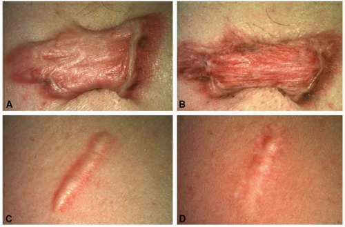
FIGURE 10-5 Keloids and hypertrophic scars after cryotherapy and triamcinolone acetonide (TAC) injections. A,C: Before treatment. B,D: After four sessions of cryotherapy and intralesional TAC injections (40 mg per mL).

Although systemic side effects of intralesional corticosteroid injections are rarely observed, cases of Cushing’s syndrome following TAC injections for keloid and hypertrophic scar treatment in children have been reported in the literature. A review by Fredman and Tenenhaus recommends a maximum dose of 30 mg of intralesional corticosteroids per month in children, while acknowledging that close observation of young patients is required after treatment.39 In adults, very few cases of iatrogenic Cushing’s syndrome after intralesional TAC injections have been reported; generally among these, extremely large doses (up to 1,200 mg) were applied within 1 month to treat multiple keloids.40 Although the possibility of adrenal complications in adults exists, it is highly unlikely in most patients after intralesional injection of the corticosteroid alone. In patients with multiple or larger lesions, alternating treatment of the different scars or scar segments can be taken into consideration as a precaution.

For older hypertrophic scars and larger keloids, the combination with cryotherapy represents a popular practical approach in daily routine and seems to be highly effective (Fig. 10-5).41,42 Marked improvement of hypertrophic scars and keloids after application of cryotherapy in combination with intralesional TAC injections has been reported.43,44,45 The exact mechanism of action of cryotherapy is not yet understood; aside from physical destruction of fibrotic tissue, it is believed to induce vascular damage, hypoxia, and ultimately tissue necrosis.2 An interval of at least 3 to 4 weeks between each treatment (approximately three to six sessions are needed) is commonly required to allow for postinterventional healing. Common side effects include permanent hypo- and hyperpigmentation, blistering, and postoperative pain.46,47,48 Based on communications among experts, cryotherapy performed directly prior to the injection of TAC appears to increase the therapeutic success rate. Though the mechanism is uncertain, this might be due to the edema formation caused by the preceding cryotherapy facilitating the injection and enabling the deposition of larger amounts of the drug.


Pressure Therapy

Since the 1970s pressure therapy has been a mainstay for the management of hypertrophic scars and keloids. Pressure garments remain a common choice for the prevention of excessive scar formation after burn injury, although the mechanism of action and efficacy remain vague. Decreased collagen production through reduced capillary perfusion, tissue hypoxia,49,50,51 and an increased rate of fibroblast apoptosis52 are presumed.

Only gold members can continue reading. Log In or Register to continue

Stay updated, free articles. Join our Telegram channel

Oct 15, 2018 | Posted by in Dermatology | Comments Off on Medical Management of Scars

Full access? Get Clinical Tree

Get Clinical Tree app for offline access