Case 1
Clinical Presentation
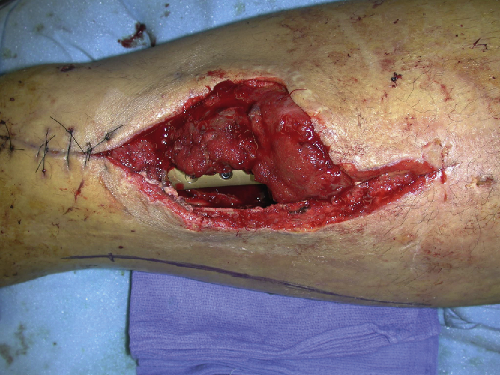
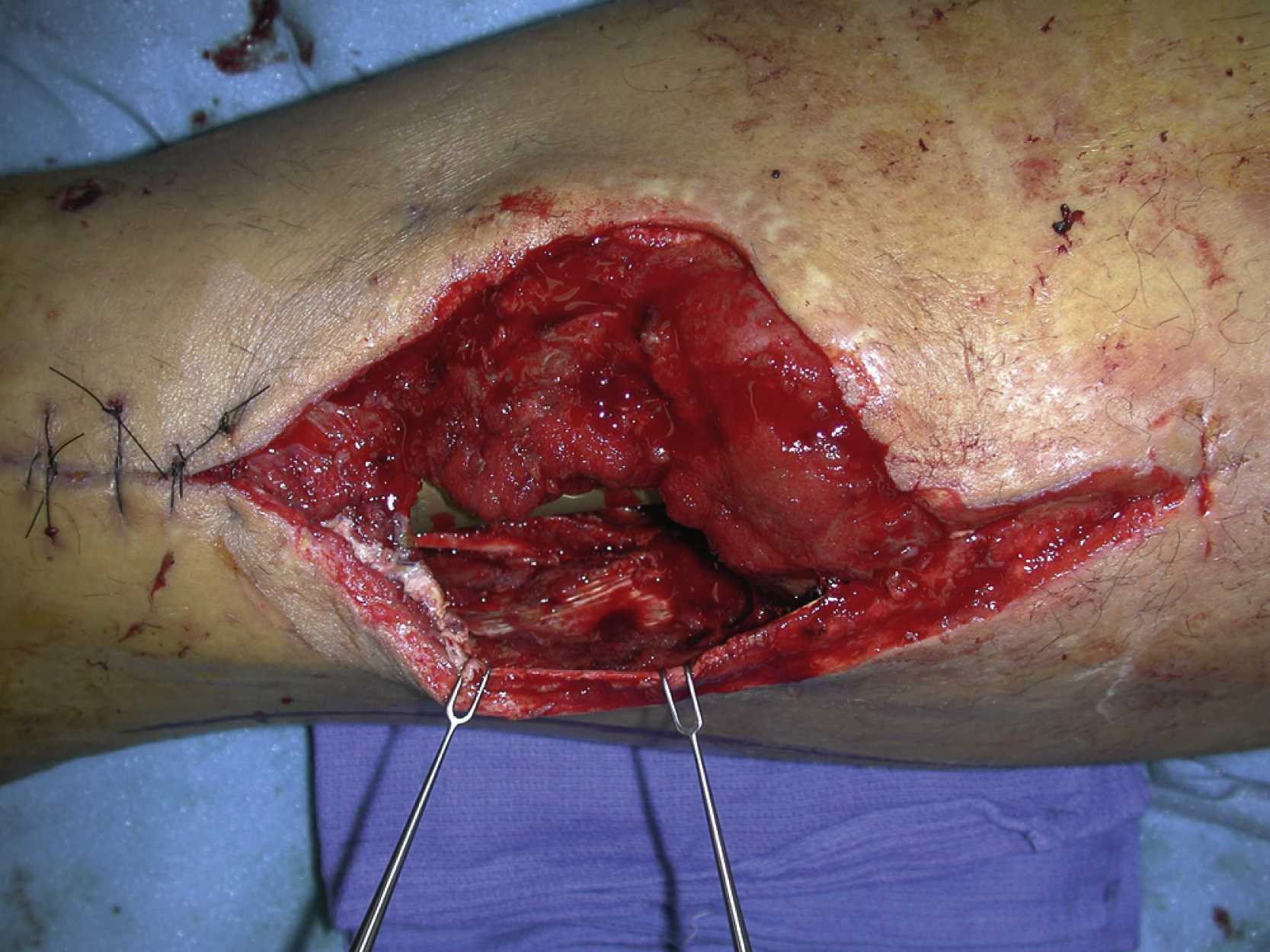
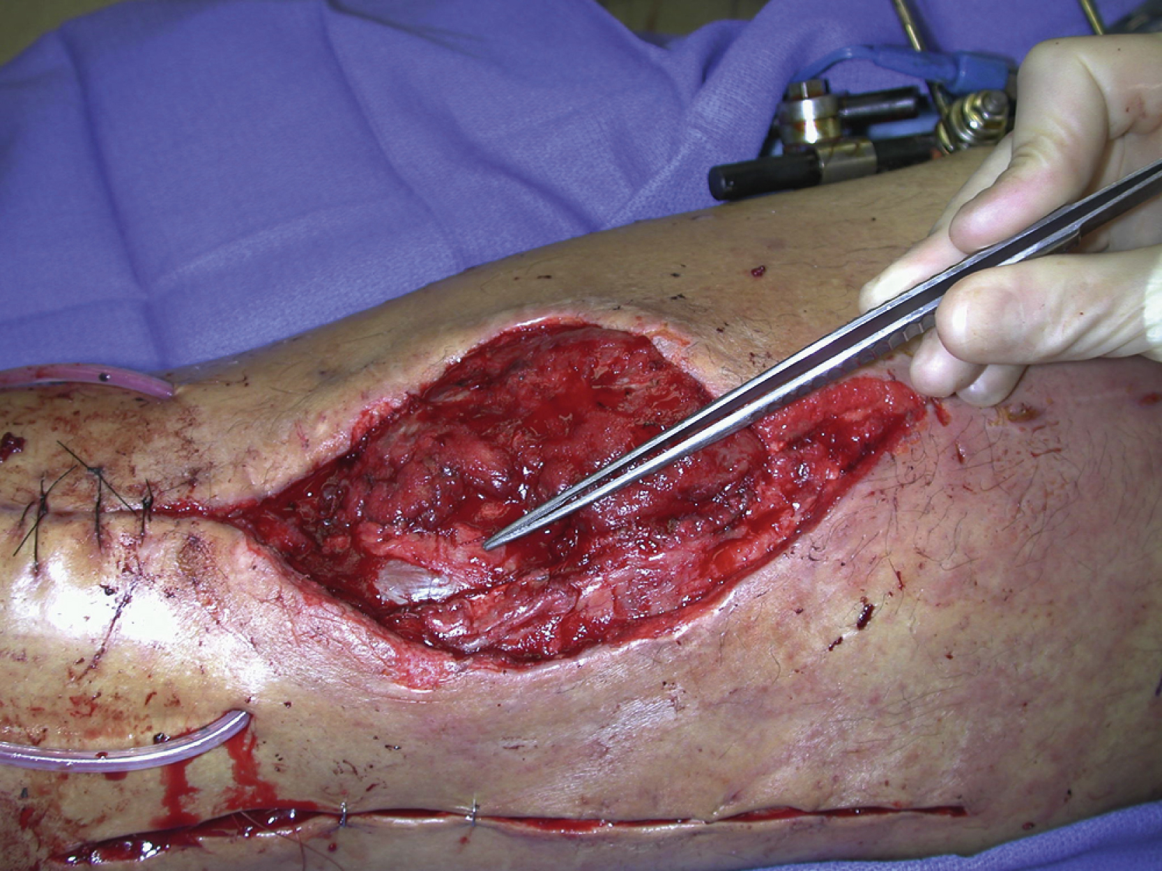
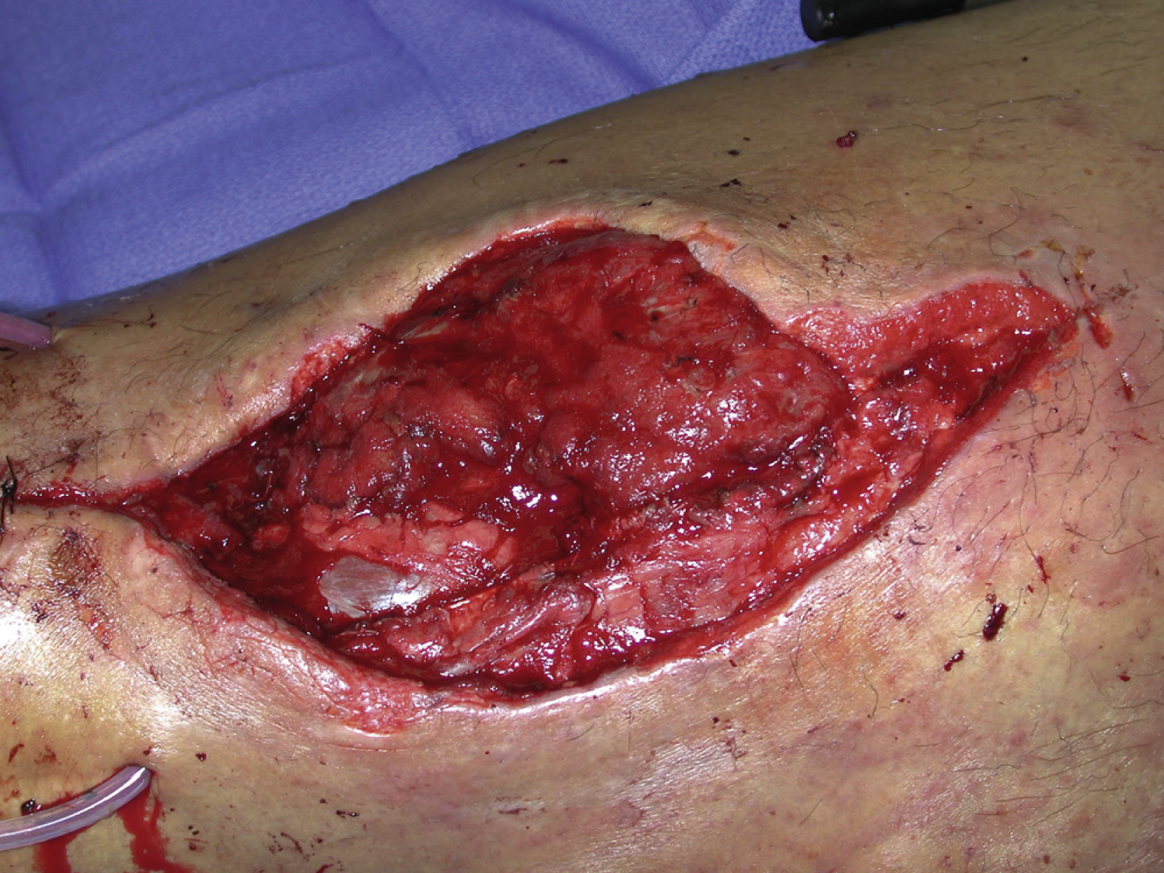
A 42-year-old White male had a complicated lateral thigh wound following an open distal femur fracture. He underwent an open reduction and internal fixation of the distal femur fracture by the orthopedic trauma service, which left a large open wound, measuring 12 × 8 cm, with the exposed fracture site and reconstructed plate ( Figs. 41.1 and 41.2 ). The plastic surgery service was asked by the primary service to perform a soft tissue coverage and definitive wound closure.
Operative Plan and Special Considerations for Reconstruction
For this relatively large soft tissue wound with the exposed femur fracture site, hardware, and potential space, a local muscle flap with a relatively large size of well-vascularized tissue, such as the biceps femoris muscle, can be selected to provide a one-stage soft tissue coverage and also obliterate the potential space. The muscle, which received a blood supply from a few branches of the profunda femoris artery, is a type II muscle flap but is considered to be reliable if the patient is free of peripheral vascular disease in the profound artery. Its long head is a large muscle that can be used to cover lower lateral thigh soft tissue defect. A skin graft to the muscle flap would be needed for the final wound closure. Adjacent skin rearrangements can also be added to facilitate the entire wound closure in addition to a skin graft to the muscle flap.
Operative Procedures
Under general anesthesia with the patient in the supine position, the right distal lateral thigh wound was debrided. All colonized tissues were removed. The open wound appeared to be fresh and clean after a more definitive debridement by the plastic surgery service.
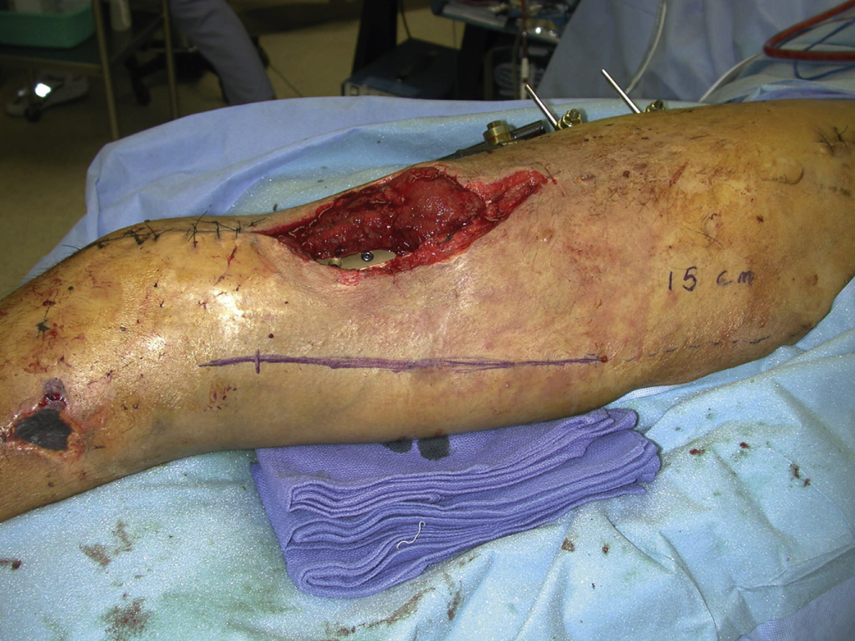
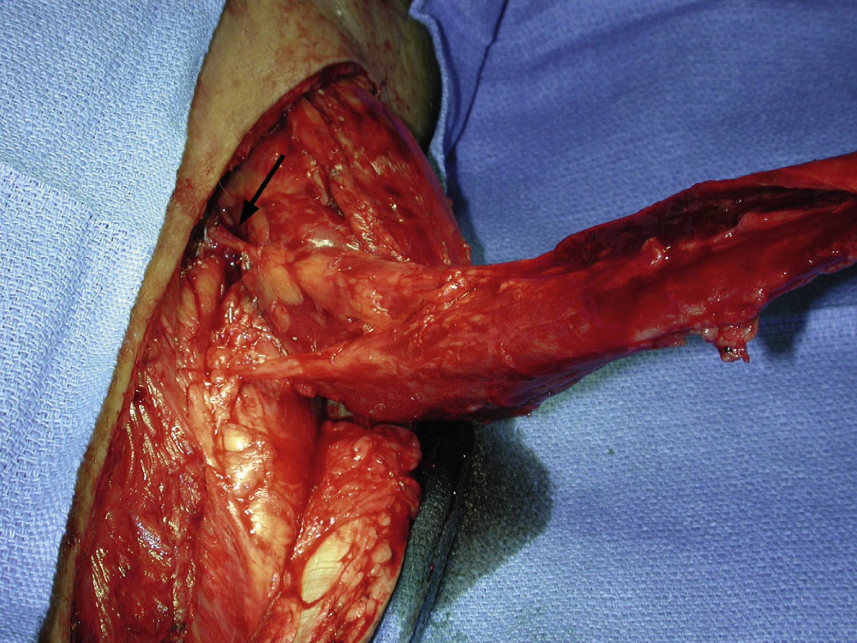
The design for the long head of the biceps femoris muscle flap was marked ( Fig. 41.3 ). The skin incision was made through the skin, subcutaneous tissues, and fascia down to the hamstring muscles. Once the long head of the biceps femoris muscle had been identified, the flap dissection was carried out to explore its insertion to the head of the fibula. Once its attachment to the fibula had been divided, the muscle was gradually elevated from distal to proximal and its minor pedicles from the profunda artery were identified. The flap was then elevated so that it could be rotated to cover all exposed structures but only one minor pedicle was divided during the flap dissection ( Fig. 41.4 ). The flap was temporarily inset into the wound to cover the entire fracture site and exposed hardware. One drain was placed under the flap ( Fig. 41.5 ). The flap was inset into the wound with several interrupted 3-0 Monocryl sutures. Additional local tissue rearrangements were performed to facilitate a complete wound closure ( Fig. 41.6 ).
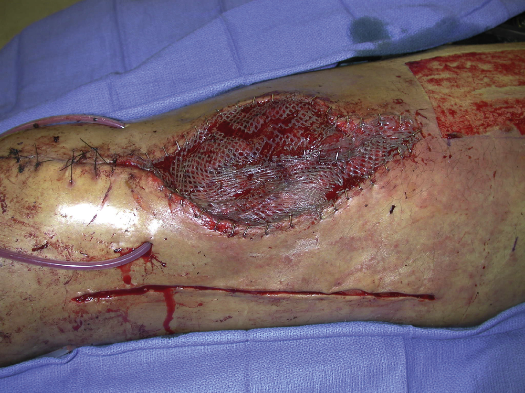
A split-thickness skin graft was harvested with a dermatome from the right anteromedial thigh. It was meshed to 1:1.5 ratio. The skin graft was placed over the muscle and the rest of the granulation wound and secured with multiple skin staples ( Fig. 41.7 ).
Follow-Up Results
The patient did well postoperatively without any issues related to the flap reconstruction and wound closure. He was discharged from hospital on postoperative day 7. The right distal lateral thigh wound healed uneventfully ( Fig. 41.7 ). He was followed by both the primary service and the plastic surgery service for subsequent postoperative care.
Final Outcome
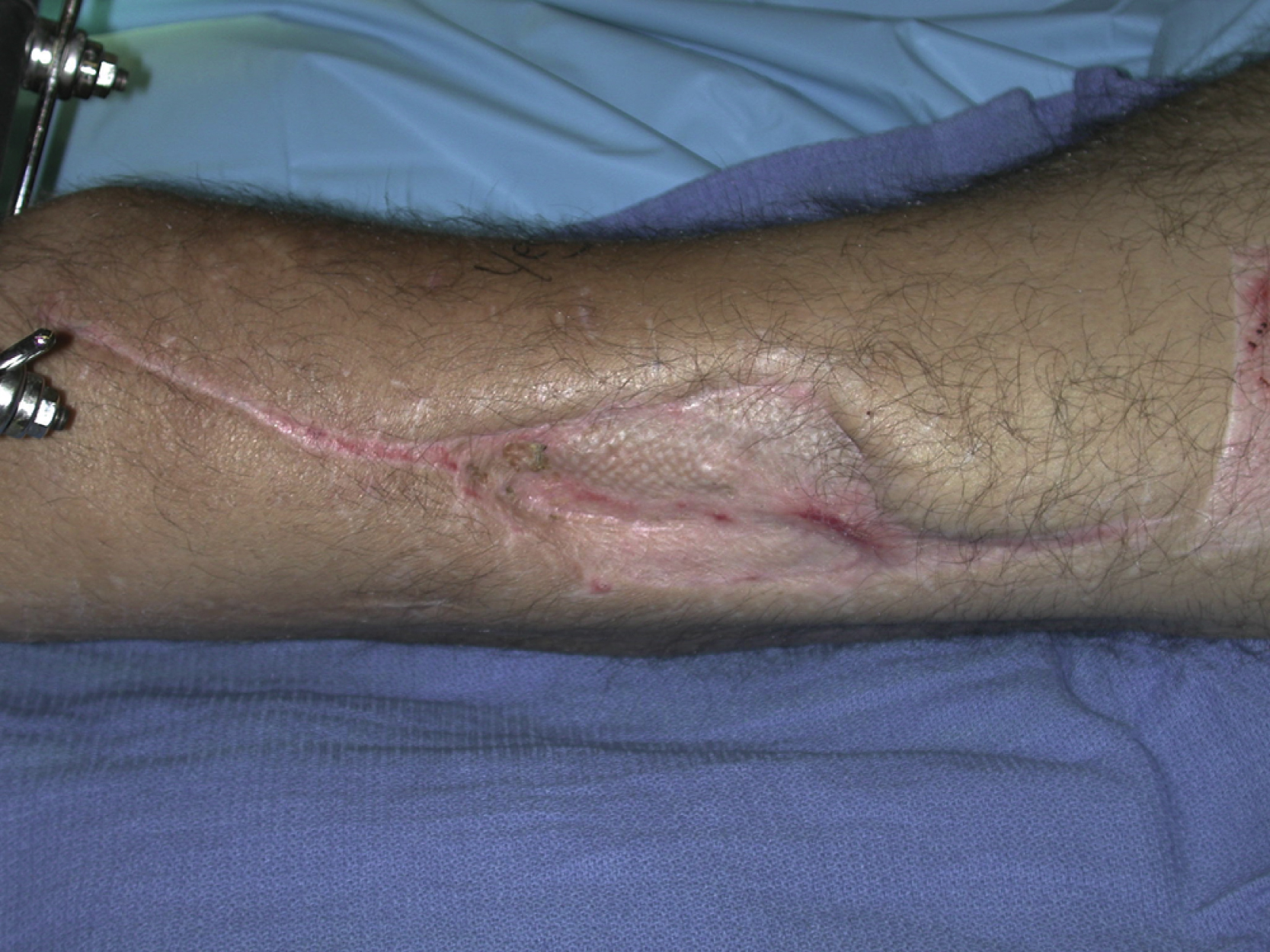
After acute care with the plastic surgery service, the patient was followed by the primary service for his routine postsurgical visits. The right lateral thigh flap reconstruction site has healed well and there has been no wound breakdown, recurrent infection, or any other long-term problems related to the flap reconstruction ( Fig. 41.8 ).
Related posts:


Stay updated, free articles. Join our Telegram channel

Full access? Get Clinical Tree


