Introduction
Recently, standardization of population screening and advances in diagnostic techniques have allowed for earlier detection of breast cancer, which allows breast cancer diagnosis to occur when the relative tumor burden is low. Earlier detection often allows for tumor excision via breast conservation, which includes appropriate ablation with maximal breast preservation. Breast conservation therapy (BCT) implies a therapeutic-surgical approach to the tumor based on the partial mastectomy in conjunction with adjuvant radiotherapy directed toward the tumor bed. This strategy is proven to be a treatment comparable to mastectomy in terms of overall survival for early stage cancer.
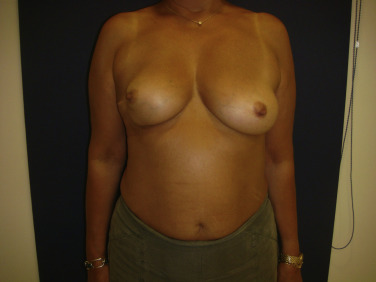
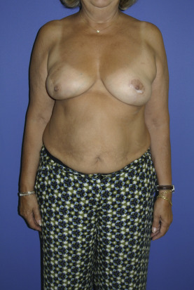
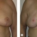
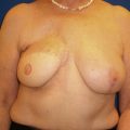
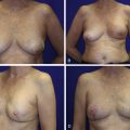
In patients with early stage breast cancer, there are several resection techniques that can be performed, the purpose of which is to make sure that margins are oncologically safe and, in addition, to achieve aesthetically acceptable results. Even with this approach, the fact remains that approximately one-third of patients will subjectively report a negative outcome related to the ablated breast. The breast asymmetry or the modification of the location and/or shape of the nipple–areolar complex ( Fig. 10.1 ) are the most frequent reasons accounting for this dissatisfaction. The location of the tumor and subsequent partial mastectomy defect can affect outcomes because certain areas are better able to tolerate the resection. The standardization of this approach has a direct consequence, which is that the number of patients with partial volumetric deficits is increasing. Plastic surgeons are now confronted with a variety of patients with partial mastectomy defects following radiotherapy, which has been a significant source of psychosocial discomfort and distorted body image. For these and other reasons ( Table 10.1 ), this increase in the number of patients with breast distortion following breast conservation alone ( Fig. 10.2 ) has aroused major interest in the development and use of local techniques for breast reconstruction.
| Improvement in oncologic detection methods → major % of tumor detection in early stage cancer status |
| Consolidation of breast-conserving therapy → lesser volume resections |
| Substitution of “whole breast radiotherapy” for radiotherapy focused on the tumor bed (major availability for local tissues in good conditions) |
| “Replace like with like” concept |
| Development of microsurgical techniques and perforator flap surgery |
| Presence of lipofilling as an additional procedure to local techniques |
Currently it is difficult to estimate the percentage of women treated with BCT who will end up undergoing reconstructive surgery, either immediate or delayed, given the multitude of factors affecting this decision ( Table 10.2 ). Before surgery, a multidisciplinary approach is highly recommended, in conjunction with the oncoplastic surgeon and the oncologist, to agree on the treatment to be applied, which will certainly affect the indications for reconstruction.
| Logistic Factors | Patient Factors | Oncologic Factors |
|---|---|---|
| Availability of plastic surgeon/oncoplastic surgeon in the reference hospital | Personal decision of the patient, oriented by plastic surgeon/oncoplastic surgeon | Possibility to perform breast-conserving therapy (tumor size and location) |
| Knowledge of oncoplastic techniques by the plastic surgeon/oncoplastic surgeon | Breast size before the surgery | Oncologically safe margins, defect size in relation to the breast size |
| Existing information in relation to breast partial reconstruction techniques | Comorbidities | Radiotherapy: need of adjuvant radiotherapy, type of radiotherapy |
Oncoplastic Surgery: Displacement Techniques versus Local Flaps
The oncoplastic techniques are born with the difficult function of combining an oncologically safe resection with an aesthetically satisfactory postsurgical result for the affected woman.
There exist multiple oncoplastic techniques, generally described according to the size and location of the tumor; thus, each case will be unique in that there will be an optimal treatment strategy based on the breast and tumor characteristics. However, the selection of each technique will depend on the expertise of the surgeon, and the indication for each procedure will be determined by the size of the defect in relation to the size of the breast before surgery.
The oncoplastic techniques can be differentiated into two large groups depending on the type of tissue handling that is carried out. Broadly speaking, patients with large and/or ptotic breasts will benefit from oncoplastic techniques based on the mobilization of breast tissue (volume displacement techniques) that, in some cases, will improve the aesthetic appearance of a woman’s breast. On the other hand, women with small breasts or who do not want an intervention on the contralateral breast will require a volume contribution through the use of local flaps incorporating the principles of volume replacement.
- •
Volume displacement techniques: oncoplastic techniques based on mobilization after resection of the remaining breast glandular tissue toward the lumpectomy bed.
Upon resection and mobilization of the tissue itself without adding new volume, the final size of the breast will be smaller than that of the contralateral breast. For this reason, to avoid residual breast asymmetry, these techniques should be accompanied by mastopexy or reduction mammaplasty procedures of the contralateral breast.
These are techniques with a relatively simple learning curve by applying well-known concepts for the plastic surgeon, with a seemingly predictable end result. In addition, they allow the reconstruction of the defect without requiring anything other than the breast tissue, so the creation of new donor defects is avoided. The inclusion of a plastic surgeon in designing the skin pattern for the oncologic resection can minimize the creation of new scars and optimize scar location; thus, the same incisions made for the tumor extirpation can be used for the oncoplastic reconstruction.
Despite optimal planning and execution, complications can occur and usually are due to excessive glandular mobilization that may lead to steatonecrosis that could mask a potential recurrence. Along the same lines, alteration of the normal breast architecture as well as mobilization of the original tumor bed may add to the complexity and difficulty of oncologic follow-up; however, this contention has not been scientifically proven.
- •
Local flap reconstruction: oncoplastic techniques based on the use of pedicled local flaps for the supply of volume from nearby tissues, without using the gland itself.
When recruiting regional tissue, donor areas adjacent to the defect will be required, which, in addition to those used for oncologic resection, will generate new scars.
There are several different local flaps that are available to correct partial mastectomy defects that will depend on the chosen donor area and on the type of flap to be mobilized to the defect zone. These include fasciocutaneous flaps or musculocutaneous flaps. In general, these techniques are indicated for women with small breasts and large tumors because there is not enough glandular tissue to perform volume displacement techniques.
In certain cases, if the same amount of previously resected tissue is provided and an adequate inset of the flap is made, an acceptable symmetry between both breasts can be achieved without the need to perform a symmetrization procedure on the contralateral breast.
For this reason, these techniques are also indicated for women who do not want an intervention on the contralateral breast; however, the ability to achieve optimal symmetry in these cases is less predictable because it will depend on the postoperative settling of the flap and how it will respond to the possible adjuvant treatment. Unlike volume displacement techniques, the normal architecture of the breast or the tumor bed is not modified.
Local Flaps for Breast Reconstruction
Introduction, Indications, and Contraindications
Despite the standardization of perforator free flaps as a gold standard for breast reconstruction, local coverage options like pedicled flaps should not be underestimated, because their use has shown to be effective in women with lumpectomy and mastectomy sequelae.
There is a wide range of local possibilities for the restoration of breast defects, from techniques based on axial vessels (for example, latissimus dorsi [LD] musculocutaneous flap) to techniques based on perforating vessels (for example, thoracodorsal artery perforator [TDAP] flap, etc.).
Therefore, local flaps are a therapeutic alternative that should be included in the surgical arsenal of any plastic surgeon specializing in breast reconstruction, because they may turn out to be the indication of choice in certain cases, presenting a series of benefits as follows:
- •
Concept of replacing “like with like”: the skin texture, thickness, and color will always be more similar to those of the original breast, if the tissue used for the reconstruction is adjacent to it.
- •
Shorter duration surgeries: pedicle flaps do not require dissection of recipient vessels, a vascular anastomosis, or the use of microsurgical dissection techniques and/or special material (except perforator flaps).
- •
Pedicle flaps have an expedited learning curve (except for perforator flaps).
The surgical technique of these flaps is based on rotation/transposition of the tissues near the breast area such that the breast quadrant with the defect to be reconstructed will play a definitive role when deciding on the various flap options available. Thus, taking into account that the vast majority of these pedicle flaps are based on the excess dermal adipose tissue present in the axillary or dorsal region, the defects located at the inferolateral quadrant will be those with the greatest accessibility for coverage by pedicled flaps. On the other hand, defects located at medial mammary quadrants will pose a surgical challenge and will have to be approached using less conventional flaps. The majority of local pedicle flaps such as the LD musculocutaneous flap or its analog in perforator surgery, the TDAP flap, are ideally suited for lateral breast defects and less likely to reach when confronted with a medial breast defect.
Before breast reconstruction with local flaps, it is necessary to complete a full assessment of the patient and anticipated breast defect, and to preoperatively plan the reconstruction using a free or pedicle flap. The risks and benefits as well as the pros and cons must be evaluated. By performing this assessment preoperatively, the likelihood of success will be enhanced, and the risk of reconstructive failure can be minimized. This fact is more evident in delayed reconstructions, in which there are prior scars in the vicinity of the proposed flap (i.e., axillary node excision that may compromise the ability to use local flaps). In other cases, especially in patients whose oncologic resection was performed years before the standardization of BCT, the larger breast deformities in the setting of prior radiotherapy may actually be indications for completion mastectomy with total breast reconstruction with a free tissue transfer rather than attempt a partial reconstruction prone to morbidity. Thus, there is a sequence of established indications and contraindications that will indicate or rule out breast reconstruction with local flaps in cases where it is required ( Table 10.3 ).
| Indications | Contraindications |
|---|---|
| Preference for autologous reconstruction, in patients who do not want to undergo long surgeries | Absolute: previous surgical procedures, especially in axillary region or in the donor zone for the local flap |
| Partial breast reconstruction (lumpectomies, quadrantectomies), especially in small- to medium-sized breasts | Relative: “whole breast radiation” radiotherapy, axillar radiotherapy → in these cases it is preferable to perform the reconstruction with healthy tissue by a free transfer, not affected by regional radiotherapy damage |
| Autologous breast augmentation (contralateral breast) | Relative: defects in medial quadrants → difficulty to achieve coverage through traditional flaps. It is possible to perform less frequent flaps (IMAP, AICAP) |
| Correction of breast deformity after partial necrosis of the previous free flap | Relative: globally distorted breast (large previous resection + radiotherapy) → It is better to complete mastectomy and proceed to free flap reconstruction |
Types of Local Flaps
As previously mentioned, there are different alternatives for breast reconstruction with local flaps.
They can be classified depending on two principal criteria:
- a.
Depending on the tissue recruited for subsequent mobilization
- •
Dermal-fat flaps
- •
Fasciocutaneous flaps
- •
Musculocutaneous flaps
- •
- b.
Depending on the flap vascularization
- •
Random pattern vascularization
- •
Vascularization through perforator vessels
- •
Vascularization through axial vessels
- •
Some of these flaps are more versatile than others and can adapt to the needs of each specific case. On the other hand, other flaps will need specific indications that will be detailed throughout the chapter. Until recently, the most globally accepted option for breast reconstruction with locoregional autologous tissue was the LD musculocutaneous flap. With the development and standardization of oncoplastic techniques and perforator surgery, new local alternatives appear that are applicable to breast reconstruction.
Thus, random and perforator flaps are a relatively simple alternative of the use of LD flap, presenting several advantages. The primary advantage is the preservation of the LD muscle, which, apart from avoiding the morbidity associated with its sacrifice, allows the patient and the surgeon to maintain it as a reconstructive option in case of a local recurrence.
In addition, its definitive result will be more predictable as there is no subsequent muscular atrophy that decreases the overall volume of the flap. These flaps also can be combined with volume displacement techniques or with fat infiltration techniques in case the defect to be covered is extensive.
Random Pattern Flaps
The random pattern flaps are adipocutaneous flaps in which the vascularization is not based on an axial pedicle but on the perforators irrigating the subdermal network. Unlike the conventional perforator flaps, in the random flaps any perforator is dissected under direct visualization, so the viability of its distal ends will be unpredictable. To minimize the risk of flap necrosis, it is important to emphasize that its indication will be limited to small-sized defects, preferably at the level of lateral quadrants of the breast. Larger defects would be better to reconstruct with perforator flaps in which the visualization and microsurgical dissection of the perforator will provide greater security in terms of flap survival and will have a lower necrosis rate than the random flaps. Even so, its use should not be underestimated, considering that they have a series of advantages with respect to the rest of local flaps:
- •
It is relatively simple to perform and does not require use of microsurgical techniques.
- •
Intraoperative time is short, without need of positional changes.
- •
It does not include LD muscle, and its dissection does not affect the thoracodorsal pedicle, for which we maintain the rest of local reconstructive options intact in case they would be necessary, avoiding the need for new donor areas on the dorsal area.
- •
It has a lesser complication rate (except for unpredictable flap necrosis) and shorter hospital stay and less postoperative pain. This helps initiate the postoperative adjuvant therapy without delays, if necessary.
The use of these local flaps is most useful in the setting of immediate reconstruction. As mentioned, possible limitations in the setting of delayed reconstruction include prior scars or incisions that can limit flap design and applicability.
Rhomboid Flap
Dermal-fat flap is indicated for the reconstruction of small defects on the lateral quadrants of the breast.
It is especially useful for the coverage of defects in which direct closure implies generating aesthetically unacceptable “dog ear” deformations or when their correction requires increasing the scar length too much along the breast surface.
To ensure a correct cosmetic result, the flap should include a sufficient amount of fat, so it should be avoided in extremely thin women.
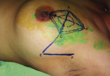
The markings are done in the supine position, transforming the lumpectomy defect into a rhomboid or diamond-shaped defect, as in the Limberg flap, so that, after excision, one of these resection margins constitutes one of the lateral borders of the flap ( Fig. 10.3 ).
During its dissection, it is necessary to extend the dissection to the muscular fascia to allow a correct axis of rotation and closure of wounds without tension; otherwise, distal tip necrosis of the flap may occur. The likelihood of total flap necrosis is rare.
Subaxillary Flap
The subaxillary based random pattern flap is an efficient therapeutic alternative for the reconstruction of small- to medium-sized defects of the superior-lateral quadrant of the breast. It is important to note that the base of the flap, which will be located at the superior aspect of the defect, must be at least 6–8 cm wide. The markings for this flap will be guided by using the pinch test in the subaxillary region, because it recruits redundant skin and fat at that level without causing a distortion in the adjacent breast contour. For this reason, this flap is not indicated for thin patients with quadrantectomy defects because the tissue available at the subaxillary level is equivalent to a maximum of 25% of the total breast volume. If there is no need for cutaneous contribution, the flap can be de-epithelialized and buried to only fill volume in the lateral mammary quadrants, as described by Chatuverdi in 2004. Similar to other random pattern flaps, the same incisions used for lymph node surgery or lumpectomy will correspond to the lateral margins of the flap; thus, cooperation between the oncoplastic surgeon and the plastic surgeon is highly recommended at the time of operation to delineate the incisions appropriately and to facilitate the technical aspects corresponding to the tumor excision, lymph node resection, and oncoplastic reconstruction.
The harvesting of the flap does not require positioning the patient in lateral decubitus, but it will be necessary to keep the arm abducted at 90 degrees to easily reach the muscular fascia and allow for the correct rotation of the flap. Once the flap has been harvested, the arm must be closed beforehand to avoid tension on the incisions and ensure a correct remodeling of the breast contour.
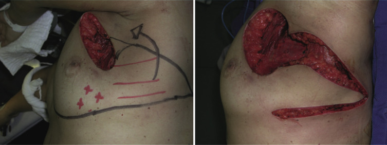
Lateral Thoracodorsal Flap
This fasciocutaneous flap with random vascularization pattern could be considered as a specular image of the subaxillary flap but inferiorly based. Therefore, it will be indicated for small- to medium-sized defects at the level of inferior-lateral breast quadrants. Initially described by Hölstrom in the mid-1980s, the design resembles an “ax” or “wedge” shape, with an approximately 7-cm wide base, located in the lateral continuation of the inframammary fold. Posteriorly, the axis of the flap will be traced toward the lateral thoracic region, where the dermal-fat excess at that level will be included in the design, being that this was previously checked by pinch test ( Figs. 10.4 and 10.5 ). In general, the dimensions of the flap do not exceed 7–9 cm of basis and 15–18 cm of length. Despite being a random flap, its main vascularization is derived from intercostal branches; therefore, during the dissection of this flap, two fundamental aspects to ensure its viability must be accounted for:
- •
Avoid performing undermining in the lateral region of the inframammary fold during oncologic resection.
- •
Include the fascia of the m. serratus anterior in the flap.
Related posts:
 Indications and Patient Selection for Oncoplastic Breast Surgery
 Oncoplastic Breast Surgery and the Effects of Radiation Therapy
 Plastic Surgeon’s Approach to Oncoplastic Breast Surgery
 Surveillance and Imaging Following Oncoplastic Breast Surgery
 Volume Displacement and Volume Replacement Techniques
 Complications of Oncoplastic Breast Surgery
Stay updated, free articles. Join our Telegram channel

Full access? Get Clinical Tree


