Clinical Presentation
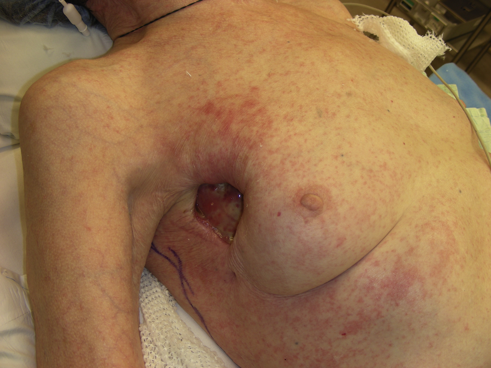
A 74-year-old White female suffered a lateral chest wound and had a partial breast defect secondary to wound debridement, complicated by the previous minimally invasive right lobectomy. Apparently, she had a seroma formation in the area, which had been managed by the thoracic surgery service. The plastic surgery service was asked to provide soft tissue coverage for the wound and to facilitate healing of the complicated wound. The lateral chest wound also involved part of the axilla and the superior lateral quadrant of the breast ( Fig. 22.1 ). Prior to the procedure the wound had been treated with local wound care.
Operative Plan and Special Considerations
Additional debridement was planned to excise all fibrotic tissues around and deep to the open wound. Such debridement should be done to healthy and normal-looking tissue either around or at the base of the wound. Attention should be paid to avoid injuries to neurovascular structures in the axilla. Because of the location and size of the soft tissue defect after debridement, a pedicled latissimus dorsi myocutaneous flap can be selected to cover this wound in one stage. The flap can be elevated from the patient’s right back and tunneled to the defect without any difficulty. In addition, a portion of the breast defect can be reconstructed at the same time. If the skin paddle of the flap is not too wide, the flap donor site can also be closed primarily.
Operative Procedures
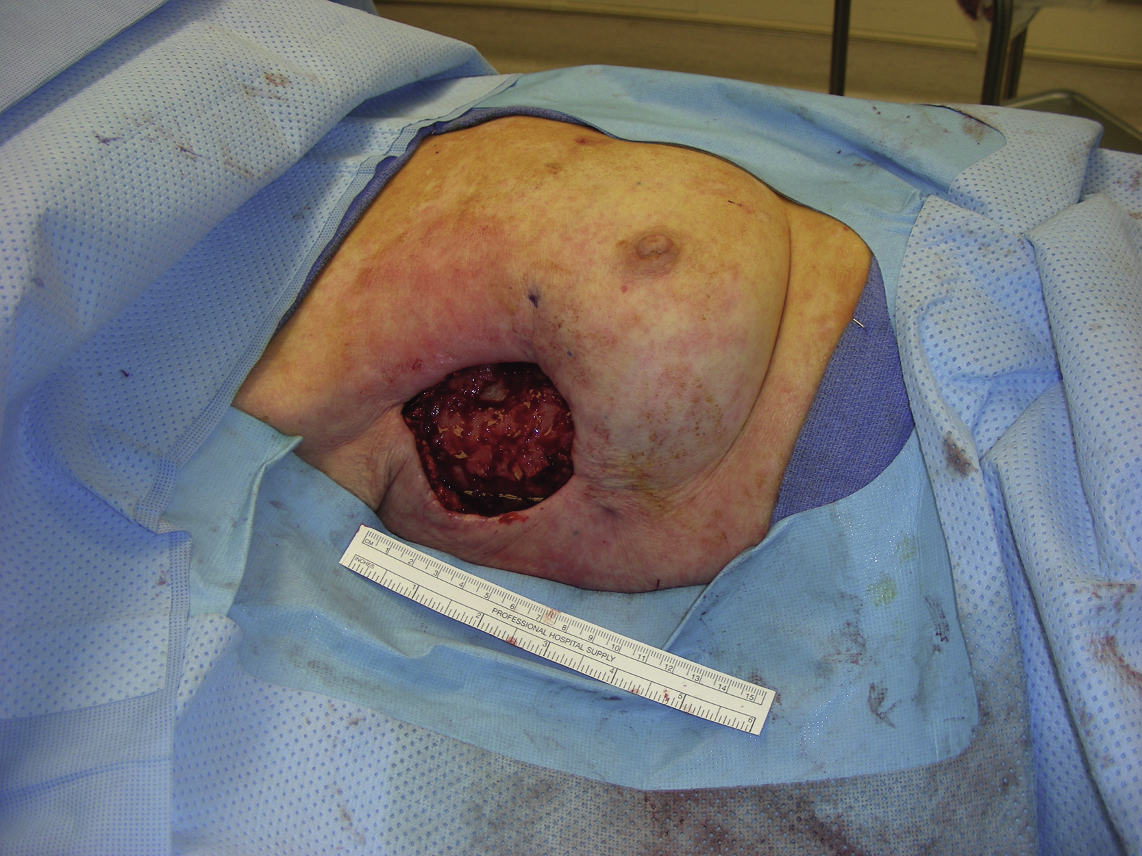
Under general anesthesia with patient initially in the supine position, the procedure started by removing all the colonized and necrotic tissues within the right axilla and chest wound. After debridement, the wound showed exposed ribs. The wound, measuring 9 × 6 cm, was irrigated thoroughly with Pulse-Vac ( Fig. 22.2 ).
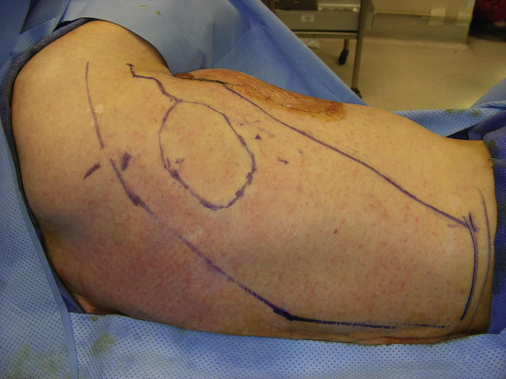
The patient was then placed in the left lateral decubitus position. The right latissimus dorsi myocutaneous flap with a 9 × 6 cm skin paddle was designed and marked. ( Fig. 22.3 ). About 10 cc of 1% lidocaine with 1:100,000 epinepherin was administered to the proposed skin incision. After the incision had been made around the skin paddle, the dissection was performed around the skin paddle down to the latissimus dorsi muscle. Once the muscle had been identified, the muscle flap was dissected more superiorly to identify the superior border of the latissimus dorsi muscle. Once the superior border had been identified, the muscle was dissected free from the chest wall superiorly, then medially, then inferiorly, and then laterally. In this way the muscle flap with its skin paddle was elevated from the chest wall. The thoracodorsal vessels were identified with a handheld Doppler. A further dissection was done around the pedicle toward the muscle’s insertion. The flap was tunneled through the lateral chest subcutaneous tissue and then placed inside the right chest wound.