Key points
- •
Patients with atopic dermatitis are more susceptible to developing irritant contact dermatitis (ICD) and allergic contact dermatitis (ACD).
- •
ICD can present with phenotypic subtypes such as asteatotic ICD, airborne ICD, acneiform ICD, chronic ICD, and acute ICD with characteristic body sites involved, predominant morphologic features, time courses, and irritant sources.
- •
ACD is a type IV delayed hypersensitivity reaction and is preceded by prior sensitization to a hapten/allergen in contrast to ICD.
- •
Allergens that can cause ACD may have different pathologic and molecular signatures.
- •
The gold standard for diagnosing ACD is through patch testing.
- •
Treatments for atopic dermatitis can affect the reliability of patch testing results.
Introduction
Immunogens are external stimuli that perturb and penetrate the skin barrier resulting in the elicitation of inflammation. This chapter will review the relationship between atopic dermatitis (AD) and two conditions that predispose patients to immunogens: irritant contact dermatitis (ICD) and allergic contact dermatitis (ACD). Special considerations with regard to diagnosis and management of ICD and ACD in the context of AD are discussed.
Immunogens, outside in and inside out
The relationship between immunogens, the skin barrier, and the immune response is intertwined with outside-in versus inside-out factors in AD pathogenesis. The outside-in hypothesis of AD pathogenesis postulates that defects in the skin barrier allow greater penetration of irritants and immunogens eliciting the atopic march; this concept includes the idea that AD predisposes infants and children to other atopic diseases such as allergic rhinitis, allergic asthma, food allergy, and allergic keratoconjunctivitis. In contrast, the inside-out hypothesis suggests that innate immunologic factors predispose toward an atopic diathesis that results in skin barrier defects.
The inside-out hypothesis argues that a predisposition toward a polarized immune response gives rise to a defective skin barrier and that innate immune abnormalities lead to an aberrantly primed immune response. Support for this hypothesis comes from the observation that AD patients have an increased Th2 response, even in nonlesional skin. Tape stripping and using acetone to create skin barrier dysfunction in mice models leads to activation of innate immunity and increased Langerhans cell (LC) access to antigens along with upregulation of Th2 cytokines ( ). In mice, overexpression of Th2 cytokines leads the skin to spontaneously develop AD and skin barrier defects ( ).
The outside-in hypothesis of AD pathogenesis is based on the observation that a percentage of AD patients have genetic defects in the stratum corneum and/or tight junctions located within the stratum granulosum, both of which are important barrier structures protecting the skin from the environment. Additional evidence for skin barrier defects initiating AD is provided by measures of epidermal dysfunction such as transepidermal water loss (TEWL) in AD. TEWL is increased in populations susceptible to AD such as neonates. Increased TEWL has been demonstrated in atopic versus normal skin, and studies have shown that even unaffected skin in patients with AD displays increased TEWL ( ). One of the best characterized genetic abnormalities associated with increased risk of AD is decreased expression of the epidermal differentiation protein filaggrin. The filaggrin (FLG) gene encodes the FLG protein, which is important for maintaining the skin barrier and preventing TEWL. Aside from structural deficiencies, decreased filaggrin can lead to increased Th2 cytokine production and production of endogenous proteases, increased interleukin-1 (IL1), and thymic stromal lymphopoietin (TSLP). These alterations in the immune milieu further erode the stratum corneum and skin barrier leaving it more penetrable to microbes and other immunogens. Nonetheless, filaggrin deficiency alone is not enough to account for most cases of AD. Although up to 40% of patients with severe AD have filaggrin mutations, only about 10% of European patients with AD are carriers. Filaggrin mutations are rare in Asian populations and are not typically found in AD patients of African descent. While patients with filaggrin mutations often have dry skin, AD, and associated atopic diseases, most patients with AD do not have an FLG mutation ( ). Other variants of genes encoding proteins in the epidermis associated with AD include filaggrin 2, hornerin, the cornified envelope precursor SPRR3, serine protease inhibitors, involucrin, loricrin as well as tight junction proteins such as the claudins ( ). In this chapter we focus on immunogens as triggering factors for dermatitis primarily from the outside-in perspective. Particularly, we focus on two types of immunogens: those related to irritant contact dermatitis (ICD) and to allergic contact dermatitis (ACD).
Irritant contact dermatitis–related immunogens
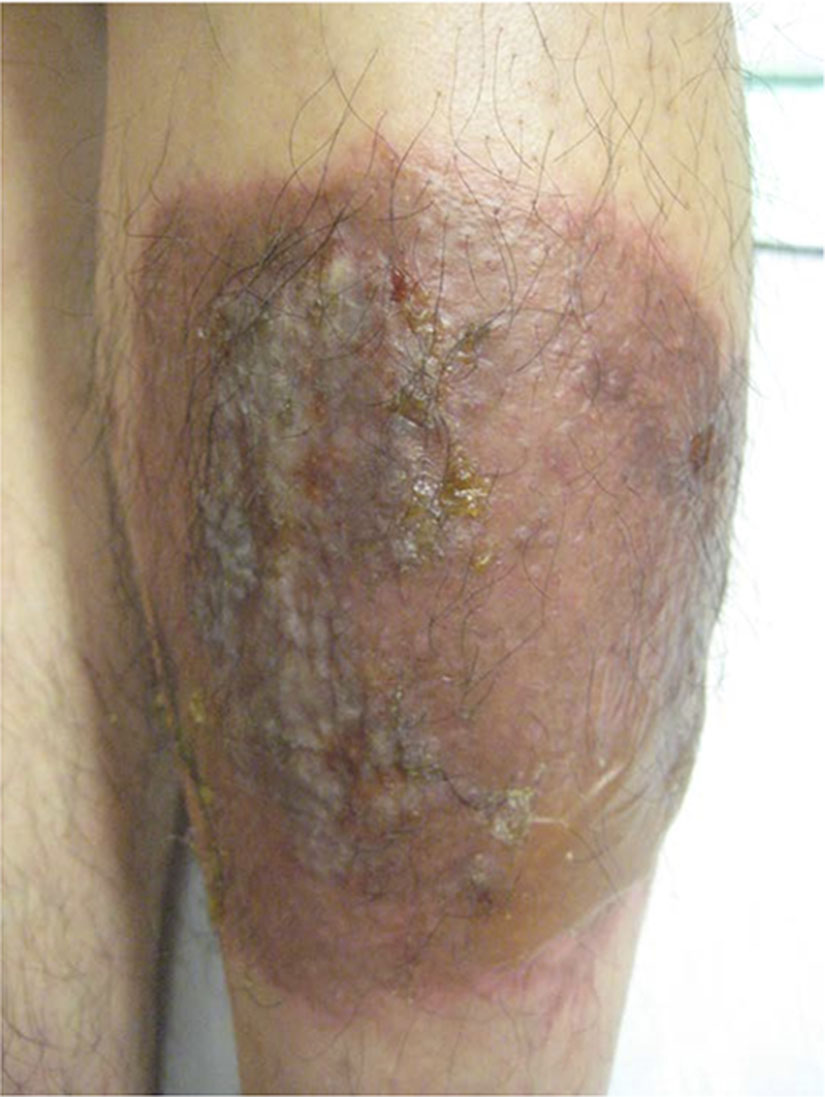
Irritants cause physical or chemical damage to the integument leading to a disruption of skin barrier integrity and the induction of inflammation. This inflammation results in ICD. No prior sensitization is necessary. ICD is a cytotoxic and inflammatory response to irritant stimuli resulting in several different clinical manifestations. Various clinical phenotypes of ICD have also been described based on the time course, predominant morphologic features, and irritant source. Examples include acute ICD, irritant reaction/chronic ICD, subjective irritation, frictional dermatitis, acneiform ICD, and airborne ICD. The prototypical ICD is acute ICD, typically due to a strong irritant, which can present with erythema, edema, vesicles, exudates with a sharp demarcation, and symptoms of burning, stinging, pain, and pruritus ( Fig. 7.1 ). In contrast, chronic ICD and irritant reactions occur to mild irritants such as water, soap, and detergents; clinically, they appear as redness, scaling, and/or erosions ( Fig. 7.2 ). Frictional dermatitis presents symptomatically with erythema, hyperkeratosis, scaling, and fissures in areas of friction. Asteatotic ICD is characterized by xerosis and superficial fissuring, and it commonly occurs on the lower extremities. Airborne ICD can occur to irritants in the air such as fibers, dust, fumes, and vapors. This dermatitis is predominant in exposed areas but can also affect covered areas by contaminating clothing. Finally acneiform ICD (due to metals, tars, metalworking fluids, chlorinated agents) can present as open and closed comedones, cysts, pustules, and nodules in a lateral distribution on the face and neck ( ).

Several mechanisms are involved to create the clinical presentation of ICD, including skin barrier disruption, keratinocyte damage, and release of proinflammatory mediators. While pure irritants do not directly induce an adaptive immune response, they can be a potent adjuvant for immune induction ( ). ICD triggers the innate immune system via similar pathways as those used by pathogens, and stimulates toll-like receptors (TLR) and cytosolic NOD-like receptors (NLR) present on both hematopoietic and nonhematopoietic cells to detect danger signals and activate inflammation. The activation of the innate immune system via TLR, reactive oxygen species, and NLRP3 inflammasomes can induce dendritic cells and in turn trigger the adaptive immune system. Indeed, a close connection between the two immune responses is increasingly recognized. Keratinocytes damaged with disruption of the skin barrier also release cytokines, upregulate major histocompatibility complex antigens, and release IL1α, IL1β, and tumor necrosis factor (TNF).
ICD in patients with AD
The incidence of ICD is increased in patients with AD likely by several different mechanisms. Several epidemiologic studies document an increased rate of irritant reactions in patients with atopy, particularly in the occupational setting ( ). Interestingly the most commonly used definition for AD in clinical trials, the Hanifin and Rajka criteria ( Table 7.1 ) for AD, includes some features of ICD such as “tendency toward non specific hand or foot dermatitis” and “intolerance to wool and lipid solvents.”
| Major criteria |
|---|
| Chronic or chronically relapsing dermatitis |
| Pruritus |
| Typical morphology and distribution: Flexural lichenification or linearity, facial and extensor involvement in infants and children |
| Personal and/or family history of atopy, asthma, allergic rhinitis, atopic dermatitis |
| Minor criteria |
| Immediate (type I) skin test reactivity |
| Elevated serum immunoglobulin E |
| Early age onset |
| Tendency toward nonspecific hand or foot dermatitis |
| Tendency toward cutaneous infections (i.e., Staphylococcus aureus and/or herpes simplex)/impaired cell-mediated immunity |
| Xerosis |
| Ichthyosis/palmar plantar hyper linearity/keratosis pilaris |
| Recurrent conjunctivitis |
| Dennie-Morgan infraorbital folds |
| Keratoconus |
| Anterior subcapsular cataracts |
| Orbital darkening |
| Facial pallor/facial erythema |
| Pityriasis alba |
| Chelitis |
| Anterior neck folds |
| Nipple eczema |
| Perifollicular accentuation |
| Itch when sweating |
| Intolerance to wool or lipid solvents |
| Food intolerance |
| Disease course influenced by environmental/emotional factors |
| White dermatographism/delayed blanch response |
Pathogenesis of ICD in AD
The defects in the skin barrier associated with AD allow for increased penetration of irritants predisposing to ICD. Rates of occupational hand dermatitis are increased in patients with AD ( ). This may be due to deficiencies in the skin barrier. The interaction between filaggrin mutations and AD in patients with occupational dermatitis was studied in Germany and found that FLG mutations are present in 15.9% versus 8.3% of ICD patients and controls, respectively ( ). The odds ratio (OR) was 2.9 (95% confidence interval [CI], 1.33–3.28) for the combined FLG -deficient and AD phenotype. After adjusting for AD, the OR of 1.62 (95% CI, 1.01–2.58) continued to indicate an increased association. In the setting of occupational ICD, of 459 patients with FLG mutations, 327 (71.2%) were atopic and 132 nonatopic. The FLG -deficient mutations in the setting of AD led to a more resistant clinical course of ICD with lower rates of recovery and higher use of topical corticosteroids ( ).
In addition to mutations in the skin barrier, patients with AD have inside-out genetic alterations in the cytokines involved in skin inflammation. One well-characterized factor is TNFα-308A. This polymorphism has been associated with susceptibility to ICD and ACD ( ). TNFα-308A carriers have increased production of TNFα and a lower threshold to irritants such as sodium lauryl sulfate (SLS). In a case control study of 478 patients with occupational ICD, carriers of the TNFα-308A variant were more prone to developing ICD of the hands ( ). TNFα is also released by keratinocytes when there is disruption of the skin barrier, which further induces inflammatory reactions and the cytokine cascade ( ). With the observation of the role for TNFα in ICD, TNFα may also be a target for treatment. It has been noted that injection of anti-TNFα antibodies may block some irritant reactions in animal models ( ).
An objective demonstration of the increased risk of ICD in AD patients is demonstrated by a decreased threshold for susceptibility to SLS-induced irritant damage to the epidermis in patients with AD ( ). It is also known that ICD can predispose patients to ACD. When skin is pretreated with the irritant sodium dodecyl sulfate (SDS), the threshold for nickel concentration necessary to induce nickel allergic contact dermatitis is lowered ( ).
Risk factors
Clinical risk factors for ICD in addition to AD include dry ambient conditions, cold temperatures, low humidity, and exposure to common irritants (e.g., detergents, friction, metalworking fluids, solvents, water, wet work, decreasing agents, disinfectants, antiseptics, acids, alkalis). Physical irritants include wood, fiberglass, plants, wool, paper, and chemical/cement/stone dust. Commonly affected areas include the hands and feet, particularly the dorsal aspect (vs. ventral due to thinner skin), face, and groin ( ).
Diagnosis
Diagnosis of ICD can only be made by adequate history, compatible clinical presentation, and negative patch test findings. In the clinical setting, skin biopsy is not reliable in distinguishing between AD, ICD, and ACD. One study examined 35 specimens from 28 patients with ICD, ACD, or AD and examined different histologic features such as presence of eosinophils, necrotic keratinocytes, parakeratosis, hypergranulosis, edema, neutrophils, crust, dermal infiltrates, erythrocyte extravasation, dermal fibrosis, and quantity of CD3, CD4, CD8, S100 staining and found a significant overlap of these findings in all three diagnoses ( ). Only presence of eosinophils in the dermal infiltrate was significantly increased in the diagnosis of AD. The authors concluded that histopathology would not be able to dependably distinguish between ACD, ICD, and AD, but it could have a role in eliminating other papulosquamous diagnoses such as psoriasis in the differential diagnosis.
Management
Management of ICD focuses on identifying the irritants and prevention. If avoidance of known irritants is not possible, patients should protect the skin barrier with emollients as well as chemical or physical barriers such as gloves or clothing to cover at-risk areas. Barrier creams and skin conditioning products can be helpful as well as mild soap-free skin cleansers for washing, after which an emollient should be used. Washing and “wet-dry” cycles should be minimized as water itself can be a mild irritant. Irritation can also occur from sweat entrapment and friction of gloves. It is important to remove gloves to reduce perspiration and sweating and/or use a liner.
Because exposure to irritants can often occur in the occupational setting, educational measures should be implemented as secondary or tertiary prevention and motivation.
For any underlying inflammation, topical corticosteroids may be of help, although benefits would need to be balanced with potential compromise of the skin barrier function. The literature regarding steroid-sparing topical immunomodulators has been mixed with tacrolimus trending toward worsening SLS-irritated skin, whereas pimecrolimus was found to be effective. Psoralen and ultraviolet A (UVA) phototherapy may also be considered for their positive effects treating ICD and ACD ( ).
Allergic contact dermatitis–related immunogens
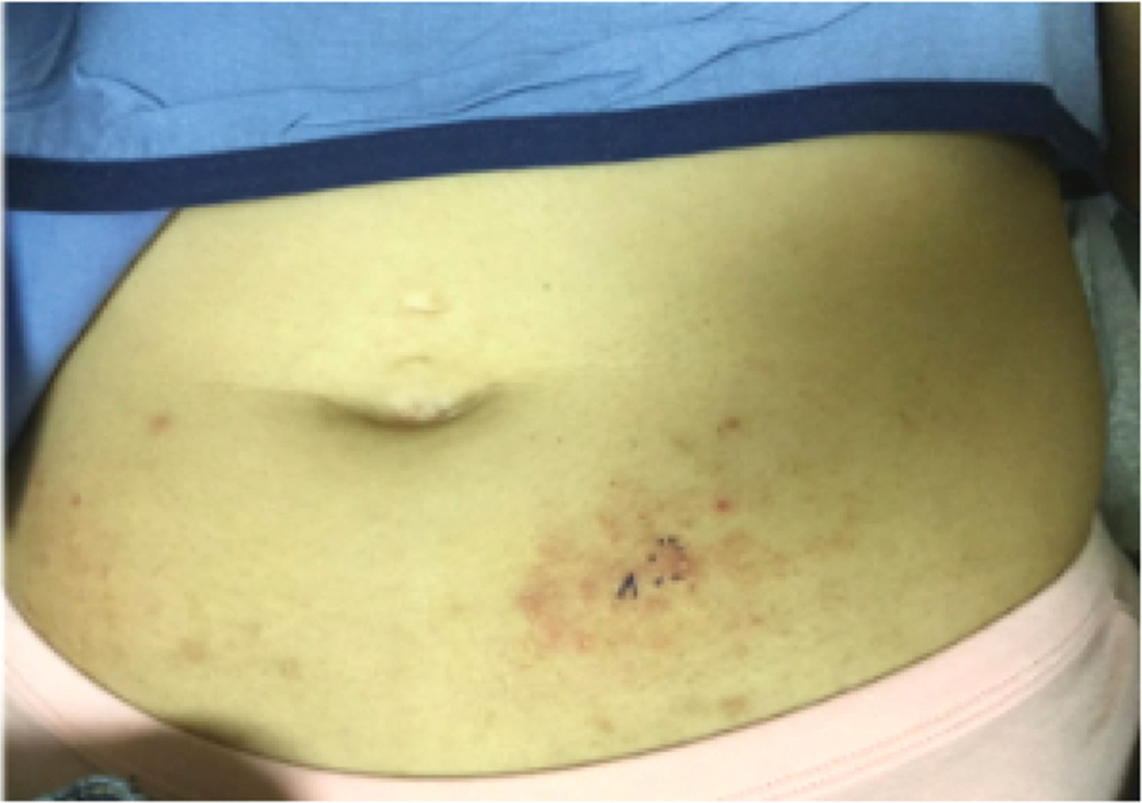
ACD can present in a myriad of ways. Erythema, scaling, edema, vesicles (especially in the acute phase), and hyperpigmentation can all be features ( Fig. 7.3 ). Distinguishing characteristics include a linear or geometric configuration. In the patient with AD, ACD can present in areas not previously affected (such as the face) or as treatment-resistant areas.
Several key differences exist to distinguish ICD and ACD ( Table 7.2 ). Key clinical differences are that ACD requires sensitization to a particular allergen and can be diagnosed with patch testing.
| Features | Allergic contact dermatitis | Irritant contact dermatitis |
|---|---|---|
| Causative agents | Low molecular weight haptens, lipid soluble | Acids, alkalis, surfactants, solvents, oxidants, enzymes |
| Genetic predisposition | More important | Less important |
| Sensitization and lag period | Necessary | Not necessary |
| Trigger | Interaction of antigen with primed T cells | Damage to keratinocytes |
| Cytokine release | Large | Moderate |
| T-cell activation | Early | Late |
| Mast cell activation | Some | Some |
| Langerhans cells | Increased | Decreased |
Related posts:


Stay updated, free articles. Join our Telegram channel

Full access? Get Clinical Tree


