Key points
- •
Issues with wound healing in patients with atopic dermatitis (AD) relate to innate inability to repair the barrier, defective angiogenesis, abnormal microbial colonization, and a prolonged inflammatory phase.
- •
Patients with AD are at a higher risk for hypersensitivity reactions such as allergic contact dermatitis to various dressings and topical medications.
- •
General measures that may help in the treatment of both AD and wound include moisturizing to restore barrier, hypochlorous acid, and adequate nutrition.
- •
Complementary and alternative treatments emerge as promising tools to treat wounds in AD patients.
Introduction
A critical function of the skin, a protein layer, is to provide a physiologic and protective barrier to the external environment. It represents the primary interface between the body and the environment. In addition to preventing loss of moisture and electrolytes from the inside, the skin is a key element in the host defense system preventing the penetration of harmful stimuli—microbial organisms, chemical irritants, toxins, as well as solar and ionizing radiation—from the outside ( ). This physiologic barrier function is primarily carried out by two structures in the epidermis: the stratum corneum (SC) and intercellualr tight junctions (TJs).
As a barrier organ, the skin encounters a variety of antigens and environmental insults on a daily basis. When immune surveillance responds inappropriately to an otherwise harmless antigen and passes this information on to the adaptive system, various pathologic conditions can develop, such as autoimmunity, allergy, chronic inflammation, and delayed wound healing ( ).
Many inflammatory skin diseases, including atopic dermatitis (AD), are perpetuated by inappropriate and/or hyperactive immune responses under the circumstances of genetic predisposition, exposure to internal or external triggers, and disruption of barrier integrity—and their interactions. In this chapter we aim to delineate the pathophysiology of skin barrier disruption in AD and discuss methods of management to repair this defect.
Atopic dermatitis pathobiology: Relationship to wound care
Previously, our understanding of the pathobiology of AD consisted of three main schools of thought: skin barrier abnormalities (outside-in hypothesis), defects in immune response (inside-out hypothesis), and altered skin resident microbiota ( ). Increased transepidermal water loss has been detected in AD patients at birth ( ), and loss-of-function mutations in filaggrin—which encodes a protein indispensable for barrier formation and function—were found in over 40% of AD patients ( ). However, recent findings showed that abnormal immunity can also lead to a loss of barrier function through downregulation of filaggrin in patients without the filaggrin gene mutation ( ).
From an immunologic perspective, AD begins with an acute phase with excessive T-helper type 2 (Th2), Th22, and Th17 cell activation ( ). Transition to the chronic phase is marked by the onset of Th1 cell activation and the continued presence of Th2 and Th22 cells ( ). Allergens and microbes such as Staphylococcus aureus and Malassezia spp. have been reported to trigger AD by inducing Th2 cytokine production ( ). Moreover, Th2 cytokine responses can be induced by keratinocytes that produce thymic stromal lymphopoietin (TSLP), which translates to Th2 polarization and production of interleukin-4 (IL4) and IL13 ( ). Their effectors—IL25 and IL33—synergize with to further promote Th2 cytokine responses ( ).
All of these processes result in two AD hallmarks: reduction in barrier proteins such as loricrin and filaggrin, and reduction in lipids (free fatty acids and ceramides) ( ), translating into a defective epidermal permeability barrier. IL22 also promotes hyperplasia, downregulates terminal differentiation, synergizes with IL17 to induce the S100 genes, and induces keratinocyte production of type 2 cytokines ( ). Whether skin inflammation in AD is initiated by skin barrier dysfunction or by immune deregulation is still in debate. Additionally, innate lymphoid cells 2 (ILC-2) were recently implicated in AD through the production of IL4, IL5, IL13, and IL6 ( ). The subject of skin barrier is further discussed in Chapter 5, Chapter 11 .
Wound healing biology
Wound healing is an intricate process regulated by sequential and overlapping phases, including hemostasis, inflammation, proliferation, and remodeling. Under physiologic conditions this process is highly efficient; however, under pathologic conditions chronic wounds may develop ( ).
Upon injury, hemostasis initiates fibrin clot formation, which serves as a scaffold and a source of growth factors and chemokines that recruit inflammatory cells to migrate into the wound bed ( ). These inflammatory cells release growth factors and IL1, IL6, tumor necrosis factor-α (TNF-α), platelet-derived growth factor (PDGF), and fibroblast growth factor-2 (FGF-2) ( ).
Fibroblasts proliferate and produce provisional matrix over which keratinocytes migrate from the wound margins ( ). Additionally, rearrangement of integrins and cytoskeletal components takes place and there is expression of proteases such as matrix metalloproteinase (MMP), which allows keratinocyte migration ( ). This process is then followed by keratinocyte proliferation. Aberrant execution of any of these steps results in healing impairment and development of chronic wounds. Notably, this model is predominantly studied in wounds that feature both epidermal and at least partial dermal injuries.
Biologic underpinning for wound healing impairment in atopic dermatitis
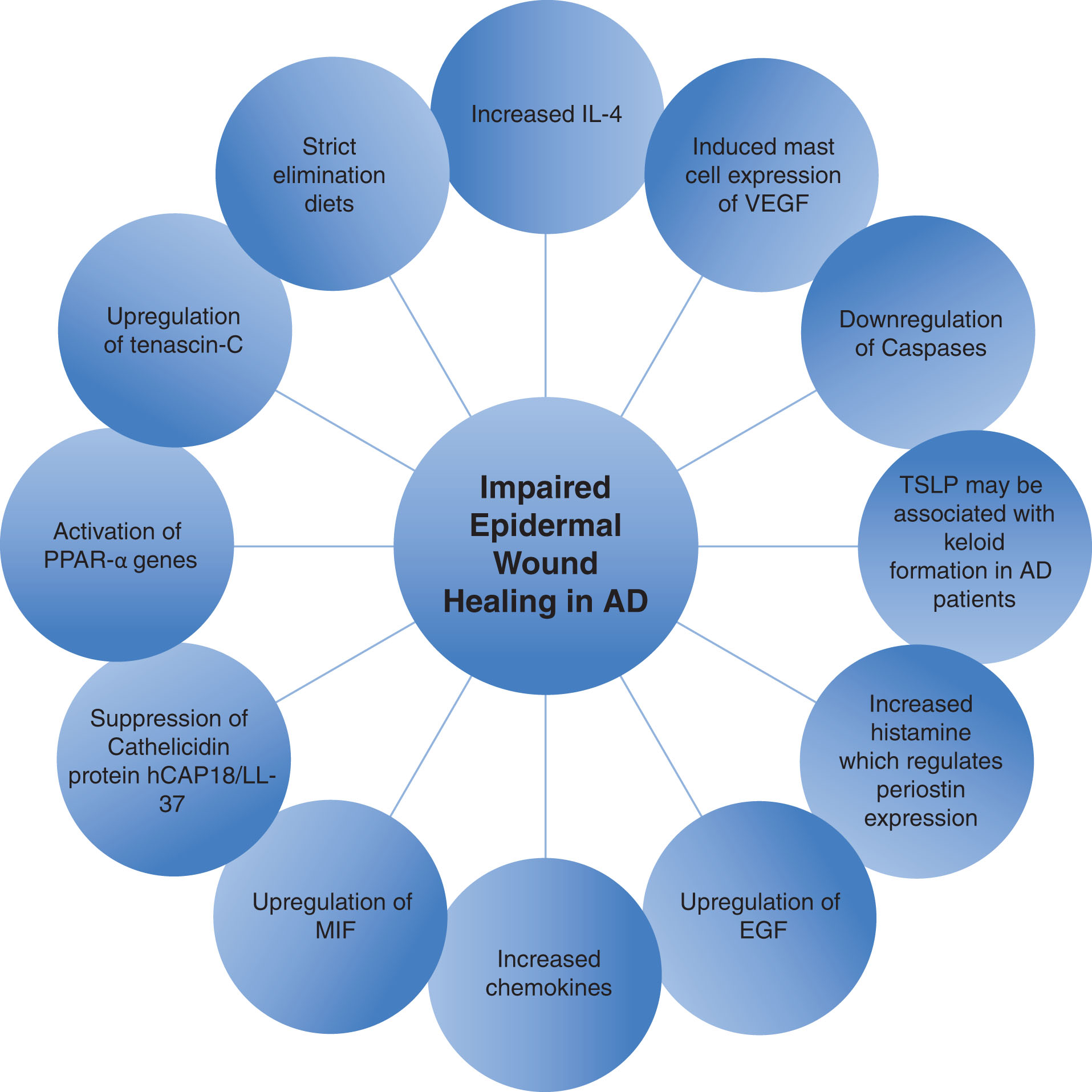
The wound healing process in AD patients is characterized by a prolonged inflammatory phase. This leads to epidermal hyperplasia and tissue remodeling, resulting in the development of spongiosis ( ). A proposed mechanism underlying the impaired wound healing in AD is depicted in Fig. 21.1 .
Wounds in AD are caused mostly by epidermal injury and disruption, rather than dermal alterations. The wound healing process in AD can rarely cause issues such as postsurgical complications. Issues with AD wound healing in general relate to innate inability to repair the barrier, defective angiogenesis, and abnormal microbial colonization, among others. These challenges are subtle but may further impact the patients’ quality of life. Therefore clinicians should pay attention to wound and exudate care in patients with AD.
Various genes and molecules may delay wound healing in AD leading to the phenotypic changes noted earlier. In this section preclinical evidence is presented in support of the role of the following elements in the potential healing impairment in AD: IL4, caspases, hyperforin, TSLP, histamine, epidermal growth factor (EGF), various chemokines, vascular endothelial growth factor (VEGF), human cathelicidin protein 18 (hCAP18)/LL-37, peroxisome proliferator-activator receptor α (PPAR-α), tenascin-C, and altered microbiome environment.
Interleukin-4
IL4 is a Th2 cytokine secreted by activated T lymphocytes and innate immune cells that affects keratinocyte differentiation ( ). IL4 reduces keratinocyte expression of epidermal differentiation complex genes, decreases the expression of defensins, alters the expression of keratins, and increases the production of the chemokine CCL26 (eotaxin 3) ( ).
Importantly, IL4 is a theraputic target in patients with AD. In clinical trials of subjects with moderate to severe AD, disease scores improved after anti-IL4Rα human antagonist antibody treatment ( ).
In an IL4 transgenic (Tg) mouse model that presents an AD-like phenotype ( ), IL4 Tg mice had delayed wound closure and reepithelialization. Additionally, the wounds showed an increased expression of proinflammatory cytokines/chemokines, elevated infiltration of neutrophils, macrophages, CD3+ lymphocytes and epidermal dendritic T lymphocytes, and larger amounts of granulation tissue ( ).
found that IL4 represses the expression of fibronectin by keratinocytes, leading to a delay in wound repair. Moreover, IL4 modulated fibronectin in an in vivo model, improving wound repair ( ). Importanly, acute IL4 stimulation resulted in modulating expression of genes involved in cell growth and survival and diminished the expression of genes involved in cell adhesion. Chronic stimulation with IL4 resulted in increased expression of genes involved in cell projection and adhesion and decreased expression of genes participating in response to wounding and defense. Taken together, IL4 appears to stimulate a disorganized epithelial structure and contribute to an increased susceptibility to microorganism invasion, which are also characteristics of AD lesions ( ).
Vascular endothelial growth factors/angiogenesis
Angiogenesis is the growth of new blood vessels from preexisting vessels and occurs physiologically in wound healing. Lymphangiogenesis can be activated in inflammation and tumor metastasis ( ). AD as well as other inflammatory skin disorders, including psoriasis, are characterized by altered angiogenesis, lymphangiogenesis, or both ( ). Mast cells in AD lesions may stimulate angiogenesis via the release of proangiogenic factors ( ). It has been established that plasma and skin concentrations of VEGF are increased in AD ( ). A possible association between polymorphisms of the VEGF/VEGFR genes and AD has been established ( ). Moreover, IL9/IL9 receptor genes are overexpressed in AD, and IL9 induces VEGF secretion from mast cells ( ).
Interestingly, mast cells express corticotropin-releasing hormone receptors, which leads to the secretion of VEGF ( ). It is well known that stress worsens AD, and stress stimulates the release of corticotropin-releasing hormone and corticotropin-releasing hormone-induced mast cell-derived VEGF ( ).
Semaphorins are a large family of secreted and membrane-associated proteins associated with axon guidance. They have been associated with angiogenesis ( ). Semaphorin IIIA was shown to improve skin lesions in the NC/Nga model of AD through the modulation of angiogenesis ( ). Skin microcirculation in AD is further discussed in Chapter 12 .
Caspases 1 and 8
Caspases (cysteine-aspartic proteases, cysteine aspartases, or cysteine-dependent aspartate-directed proteases) are protease enzymes playing essential roles in programmed cell death. Caspase-1, also known as IL1-converting enzyme, proteolytically cleaves the precursors of IL1β and IL18 and forms part of inflammasome. Caspase-8 is involved in the programmed cell death induced by Fas and various apoptotic stimuli.
Caspases play important roles in both chronic wound healing response and the pathogenesis of AD, therefore better understanding of caspases’ function may ellucidate the wound response in AD ( ). Downregulation of epidermal caspase-8 recapitulates a wound even in the absence of trauma to the skin ( ). Moreover, in humans, systemic deficiency of caspase-8 causes eczema ( ).
Christopher et al. found that deletion of epidermal caspase-8 in a mouse model shared many features of AD, including acanthosis, scaling, elevated serum immunoglobulins, mast cell infiltration, and spongiosis ( ). A C8 knockout (KO) mouse model presented with maximal levels of Th2 markers relative in young mice, which waned as the animal aged. These genes included IL4, IL5, IL9, and IL13 and chemokine receptor 4 ( ). Overexpression of caspase-1 in the epidermis has been reported to phenotypically mimic caspase-8 KO mice ( ).
Caspase-1 was found to be upregulated in both wounds and the caspase-8 KO mouse. Caspase-1 plays an important role in the inflammasome conformation and inflammatory response stimulating epithelial stem cell proliferation and tissue remodeling (through TSLP production and p38 mitogen-activated protein kinase activation) ( ).
There are various similarities between the wound signature in AD and the wound healing response seen in caspase-8 deficiency. Caspase-8 KO mouse models have many features of AD, including acanthosis, scaling, elevated serum immunoglobulins, mast cell infiltration, and spongiosis ( ). concluded that an unknown factor that normally would induce the expression of epidermal caspase-8 following wound closure may be impaired in AD, resulting in the sustained repression of caspase-8.
Thymic stromal lymphopoietin
TSLP is a Th2 cytokine that recently has been described in the pathogenesis of fibrosis. TSLP roles in systemic sclerosis ( ), idiopathic pulmonary fibroblasts ( ), and IL13-induced AD ( ) have been described.
Subcutaneous injection of TSLP-induced skin fibrosis in a murine model ( ) and in the transgenic IL13 AD mouse model, the neutralization of TSLP, reduced collagen deposition and the inflammatory response ( ). proposed that TSLP is an inducer of extracellular matrix synthesis and activator of the CXCR4/SDF-1 axis in skin fibrosis, especially in keloid pathogenesis. Although keloids in AD patients are rare, the data suggest that its formation in those patients is mediated through TSLP; however, more research needs to be done to establish a relationship in human pathobiology.
Role of the microbiome
proposed that Th2 cytokines, as well as staphylococcal lipoteichoic acid (LTA), may contribute to delayed wound healing and pathology of AD by deregulating MMP production and altering cell migration. It is well known that skin of AD patients is chronically colonized with S. aureus ( ). Moreover, MMPs are required for normal migration and wound closure, and overexpression is observed in chronic wounds. MMP-1, -9, and -10 are expressed at the wound edges and are required for keratinocyte migration ( ).
Th2 cytokines are overexpressed in AD, and inhibit MMP expression and keratinocyte migration. Overexpression of MMPs induced by LTA may contribute to skin disease as well. Recent studies have described MMP expression in AD skin ( ).
Periostin and histamine
Mast cells contribute to the healing process and mast cell–deficient mice have impaired wound healing ( ). Histidine decarboxylase KO mice resulted in delayed would healing, and exogenous histamine administration restored the normal healing process ( ). Interestingly, H 1 R antagonists (but not H 2 R antagonists) were found to reduce wound closure in mice ( ), and H 1 R antagonist inhibits the synthesis of collagen 1 by dermal fibroblasts ( ). Moreover, there is an increase in mast cell number in fibrotic tissues, including scleroderma skin and keloids ( ). cultured dermal fibroblasts derived from wild type (WT) or periostin KO (PN[-/-]) mice and stimulated them with histamine. They found increased expression of colagen 1 mRNA after 48 hours of coincubation with histamine and concluded that histamine-mediated tissue remodeling may require the expression of periostin as a second messenger to elicit tissue remodeling ( ). studied how periostin regulates the proliferation and differentiation of keratinocytes. Fibroblasts secrete periostin, which stimulated IL1α-induced IL6 production and activated IL6 production through the nuclear factor (NF)–κB pathway ( ). Histamine is a major proinflammatory mediator that is increased in AD patients, therefore understanding the histamine-periostin pathway may explain the wound healing physiology in these patients.
Epidermal growth factor
The EGF pathway is essential for keratinocyte proliferation and differentiation and for proper wound healing ( ). Upregulation of this pathway has been found in chronic inflammatory skin disorders, including psoriasis and AD ( ).
described that EGF treatment in human keratinocytes and HaCat keratinocytes resulted in a decrease of IL17A and IL6 expression in the skin. In vivo, EGF may attenuate IL6 production in the skin and reduce local Th17 response. This suggests a protective role for EGF in AD and a new role for EGF in modulating IL17 responses in the skin.
Chemokines
Fibrogenic and fibrosis-associated cytokines (TGF-β1, IL11, and IL17) as well as the chemokine superfamily have been shown to regulate atopic skin inflammation ( ). CCL11, CCL17, CCL22, CCL26, CCL27, and CX 3 CL1 interactions are directly correlated with disease activity and severity ( ).
identified chemokine-mediated pathways regulating tissue remodeling in AD. They analyzed the chemokine receptor expression of human dermal fibroblasts and showed that among the CCR3 ligands, the chemokine CCL26 is the most highly expressed in AD, and that the keratinocytes were the predominant source ( ).
Macrophage migration inhibitory factor
Macrophage migration inhibitory factor (MIF) is a lymphokine involved in cell-mediated immunity, immunoregulation, and inflammation and plays a role in the regulation of macrophage function in host defense through the suppression of antiinflammatory effects of glucocorticoids. MIF has been identified as a key effector of estrogen effetcs on wound repair ( ). Moreover, endothelial MIF expression is upregulated in AD ( ). Therefore this cytokine may play a role in the healing process of AD skin.
Cathelicidin protein hCAP18/LL-37
hCAP18/LL-37 is an endogenous antimicrobial peptide (AMP) that plays a crucial role in barrier protection. Wounding of the skin induces hCAP18/LL-37 expression in normal skin ( ). In AD lesions, the expression of hCAP18 mRNA was suppressed following wounding. It is hypothesized that the abundant inflammatory response in AD neutralizes the expected induction of hCAP18 ( ). Inability to upregulate hCAP18 in eczema following injury is likely to affect antimicrobial protection and tissue repair in AD.
Peroxisome proliferator-activated receptors
PPARs are ligand-activated transcription factors that belong to the nuclear receptor superfamily. The PPAR family genes are activated by various fatty acid metabolites produced during inflammatory responses and regulate other signaling pathways, including NFκB, through ligand-dependent transrepression ( ). Skin injury was found to stimulate PPAR-α reexpression in keratinocytes during the healing process, and PPAR-α −/− mice have a transient delay in initial wound healing ( ). Interesntingly, PPAR-α–deficient mice display many features similar to AD, including increased epidermal thickening, dermal inflammatory cell infiltrate, and other atopic features such as lung inflammation and hyperactivity ( ). Understanding the PPAR-α pathway and its regulators may help scientist to better discern the healing process in AD patients.
Tenascin-C
Tenascin-C is part of the tenascins family, which are expressed in human tissues during organogenesis as well as embryogenesis and wound healing ( ). Upregulation of the tenascin-C expression was found to be specific to AD lesions, and tenascin-C was markedly upregulated by IL4 and IL13, and moderately upregulated by interferón-gamma ( ). Human keratinocytes are a major source of tenascin-C, especially during the early phase of wound healing ( ). The high expression of tenascin-C in the skin lesion of AD patients might reflect the process for remodeling or overrepairing the inflammatory lesion by keratinocytes. Taken together, these pathways that may be dysregulated in AD may help to understand some of the phenotypical observations that have been made pertaining to wound healing impairment in AD. However, better translational evidence is needed to provide a comprehensive model for wound healing impairment in AD.
Surgical complications in atopic dermatitis
AD management is often complicated by colonization of S. aureus ( ). Colonization with this bacterium is a risk factor for certain infections such as skin cellulitis and osteomyelitis. Thus atopic patients are at increased risk of infection when facing surgical procedures that break the protective skin barrier ( ).
Among patients undergoing cardiac surgery, 70% to 80% of wounds in those with mediastinitis following conventional median sternotomy have positive cultures for S. aureus ( ). Thus it has been suggested that AD patients undergoing sternotomy are at an increased risk of mediastinitis given the S. aureus burden associated with AD and the infectious risk this poses postoperatively ( ). Of note, no postoperative mediastinitis has been observed in AD patients who underwent thoracotomy, suggesting that may be a more favorable surgical approach in patients with AD requiring cardiac intervention ( ).
AD has additionally been found to be an independent risk factor for surgical site infection following anterior cruciate ligament reconstruction (ACLR) ( ). This is thought to be due to the aforementioned susceptibility of S. aureus colonization of atopic skin lesions ( ). Rates of surgical site infection following ACLR are low, ranging from 0.14% to approximately 2.32%. Nonetheless, this rare complication has the potential to cause significant repercussions, as it may require further surgical intervention or even graft removal ( ).
Wound care in atopic dermatitis
General measures for patients with wounds and atopic dermatitis
An impaired barrier predisposes to skin injuries, which can progress to nonhealing wounds. The pillar of wound prevention is barrier repair through moisturization. Moisturizers have broad utility within dermatology, especially for AD, since they help alleviate symptoms and improve barrier function ( ). Therefore moisturizers represent a pillar in wound prevention.
Microorganisms colonize human skin and resultant wounds as well as the mouth, digestive, and reproductive tracts. As a result, the number of bacteria colonizing the human body far exceeds the number of our own cells ( ). Moreover, it is well known that AD patients are chronically colonized with S. aureus ( ), and the presence of S. aureus is correlated with flares. Failure to heal of wounds has been associated with biofilms, as they have been shown to prevent wound contracture and epithelialization, disrupt the host immune response, and induce chronic inflammation ( ).
Hypochlorous acid (HOCl) exhibits antiinflammatory effects through blockade of NFκB signaling in keratinocytes ( ) and decreases sensory nerve transmission by inhibiting intracellular calcium signaling, reducing pain and itch symptoms ( ). HOCl also reduces serum levels of IL1β, IL6, and TNF-α ( ) proinflammatory cytokines elevated in skin and serum ( ). Moreover, HOCl decreases sensory nerve transmission by inhibiting intracellular calcium signaling, thus reducing pain and itch symptoms ( ). found that stabilized HOCl solution had dose-dependent favorable effects on fibroblast and keratinocyte migration. Due to the antiinflammatory properties, HCOl is a useful solution to be used in patients with AD and chronic wounds.
Drug delivery vehicles
Various drug delivery systems may be utilized to treat chronic wounds. In patients with AD and wounds, clinicians have an opportunity to leverage the vehicles to benefit patients with AD. The most common examples are ointments and hydrogels, which can improve the impaired barrier for these patients.
In general, the barrier function of the skin poses a drug delivery challenge. Optimal drug delivery systems should feature nontoxicity, good biocompatibility and biodegradability, drug stability, and optimized drug absorption and bioavailability ( ). Relative to other drug delivery vehicles commonly used in AD such as ointments, hydrogels have demonstrated bioequivalence, higher ease of use, and patient satisfaction; this may prove to be a useful tool in wound management in AD. Hydrogels are composed of a network of three-dimensional hydrophilic polymers that can withstand swelling without losing their structural integrity. They can be used as drug delivery systems given their biocompatibility, flexibility, and ability to hold high water content ( ).
A hydrogel formulation containing desonide has been found easy to use, quick to disappear, comfortable, soothing, nondrying, and not greasy or shiny on the skin by AD patients. Desonide hydrogel 0.05% was found to be equally effective to desonide ointment 0.05% at reducing AD symptoms. However, when compared to desonide ointment, desonide hydrogel has higher rates of patient satisfaction for absorbability and lack of greasiness. This hydrogel preparation has consistently been rated higher by patients than other vehicles, has higher rates of patient adherence, and has been found to be efficacious in a shorter time frame than other vehicles. Furthermore, hydrogel formulations improved skin hydration when compared to a moisturizing lotion ( ).
Dressings and textiles
The treatment of chronic wounds typically involves the application of various dressings, and often these products contain active medications. Additionally, people with AD may exhibit increased percutaneous drug absorption due to their skin’s impaired barrier function. Therefore patients with AD are at a higher risk for hypersensitivity reactions such as allergic contact dermatitis.
Many dressings when in contact with fluids, including wound exudate, change their composition into hydrogels. This property can help hydrate and improve the defective barrier seen in patients with AD.
Wound care regime in AD may include wet wrap therapy, which helps to rehydrate and soothe the skin. Fabric wraps are to be soaked in warm water and applied to the affected areas of the skin. A dry layer is then applied over the wet one, such as pajamas, which can be worn as a top layer to maintain the wet dressings undisturbed. This process is best done after the patient has bathed and moisturized the skin. The wet wraps can be left on overnight ( ).
Hydrocolloid dressings containing ceramides may have a positive therapeutic effect in AD. The SC functions as a watertight protective barrier from external insults, partially via composition of various lipids. Ceramide, a sphingolipid, is a major component of the lipid bilayer, accounting for almost half of the lipids in the SC by weight. This sphingolipid is intimately involved with the skin’s barrier function ( ). Low levels of ceramides in the SC are associated with the dry skin and barrier-disruption exhibited in AD. Furthermore, insufficient levels of ceramides can lead to increased susceptibility to infection, increased transepidermal water loss (TEWL), and skin inflammation. These deleterious processes may eventually trigger erosions following excoriations and other minor trauma, which would subsequently be difficult to heal given the skin’s disrupted barrier function ( ).
Topical ceramide can be utilized to improve skin barrier function ( ). More recently it was found to accelerate wound healing as well ( ). Hydrocolloid dressings containing 0.3% ceramide-2 showed reduction in wound size and greater regeneration of epidermal and dermal layers in mice models relative to control (thickness of regenerated epidermis/dermis was 3.27 times thicker, on average, for ceramide group compared to control group). Ceramide dressings also helped reduce the wound-related increase in TEWL, suggesting they may be of use in the management of wounds in AD ( ). Dressings and textiles specially coated with zinc may also have beneficial effects on the management of AD. Textiles that capitalize on zinc’s intrinsic antibacterial and antioxidative properties, such as Benevit Zink+, have demonstrated rapid improvement of AD symptom severity with regard to pruritus and sleep quality when utilized overnight ( ).
Cutaneous superinfection of atopic lesions with S. aureus plays a significant role in inflammation and exacerbations seen in AD. In fact, AD severity can be correlated with cutaneous S. aureus colonization ( ). Therefore treatment of AD and AD-associated wounds includes antimicrobial components in an effort to reduce the S. aureus burden, leading to a positive effect on the disease overall ( ).
Silver products have historically been used as antimicrobial wound dressings, given silver’s antiseptic properties and the relatively low drug resistance associated with it ( ). Newer developments show these antimicrobial effects are exhibited by silver-coated textiles as well, such as Padycare textiles (micromesh with 20% woven silver filaments). In vitro studies of these textiles demonstrate their ability to significantly reduce the bioburden of S. aureus , Pseudomonas aeruginosa , and Candida albicans ( ). Importantly, the antimicrobial efficacy of silver-impregnated textiles is reduced with repetitive washing (from >99% reduction in Escherichia coli growth to 81.6% [deionized water only] to 75.1% [deionized water and detergent] reduction in E. coli growth) ( ).
Chitosan functionalized textiles have similar antibacterial properties, particularly against S. aureus , Methicillin-resistant S. aureus (MRSA), and vancomycin-resistant S. aureus (VRSA), making it a prime candidate for possible management of wounds in AD ( ). Chitosan is a cationic biopolymer with unique biologic properties that is found widely in nature, composed of fungi and the exoskeletons of insects as well as marine invertebrates. As a free molecule, chitosan inhibits bacterial growth and adhesion. Chitosan appears to bypass the antibiotic resistance mechanisms typical to the Staphylococcus spp., allowing it to have significant bactericidal activity on MRSA and VRSA. In vivo studies with AD patients demonstrated that cotton-based clothing with a chitosan antimicrobial finish aided in modulating the cutaneous bacterial profile by reducing S. aureus counts and increasing coagulase-negative Staphylococci . Therefore chitosan functionalized textiles may be a viable alternative to traditional antibiotic regimes in AD wound management ( ).
Other textiles have been implemented in AD management, including silk. Silk inherently reduces irritation in AD given its unique smooth texture ( ). Additionally, products such as Dermasilk combine the smoothness of silk with an antimicrobial coating, making it an optimal textile for AD patients with wounds at risk for infection ( ). Nonetheless, it is important to consider costs when recommending such specialty dressings and textiles, as they may be high.
The role of nutrition in wound care
The role of nutrition in wound healing of eczematous patients is a concept that has not been studied directly. However, with the current knowledge of the role of nutrition in the pathogenesis of AD and to promote wound healing, clinicians should consider educating patients about the importance of (1) adequate nutrition and clarifying the common misconceptions about food allergies, and (2) the role of micronutrients, polyunsaturated fatty acid (PUFA), and healthy microbiome to attenuate inflammation and to promote wound healing in AD.
Adequate nutrition
Evaluating AD patients’ nutritional status and ensuring sufficient calories from a balanced diet of carbohydrates, fats, and protein, and supplementing protein, fluid, and vitamins A and C, is needed for adequate wound healing. Strict elimination diets are more likely to lead to nutritional deficiencies ( ) and delayed wound healing ( ). Therefore, to prevent nutritional deficiency and the subsequent delayed wound healing in AD patients, it is important to clarify the common misconceptions associated with food allergies. Nutrition is further discussed in Chapter 8 .
Vitamins and minerals
While various vitamin and mineral deficiencies have been shown to play a role in the pathogenesis of AD and wound healing, the evidence for their use in the treatment or prevention of AD in pediatric and adult patients has been unconvincing ( ).
Zinc is an essential cofactor for metalloenzymes involved in cellular function. Deficiency of zinc has been correlated with impaired wound healing ( ), but there is no proven benefit of oral zinc supplementation in AD treatment. Zinc deficiency disturbs all phases of wound healing. In the inflammatory phase, there is reduced immunity and augmented susceptibility to infections. In the proliferative phase, there is decreased collagen synthesis and tensile strength. Finally, in the remodeling phase, there is a diminishing of fibroblast proliferation, collagen synthesis, and epithelialization ( ).
Oxidative stress has been proposed as another pathophysiologic factor contributing to AD, therefore suppression of oxidative stress via the supplementation of antioxidants may be an effective treatment strategy. Selenium is strongly involved in antioxidant functions; however, its direct wounding healing role in still unclear ( ).
Several vitamins, including A, C, and E, are known to protect cells against free radical damage and have been hypothesized to play a possible role in the prevention and management of AD ( ). Various mechanisms of the role of vitamin D in AD have been proposed, including immunomodulation, reduced cell proliferation, and improved epidermal barrier function ( ). Cathelicidin is an AMP induced by vitamin D that promotes wound healing. Vitamin D and its receptor signaling control structural integrity of epithelial barriers with implications for wound healing ( ).
While recommendations for micronutrient supplementation are unknown in the treatment of AD and the specific roles micronutrients play in wound healing, dermatologists should remind their patients that maintaining a healthy, wholesome diet with less processed foods has been shown to benefit patients with AD.
Polyunsaturated fatty acids
For wound healing in AD patients, adequate PUFA dietary intake may help to rebuild the structural integrity of the damaged skin barrier and support synthesis of signaling molecules, which influence the inflammatory response in the skin ( ).
There are a multitude of ceramide species found in both the dermis and the epidermis. Biochemically, n-3 and n-6 are essential fatty acids that are particularly significant in maintaining a healthy skin barrier as they contribute to the formation of ceramides ( ). However, a deficiency is difficult to produce unless a fat-free diet is maintained.
Immune dysregulation plays an important role in the pathogenesis of AD and is needed for proper wound healing regulation of the immune system and attenuation of inflammation. There are diverse advantageous effects on the immune response that have been attributed to the long-chain n-3 fatty acids eicosapentaenoic acid (EPA) and docosahexaenoic acid (DHA) ( ). Through different mechanisms, these n-3 fatty acids have an inhibitory or regulatory effect on the immune response. One example is the regulatory effect of these n-3 fatty acids on antigen presentation and adhesion molecule expression, as part of the Th1 and Th2 response, as well as on proinflammatory cytokine and eicosanoid production ( ). Protectins and resolvins are n-3 PUFA-derived molecules, which play an important role in switching from initiation to the resolution phase of inflammation.
The biochemical effects of PUFAs are known to benefit the integrity of skin barrier by optimizing skin lipid biology and are known to attenuate multiple inflammatory cascades within the immune system.
Probiotics
Gut probiotics and commensals may moderate skin wound healing through effects on systemic immunity and enhanced nutrient absorption. The differences in gut microbiota between those with atopic diseases and controls have been described. AD patients have higher proportion of Clostridia, Clostridium difficile, E. coli , and S. aureus and decreased Bifidobacteria, Bacteroidetes , and Bacteroides in the gut microbiome than in healthy controls ( ).
Certain bacterial strains have the potential to attenuate skin inflammation when administrated systemically by changing the composition of gut microbiome. For example, a study found Lactobacillus paracasei modifies the composition of intestinal microbiota by increasing Bifidobacteria and Lactobacilli microbiota and also reinforces skin barrier and decreases skin sensitivity while significantly increasing Th1 cell-dependent immune responses ( ). Also, it was found that supplementation of probiotics such as Lactobacillus reuteri encourages an upregulation of antiinflammatory IL10, which induces Foxp3+ Treg lymphocytes to minimize tissue damage at the wound edge ( ).
Aside from manipulating the immune system, the gut microbiota and orally introduced probiotics may achieve improved absorption of nutrients essential for skin wound healing, especially vitamins, minerals, and cofactors for enzymes involved in tissue repair ( ). Additionally, several Lactobacilli spp., including Lactobacillus coryniformis and Lactobacillus rossiae , have been shown to produce vitamin B 12 , which is valuable for wound healing ( ). L. reuteri and Lactobacillus acidophilus were shown to increase absorption of dietary vitamin D and E, known to be important for wound healing ( ). Though a therapeutic potential of probiotic bacteria for wound healing in disorders exists, more definitive studies are needed to explore the benefit of probiotics in wound healing for AD patients. The microbiome and probiotics are further discussed in Chapter 6, Chapter 24 .
Complementary and alternative medicine in wound care
Complementary and alternative medicine (CAM) is commonly practiced throughout the world. In China, CAM represent up to 40% of all health care practice ( ). Up to 80% of populations in Asia and Africa utilize CAM as their primary form of medicine. Despite CAM-prevalent utilization around the world, there is a need of clinical research to elucidate mechanisms of action and validate its use.
Due to the worldwide prevalence of chronic wounds and their notorious difficulty in treatment, CAM regimens have been clinically evaluated for their efficacy. Moreover, there are an increasing number of trials studying CAM to treat patients with AD. Interestingly there is a growing interest in CAM as a primary, maintenance, or simultaneous treatment for AD ( ). Patients with AD and their family members may choose CAM as their primary approach to relieve their illness or at least to improve the duration and quality of symptomatic relief.
Standard treatment for chronic wounds and AD can be expensive, painful, and frustrating; moreover, it may be associated with various adverse effects. Among other widely practiced alternative wound healing methods and AD treatments, the topical application of honey, vitamins, and herbs as well as dietary modifications are some examples of CAM therapies with some evidence supporting their use in clinical studies. Additional CAM treatments are discussed in Chapter 26, Chapter 27 .
Curcumin
Turmeric (Curcuma longa) is a popular bright yellow spice used worldwide in traditional medicine and for cooking and dye. Curcumin is the active constituent of turmeric. The use of curcumin in medicine has been heavily studied, particularly with regard to its role in modulating inflammation ( ). In some Eastern countries, curcumin’s antiinflammatory effects are exploited as a part of AD management ( ).
Curcumin’s role in AD has also been described. Curcumin modulates protein kinase C-θ pathway in vitro, leading to the inhibition of T-cell activation ( ). In mice, it was also found to ameliorate ovalbumin-induced AD pathology through inhibition of TSLP, IL33, IL4, IL5, L13, IL31 and reduced STAT-6 phosphorylation ( ).
In mice, oral administration reduced the production of proinflammatory cytokines by keratinocytes, improving cutaneous signs of AD such as inflammation and dermoepidermal thickening ( ). Importantly, in humans, daily applications of a combination herbal extract cream (Herbavate) that contains C. longa was found to alleviate AD symtoms ( ).
Curcumin has been studied in wound healing. Animal model studies have demonstrated topical application of curcumin accelerates wound healing and reepithelialization rates when compared to controls ( ). Topical application of curcumin was found to have a more robust effect on wound healing when compared to oral administration. This is likely due to the higher availability of curcumin at the wound site when applied topically ( ). However, topical application may lead to yellowish or brown discoloration, and patients should be warned in advance about this potential side effect.
Curcumin is thought to play an active role in wound healing, particularly during the inflammatory and proliferative phases ( ). Curcumin has been observed to reduce inflammation through the suppression of TNF-α, an inflammatory mediator involved in the inflammatory phase of wound healing (see Introduction). In the proliferative phase, studies have shown that curcumin may increase hydroxyproline and collagen synthesis, accelerate the maturation of collagen fibers, and reduce the number of MMPs. Curcumin has also been shown to promote the differentiation of fibroblasts into myofibroblasts, thus aiding in wound contraction and resulting in a shorter epithelization period ( ).
Although promising for the treatment of both AD and wounds, curcumin has been associated with allergic contact dermatitis and has emerged as a contact allergen ( ). Historically, it was thought that patients with AD were less likely to develop contact dermatitis; however, more recent evidence has shown that patients with AD have similar if not higher rates of positive patch test results to common contact allergens ( ). Therefore caution needs to be taken when using topical curcumin for wounds in patients with AD due to possible increased risk of contact allergic dermatitis.
Glycerol
Glycerol is a trihydroxy alcohol widely used in dermatologic preparations. Topical glycerol application aids in increasing epidermal hydration, which is of utmost importance in the management of skin diseases characterized by xerosis and impaired epidermal barrier function, such as AD ( ). Glycerol additionally promotes the process of wound healing ( ) and has antiirritant and antimicrobial properties, among other functions. Gram-negative bacteria in particular appear to be more susceptible to glycerol than gram-positive bacteria ( ). Glycerol’s wound healing properties combined with its antiirritant, antimicrobial, and hydrating qualities suggest this topical agent may be of potential use in treating wounds in AD.
Hyperforin
Hyperforin is a major active constituent of St. John wort ( Hypericum perforatum L.) extract, a traditional herbal medicine with wound healing properties ( ) and potential antiinflammatory properties in AD ( ). proposed that hyperforin/2-hydroxylpropyl-beta-cyclodextrin (HPβCD) is a potent targeted therapeutic agent that can be used to promote epidermal wound healing and treat AD. They measured the stretch-induced Ca 2+ responses ex vivo in atopic skin and found that the Ca 2+ response was impaired in AD. The effect was recovered after the application of hyperforin/HPβCD, suggesting the therapeutic properties and potential to promote epidermal wound healing and treat AD ( ).
Summary
The wound healing process in AD patients is overall altered. Various genes and molecules have been described in atopic skin that may be involved in the delayed healing process. Issues with healing may relate to innate inability to repair the barrier, defective angiogenesis, and abnormal microbial colonization, among others. In general, these challenges are subtle but impact patients’ quality of life. Wound healing in AD can rarely cause issues such as postsurgical complications.
Special drug delivery vehicles commonly used in AD such as ointments and hydrogels prove to be useful tools in wound management in AD. Caution needs to be taken when considering special dressings and textiles in patients with AD due to the increased risk of development of allergic contact dermatitis.
Also, adequate nutrition is needed to address both AD and enhancement of skin healing properties. PUFA, healthy microbiome, and wholesome diet are needed to attenuate inflammation and to maintain a healthy SC. CAM interventions are receiving increased attention among physicians and researchers, and its role is being investigated for both wound healing and AD.
Further readings
Related posts:

Stay updated, free articles. Join our Telegram channel

Full access? Get Clinical Tree


