Key points
- •
Although the original term atopy provides a general framework of understanding about allergic clinical conditions, this big umbrella now includes a group of patients who have heterogeneous clinical and laboratory-defined characteristics. Similarly, atopic dermatitis , the skin atopy, characterizes a group of patients with heterogeneous clinical and laboratory-based findings.
- •
Since the immune milieu in children is a dynamic system, atopic dermatitis manifested in childhood may have a different pathomechanism than those occurring in adulthood.
- •
The intrinsic subtype of atopic dermatitis, with its absence of an elevation in serum immunoglobulin E (IgE), may differ from the extrinsic subset of atopic dermatitis in pathophysiology, as IgE is a direct product of the important Th2 cytokine interleukin-4 (IL4).
- •
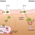
The atopic dermatitis patients with filaggrin gene mutation may manifest skin disease differently than those patients with intact skin barrier proteins, since skin barrier defect favors easy entry of pathogens and allergens, leading to triggering of cutaneous inflammatory processes.
- •
The potential pathophysiologic differences of onset age, IgE, and skin barrier protein mutation, together with varied clinical responses to targeted immune modulators, point to the need for redefining atopic dermatitis or for defining specific subsets according to their verified pathomechanisms. Refining atopy will facilitate more accurate and personalized management of atopic dermatitis.
Introduction
In this chapter, we intend to discuss redefining the term atopy . Before we begin the process, however, we should first clearly delineate the meaning of the original term. Thus we first ask, What is atopy? and What defines atopy?
According to Merriam-Webster Dictionary online, atopy (2019) is “a genetic disposition to develop an allergic reaction (such as allergic rhinitis or asthma) and produce elevated levels of IgE upon exposure to an environmental antigen and especially one inhaled or digested.”
Another definition of atopy provided by is “a personal and/or familial tendency, usually in childhood or adolescence, to become sensitized and produce IgE antibodies in response to ordinary exposure to allergens, usually proteins.”
The , an educational and training organization of nearly 7000 members of allergists and immunologists and a trusted information source for patients, states atopy is “the genetic tendency to develop allergic diseases such as allergic rhinitis, asthma and atopic dermatitis (eczema). Atopy is typically associated with heightened immune responses to common allergens, especially inhaled allergens and food allergens”.
Historically, atopy was first described by Coca and Cooke in their article on the classification of hypersensitive phenomena. The term as they introduced it is derived from the Greek words a and topos , meaning “without” and “place,” respectively. They aimed to designate a terminology place for disorders such as hay fever and asthma ( ). While the abovementioned definitions provide a general framework of understanding atopy, detailed evidence, as delineated later in this chapter, points to a need to redefine or to subdivide this diverse group of patients who were currently categorized in the single and large disease entity of atopy. These challenging data include heterogeneous clinical manifestations, existence and nonexistence of skin barrier protein defect, presence and absence of elevated levels of serum IgE, different degrees of altered immune milieu, and favorable and unfavorable clinical responses to targeted immunomodulatory therapy. In this chapter we provide evidence to argue for such a need for redefinition or subdivision, with the focus on skin perspective.
Clear definition of atopic dermatitis
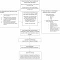
The current lack of uniform clinical definition and objective test that could unequivocally confirm the diagnosis of atopic dermatitis has led to significant differences in determination of disease prevalence, performance of prediction models, and risk factors ( ). The diagnostic criteria proposed by was deemed too complicated, and other diagnostic criteria were used in different geographic locations ( ). Since diagnostic criteria define disease entity, the effect of imperfect definition could result in misleading laboratory and clinical research data.
Childhood-onset versus adult-onset subsets
The immune system in childhood has many differences compared to that in adulthood ( ). First, some immune components are not fully mature during childhood. In particular, many changes occur during the first year of life ( ). Another difference is that adulthood, compared to childhood, has encountered more immune challenges and developed a lot more immune memories to these challenges, whether they are pathogenic or allergic in nature. As a result, the proportion of antigen-encountered lymphocytes becomes expanded in adults compared to that in childhood, which has a larger percent of naïve lymphocytes ( ). In addition, we must consider a naturally occurring immune cell “aging” process that renders immune cells more “permissive” later in adult life ( ). Immunologic parameter studies also point to a dynamic immune milieu occurring in infancy and childhood ( ). Some known differences between the childhood and adult immune milieu are depicted here. Newborns predominantly rely on maternal circulation-containing antibodies for their immune defense, but over the first few months of life their thymus pours out substantial amounts of T cells, which become various subsets over the next few years ( ). The CD4+ and CD8+ T cells in the peripheral blood increase from childhood to adulthood with an interesting decrease of B cells ( ). The reduction of peripheral B cells in adulthood may simply reflect a greater entry of those cells from peripheral blood into the solid lymphoid organs ( ). On the contrary, monocytes, NK cells, and regulatory T cells are decreasing from early childhood to adulthood ( ). The differences between childhood and adult immune milieu are manifested not only in cell number but also in cell function. The innate immunity in the early postnatal period, for example, is represented by NK cells predominantly in cytokine-producing mode rather than in cytotoxic activities ( ). Responses to toll-like receptor stimulation are lower early in life ( ). IL10, one of the Th2 cytokines important in atopic dermatitis development, is higher in early infancy. Interestingly, the IL10 level decreases to a level below the adult’s during infancy but then increases again until it reaches adult level ( ). So in theory, immune-mediated diseases that have onset during childhood differ from those that have onset during adulthood in terms of the immune system’s ability to handle certain challenges and how the immune system responds to challenges, at least from an immunologic perspective. Even if childhood atopic dermatitis persists into adulthood, the different adult immune milieu could still modify the disease to some extent. Therefore appropriate treatments for childhood-onset disease may need to tailor-suit the immature and dynamic nature of immune milieu in the childhood, and the same for the adult-onset disease.
Intrinsic versus extrinsic subsets
Although, historically, all patients affected by atopic dermatitis are considered uniformly extrinsic, with a common finding of elevated serum IgE level, subsequently the concept of intrinsic atopic dermatitis, which characterizes a subgroup of patients who do not have abnormally high total serum IgE level or antigen-specific IgE, emerges ( ). The robustness of this intrinsic form of atopic dermatitis is unsettled. While some academicians have named this intrinsic form of disease “nonallergic atopic dermatitis” or “atopiform dermatitis,” other academic physicians did not utilize the same criteria to define this subtype of dermatitis in their clinical investigations ( ). Some academic physicians argued that this intrinsic form of skin inflammation may not necessarily be atopic at all and that the term atopiform dermatitis would be more appropriate since atopiform will not indicate the disease as atopic or atopy, per se ( ).
Since the Th2 cytokine IL4 is a prerequisite for IgE production in humans and a high IgE level could point to an upregulation of IL4, patients with intrinsic atopic dermatitis and a normal level of IgE may not have a prominently altered immune system that manifests with IL4 upregulation ( ). So in theory, the patients affected by intrinsic atopic dermatitis may have had no altered internal immune milieu or have an altered immune milieu other than IL4 upregulation. From the clinical data, the intrinsic patients are primarily female and are the significant majority in infant atopic dermatitis ( ). On the genetic level, a higher percentage of patients with extrinsic atopic dermatitis have IL4/IL13 receptor polymorphism, whereas a higher percentage of patients with intrinsic counterpart have β 2 -adrenergic receptor polymorphism, probably implicating weakened adrenergic responses in intrinsic patients ( ). In a study completed in 2000, the phenotypes of epidermal dendritic cells between these atopic subtypes were found to have some differences. Specifically, the expression of Fc epsilon RI in CD1a+ dendritic cells of extrinsic atopic dermatitis is significantly higher than that of intrinsic counterpart ( ).
The study of in vivo cytokine mRNA expressions did indeed reveal some differences between the skin of patients with intrinsic atopic dermatitis, compared to that of patients with extrinsic atopic dermatitis. The number of dermal eosinophil infiltration and the cytokine levels of IL5, IL13, and IL1β were higher in the skin of extrinsic atopic dermatitis patients than that of intrinsic atopic dermatitis patients, although these cytokine levels from both groups of patients were higher than normal subjects without atopic dermatitis. Interestingly, IL4 and IL10, the two major Th2 cytokines involved in atopic dermatitis development, were equally elevated in the skin of both groups of atopic dermatitis patients compared to normal subjects without atopic dermatitis ( ). In addition, peripheral blood count of eosinophils, serum eosinophil cationic protein level, and IL5 were higher or more detectable in patients with extrinsic atopic dermatitis, compared to those with intrinsic atopic dermatitis, which further supports the eosinophil differential between these two subsets ( ).
Another study pointed out that intrinsic atopic dermatitis patients tend to have higher sweat concentrations of nickel and a correspondingly higher frequency of positive patch test to nickel, compared to the extrinsic atopic dermatitis patients. These intrinsic patients also have greater frequency of having cobalt allergy by patch test ( ). From the perspective of contact allergen reaction, it is probably not appropriate to name an intrinsic form of atopic dermatitis as nonallergic atopic dermatitis ( ). A study completed in 2013 showed that the patients affected with the intrinsic form of atopic dermatitis exhibited higher Th17 immune activation compared to those with extrinsic counterpart, although the Th2 immune activation is similar between the subtypes ( ). The presence of clinical and immunologic differences between these two subtypes of dermatitis suggests a somewhat different pathophysiology between them. However, to definitively sort out these differences, the academic community would need to reach consensus criteria on various subtypes to guide subsequent clinical and translational investigations that will lead to improved understanding of the disease. The differential characteristics between these two subsets are depicted in Table 2.1 .
| Characteristics | Intrinsic atopic dermatitis | Extrinsic atopic dermatitis |
|---|---|---|
| Onset | Mostly infant onset | Variable |
| Patient gender | Mostly females | Both males and females |
| Serum IgE level | Normal | Upregulated |
| Tissue eosinophilia | Less numerous than extrinsic | More numerous than intrinsic |
| IL4/IL13 receptor | ||
| Polymorphism | Lower percentage than extrinsic | Higher percentage than intrinsic |
| β 2 -Adrenergic receptor | ||
| Polymorphism | Higher percentage than extrinsic | Lower percentage than intrinsic |
| FcεRI in CD1a+ | ||
| Dendritic cells | Lower upregulation than extrinsic | Higher upregulation than intrinsic |
| IL4 (skin) | Upregulated, equal to extrinsic | Upregulated, equal to intrinsic |
| IL10 (skin) | Upregulated, equal to extrinsic | Upregulated, equal to intrinsic |
| IL1β (skin) | Upregulated, lower than extrinsic | Upregulated, higher than intrinsic |
| IL5 (skin) | Upregulated, lower than extrinsic | Upregulated, higher than intrinsic |
| IL13 (skin) | Upregulated, lower than extrinsic | Upregulated, higher than intrinsic |
| Th17 immune | ||
| Activation | Higher than extrinsic | Lower than intrinsic |
| Contact allergens | More frequent to nickel and cobalt than extrinsic | Less frequent to nickel and cobalt than intrinsic |
Filaggrin-defect versus filaggrin-intact subsets
The key genetic mutation in association with atopic dermatitis is the loss-of-function mutation of the key skin barrier protein filaggrin, a stratum corneum component (see Chapter 11 ). Although this is highly relevant, and it is highly prevalent in Europe, affecting nearly 50% of the atopic dermatitis patients, it is not so prevalent in Asia or Africa. Only 10% to 20% atopic dermatitis patients in Asia have one or more filaggrin mutations ( ). In patients of African descent, the picture is somewhat muddy. Although atopy is disproportionally affecting black children, the rate of filaggrin mutation is not clearly determined. Recent studies have provided scientific evidence that loss-of-function mutation in certain filaggrin gene regions uncommon in the white patient population may be responsible for this population, and a modified method may be superior in delineating the mutations in this population ( ). Thus far we do not have comprehensive information on genetic mutations of skin barrier proteins other than filaggrin occurring in atopic dermatitis patients who have intact filaggrin. Since filaggrin defect, with the resulting skin barrier compromise leading to easy penetration of pathogens and allergens into the skin, would play an important role in the pathogenesis of atopic dermatitis, in theory the atopic dermatitis occurring in patients with filaggrin mutation would have a different pathogenic mechanism than those occurring in patients with intact skin barrier functions, at least from the skin barrier perspective. In fact, there is evidence to suggest that filaggrin defect confers distinct disease risks. Furthermore, the frequency of R501X mutation of the filaggrin gene in white subjects was three times higher in patients with atopic dermatitis and history of eczema herpeticum (25%) than in patients with atopic dermatitis and no history of eczema herpeticum (9%; ), suggesting that a difference in gene location of filaggrin mutation could confer a different disease phenotype. In addition, atopic dermatitis patients with filaggrin mutation tend to have more severe skin disease, earlier onset of disease, poorer skin hydration condition, recurrent bacterial infections, and increased risk of sustained molluscum contagiosum skin infection ( ).
Nonuniform clinical responses to immune modulatory therapeutics
One possible way to analyze pathogenic mechanism is through observation of clinical responsiveness to targeted immunomodulation therapies, such as biologics and small molecule inhibitors. In many prior reports on clinical trials for atopic dermatitis, subsets of patients can respond favorably or negatively to immunologic therapeutics. In one study, not all atopic dermatitis patients treated with interferon-gamma (IFN-γ), a prototypic Th1 cytokine that counters Th2 immune milieu, had experienced significant improvement, with percent improvement as low as 27% ( ). In another study in which atopic dermatitis patients received omalizumab, an antibody-type biologic medication targeting IgE, only a subset of patients with intact filaggrin and higher serum levels of phosphatidylcholines had a good clinical response ( ). Efalizumab, a T-cell activation and migration inhibitor, has also been examined for its efficacy for treating atopic dermatitis. A 12-week pilot trial of this subcutaneously administered biologic for 10 patients resulted in 6 patients who had at least 50% improvement in Eczema Area and Severity Index (EASI) score, and 1 patient with EASI score worsening as a result of this treatment ( ). In a 13-week study of systemic pimecrolimus for moderate to severe atopic dermatitis, the average improvement of EASI score was a little over 60% when patients received the highest trial doses (30 mg twice daily; ). More recently, atopic dermatitis patients who responded favorably to biologic treatment of dupilumab, a humanized antibody against IL4Rα, the cellular receptor for IL4 and IL13, have upregulation of IL4, IL13, or both ( ). Even with dupilumab, not all patients with atopic dermatitis received the same degrees of improvement ( ; ). Therefore distinct therapeutic responses to specifically targeted molecules could assist researchers in defining the clinical subsets. Additional target therapies are discussed in a later chapter.
Future directions
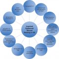
How should we then approach this singly grouped disease with apparently heterogeneous clinical characteristics? Several suggestions are provided here, including a clear definition of clinical subsets, consensus on diagnostic criteria on subset determination, characterization of clinical trial findings according to patient subsets, and utilization of artificial intelligence and Big Data (omics) analytics to facilitate the disease subset determination process ( ). One academic group pointed out that there are overlapping genes and candidate genes derived from multiple omics data, including filaggrin, SPINK5 , S100A8 , and SERPINB3 in relationship to atopic dermatitis pathogenesis, and that there are overlapping immune pathways involving atopic dermatitis by Th2, NFκB, macrophage, fibroblast, and endothelial cell. These medical researchers have advocated that by integrating omics layers that often have complementary and synergistic effects, such as genome, epigenome, transcriptome, proteome, metabolome, lipidome, exposome, and microbiome data, we may be able to fully capture the information flow underlying atopic dermatitis manifestation ( ). Exposome is defined as the sum of external factors that a person is exposed to during a lifetime ( ). Ultimately, clinical researchers who devote their efforts in this disease and physicians who care for this group of patients would also need to come to a consensus on how to redefine the disease entity or to define subsets for this group of diseases. Without a consensus to follow there will be a continuous variation in the diagnosis and treatment for these patients that will not serve our patients well. Variation on definition of atopic dermatitis that has resulted in a substantial difference in disease prevalence estimation, prediction models’ performance, and associated risk factor determination would need to be corrected ( ). Once atopy is redefined and its subsets are delineated, clinicians will have a better understanding of the disease and will be able to provide the most suitable treatments accordingly. A recently published study, which was conducted on more than 30,000 participants evaluated from birth into midlife, identified 4 clinical subtypes of AD with regard to disease activity that continue into adulthood: high probability (2-3%), increasing (2-6%), decreasing (4%), and low probability (88-91%). These findings will open new opportunity for further defining AD subtypes that will help better management of the disease ( ).
Summary
As the medical and scientific communities examine this group of patients categorized under the big umbrella of atopy, the data we gathered point to a need to redefine or to determine distinct subsets within this umbrella. Several aspects of the disease considered as the basis for subset determination include childhood onset versus adult onset, intrinsic versus extrinsic, filaggrin defect versus filaggrin intact, and excellent clinical response versus poor response to targeted immunomodulation therapy. In this new era of precision medicine (i.e., to deliver the right health care to the right person), a one-size-fits-all approach to atopic dermatitis management may no longer be the best practice. A personalized medicine strategy should be in our consideration as we proceed to provide the best care for patients affected by atopic dermatitis in this century and beyond ( ).
Further readings
Related posts:

Stay updated, free articles. Join our Telegram channel

Full access? Get Clinical Tree


