Gold Weight Lid Load
SURGICAL INDICATIONS
The facial nerve or its branches may be injured by trauma, surgery, tumor, stroke, infection, and idiopathic causes. This results in inability to close the eyelid completely, decreased or absent blink reflex, tearing, and sagging and ectropion of the lower lid. Loss of eyelid function may be partial or total, temporary or permanent depending on the specific cause and severity.
Patients with lost normal eyelid function generally present with a red, irritated eye and tearing. For them, corneal protection and restoration of normal appearance are top priorities. To this end, many surgical techniques have been devised. These include tarsorrhaphy, the use of a silicone encircling band, a tantalum wire and mesh implant, a stainless steel palpebral spring, a gold weight lid load, muscle transfers and grafts, facial nerve repair, cross-face nerve grafting, autogenous nerve grafting, and nerve crossover. Each of these procedures has its own indications, merits, contraindications, and complications. Gold weight lid load or gold implantation is perhaps one of the simplest procedures to perform. It is also easily reversible and has few serious complications.
Gold weight implantation is indicated in patients with acute and severe loss of normal eyelid function for whom corneal protection is of immediate concern. It is also indicated in patients in whom recovery of normal lid function is slim and for whom a maximal medical regimen is necessary or barely adequate to maintain the health of the cornea.
Gold weight implantation has certain advantages over other procedures. As a fairly straightforward and reversible procedure, it produces a simulated blink and good cosmetic result. More importantly, it can be used as a secondary procedure when other procedures have proved unsatisfactory; for example, a silicone band has eroded through the eyelid tissues or a palpebral spring has fatigued. These latter devices must be first removed before the gold is implanted. Similarly, when a patient with a lateral tarsorrhaphy becomes dissatisfied with the cosmetic appearance, the tarsorrhaphy may be opened and a gold implant performed secondarily. In selected patients with dysthyroid ophthalmopathy, a gold implant is a viable alternative to other irreversible eyelid lengthening procedures.
Gold weight implantation is contraindicated in patients with an anesthetic cornea. It is relatively contraindicated in patients with very thin and pale skin or with an atrophic orbicularis muscle. In these patients, the color of the gold weight may show through as “discoloration” of the eyelid and the bulkiness of the gold implant more noticeable. Finally, if the patient’s incomplete closure or blinking is due to congenital causes or scarring of the eyelid tissues, these causes must be corrected and gold weight implantation alone is not the solution.
In all patients undergoing a gold implantation, there will be a mild to moderate amount of postoperative ptosis. The patient must understand this and be willing to accept such an outcome.
The gold (or platinum) weights or implants are commercially available. The implants come in 0.6 to 1.0 mm in thickness, 4.5 mm width, and in lengths that vary according to their weight. The weight ranges from 0.6 to 2.0 g in 0.2-g increments. Heavier implants can be specially ordered from various manufacturers. The implants are curved to match the tarsal plate curvature. They are purified and polished so that problems with extrusion and erosion are minimized. Some of the implants have three holes and grooves for suture fixation superiorly. Some manufacturers also have a sizing weight set with double-stick adhesive strip, which makes preoperative determination of weight required much easier.
Preoperative evaluation
A medical history and a thorough ocular examination are necessary. In addition to recorded visual acuity and slit-lamp examination, lacrimal function should be carefully studied. The blinking reflex, including its frequency and completeness, should be noted. The basic and total secretion and the tear breakup time should be measured. Asymmetry in the eyebrow, the nasal labial fold, and the forehead wrinkles should be studied. To assess the severity of the seventh nerve palsy, the patient is asked to elevate the eyebrow or balloon up his mouth. To assess lagophthalmos, the patient is asked to close his eyes gently and the opening is measured with a millimeter ruler. Following this, the patient is asked to close the eyelids forcefully. If there is an opening, it is measured again. Bell’s phenomenon is observed when the examiner digitally opens the eyelid during forceful closure. The orbicularis muscle function can also be assessed subjectively.
For patients who have not had any previous eyelid surgery, determining the weight required for complete lid closure is a straightforward process. A gold weight is selected and a small strip of adhesive tape is used to anchor it to the affected upper eyelid. The upper border of the weight is positioned just below the natural lid crease. The tape is placed horizontally so that it will not mechanically interfere with lid opening. If one uses the manufacturer’s sizing set, apply the adhesive strip to the upper eyelid according to their instructions.
The patient is asked to open and close the eyelid in both the sitting and supine positions. The lid position and completeness of closure are assessed. The ideal outcome is a minimal ptosis in the sitting position with complete lid closure in the supine position. If the weight selected is too heavy, resulting in a severe ptosis, or too light, resulting in incomplete lid closure, it is gently removed from the eyelid and another weight is taped to the lid in a similar manner. This can be done repeatedly and quickly without difficulty. When the correct weight is found, the patient is instructed to wear it for 30 to 45 minutes before rechecking it position. It is frequently found that the next heavier weight is necessary for compete lid closure. Therefore, it is prudent to have at least two weights sterilized and available for the operation.
The patient’s natural upper eyelid crease is identified and marked. (For Asians without an upper lid crease or anyone who does not want to have a lid crease, a supraciliary incision 2 mm above the lid margin may be used.) A small amount of 0.2 to 0.4 ml of 2% lidocaine (Xylocaine) with 1:100,000 epinephrine is injected subcutaneously along the marking.
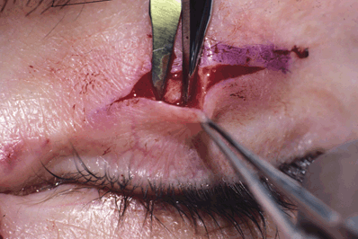
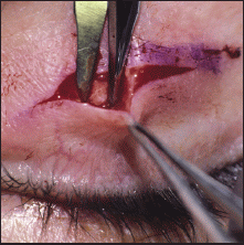
Figure 18-1. A 15- to 20-mm skin incision is made along the crease marking centrally with a No. 15 blade. The skin edges are retracted with 0.5 forceps, and the orbicularis muscle is opened with blunt-tipped scissors. A pocket between the orbicularis muscle and the tarsal plate is created by blunt and sharp dissection 4 to 5 mm inferiorly. This dissection should stay at least 2 mm superior to the lid margin to avoid injury to the lash roots and to prevent the gold weight from sinking down to the lid margin. Depending on the size of the selected implant, the pocket is extended 4 to 8 mm in each direction, medially and laterally. Undermining the orbicularis muscle superiorly allows complete covering of the implant by the muscle layer. Care should be taken so that the expansion of the aponeurosis or the aponeurosis itself is not damaged.
The sterilized gold weights are soaked in Neosporin (polymyxin-neomycin-gramicidin) solution.
Related posts:
Stay updated, free articles. Join our Telegram channel

Full access? Get Clinical Tree


