Embryogenesis
Embryogenesis is a dynamic multiple-step process that begins when an oocyte from a female is fertilized by the sperm from a male (the pre-embryonic period). The first 2 weeks after fertilization focus on rapid proliferation and differentiation of the embryo with subsequent implantation of the egg into the wall of the uterus. Also, the development of the amniotic cavity and the embryonic disc gives rise to the three germ layers of the embryo during this period. The start of the third week marks the beginning of the embryonic period (weeks 3–8) and is characterized by the formation of the primitive streak, notochord, and the three germ layers (ectoderm, endoderm, and mesoderm) from which all embryonic tissues and organs develop . The ectoderm gives rise to structures such as the epidermis and nervous system. The linings of the respiratory system and gastrointestinal tracts as well as certain glandular organs arise from the endoderm layer. The mesoderm is the source of muscle, bone, connective tissue, and blood vessels. The embryonic period is of particular importance because most organ systems develop during this time, and by the end of the eighth week the embryo has a distinctly human appearance. However, because the origins of major structures are established during this critical phase, congenital abnormalities may first appear at this time if there is any teratogenic exposure to the embryo.
The fetal period spans from the ninth week after fertilization to the birth of the fetus. This period is characterized by rapid growth and maturation of the developing organ systems. Head growth slows significantly in comparison to growth of the fetal body.
This chapter focuses on aspects of embryogenesis that pertain to facial plastic and reconstructive surgery. Embryology of the branchial/pharyngeal arches and their derivatives, orbit/eyelid complex, auricle, face, nose, and palate will be discussed in detail. A description of facial anatomy will follow.
Embryology
The Branchial/Pharyngeal Apparatus
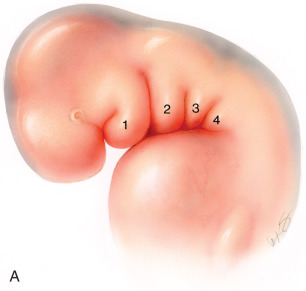
The branchial, or pharyngeal, apparatus greatly contributes to the formation of the head and neck, and begins to form during the fourth week of gestation . This apparatus consists of branchial arches, pharyngeal pouches, branchial grooves, and branchial membranes. By the end of the fourth week of gestation, four well-defined arches are visible on the external surface of the embryo. The fifth arch (also referred to as the sixth arch, depending on the theory one follows) is also present but is not externally visible ( Fig. 1.1A ). Each arch is composed of mesoderm-derived mesenchymal tissue and contains an aortic arch artery, a branchiomeric nerve, a cartilaginous bar, and a muscle component . These arches are separated by prominent grooves, or clefts, which are derived from the ectoderm, and pharyngeal pouches that are lined by endoderm. As the grooves develop, they push medially through the surrounding mesenchyme and approach the medially positioned pharyngeal pouches.
The first branchial arch is often referred to as the mandibular arch and plays a significant role in the development of the face. This arch develops one small prominence, which forms the maxilla, zygoma, and squamous portion of the temporal bone, and one large prominence, which forms the mandible. Important structures that arise from this arch are the muscles of mastication, the maxillary artery, and cranial nerve V3. Other derivatives from the first arch are listed in Table 1.1 .
| Arch | Cranial Nerve | Skeletal Structure | Muscles | Ligaments |
|---|---|---|---|---|
| I (mandibular) | Trigeminal (V3) | Meckel’s cartilage: malleus head and neck, incus short process and body, mandible | Muscles of mastication, tensor tympani, tensor veli palatini, stylohyoid, anterior belly of digastric | Anterior ligament of malleus, sphenomandibular ligament |
| II (hyoid) | Facial (VII) | Reichert’s cartilage: malleus manubrium, incus long process and lenticular process, stapes, styloid process, lesser cornu of hyoid, upper part of hyoid body | Muscles of facial expression, stapedius, stylohyoid, posterior belly of digastric, buccinator | Stylohyoid ligament |
| III | Glossopharyngeal (IX) | Greater cornu of hyoid, lower part of hyoid body | Stylopharyngeus, superior and middle constrictors | |
| IV | Superior laryngeal (X) | Thyroid cartilage, cuneiform cartilage | Inferior constrictor, cricopharyngeus, cricothyroid | |
| V/VI | Recurrent laryngeal (X) | Cricoid, arytenoid, and corniculate cartilages, trachea | Intrinsic laryngeal muscles (except cricothyroid) |
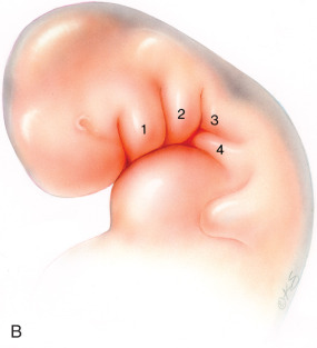
The second branchial arch is also known as the hyoid arch. Around the fifth week of development, this arch will overgrow the third and fourth arches, resulting in the formation of the cervical sinus of His ( Fig. 1.1B ). This sinus and the second, third, and fourth branchial grooves subsequently obliterate, resulting in the smooth contour of the neck. Failure of this area to fully obliterate may possibly lead to the formation of a branchial sinus . Table 1.1 lists the second through fifth arch derivatives.
Corresponding to each branchial groove are pharyngeal pouches, which represent outpouchings of the primitive pharynx. These pouchings are lined by endoderm and also push through the surrounding mesenchyme during the fourth and fifth weeks of the embryonic period. Each pouch contains a ventral and dorsal wing, and gives rise to several important organs such as the parathyroid glands and thymus .
Embryology of the External Ear
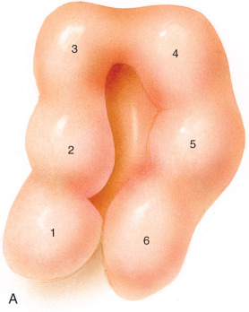
The first sign of the developing ear is seen by the presence of the otic disc, which appears as a thickening on the surface of the ectoderm at the end of the third week of gestation . The disc soon invaginates and is called the otic pit. Six small elevations known as hillocks develop at the dorsal ends of the first and second arches ( Fig. 1.2 ). The first branchial groove, which is located between the first and second arches, deepens and canalizes to form the external auditory canal. The first three hillocks arise anteriorly from the mandibular arch, and the other three hillocks develop from the hyoid arch . These hillocks will gradually fuse to form the auricle of the external ear. Although there is much controversy, it is generally accepted that the tragus, helical crus, and helix are formed from the mesodermal components of the first arch, corresponding to the first, second, and third hillocks, and that the fourth through sixth hillocks from the second arch give shape to the antihelix, antitragus, and lobule, respectively. By the 20th week of development, an anatomically complete ear can be seen. The final shape of the auricle is determined by the intrinsic and extrinsic muscles of the ear that cause plical folding of the cartilage .