Case 1
Clinical Presentation
The plastic surgery service was asked by the surgical oncology service to evaluate a 53-year-old White male with esophageal discontinuity after esophagectomy for cancer and failure of two previous esophageal reconstructions. The patient’s condition began 5 years ago when he was diagnosed with a thoracic esophageal cancer (stage III) during esophagogastroduodenoscopy (EGD). The EGD caused perforation of the cancer into the chest. He underwent an emergency thoracoabdominal esophagogastrectomy. The patient did reasonably well until he developed a gastric outlet obstruction and empyema 1 year later requiring further thoracic and abdominal surgery. Systemic chemotherapy and local radiation to his left neck completed the cancer treatment.
An anastomotic recurrence was revealed 2 years after his initial surgery. The patient underwent a thoracotomy with completion esophagectomy. Immediate gastrointestinal continuity was restored with a substernal colonic interposition utilizing the transverse colon and a portion of the descending colon supplied by the left colic artery. The distal half of the colon interposition was necrosed and was resected on postoperative day 1. A cervical tube esophagostomy was placed. The remaining colonic conduit was sutured to the anterior abdominal wall and a tube colostomy was created. The patient recovered over the next 2 years and remained cancer-free for at least 5 years after his initial esophagectomy. He strongly desired and sought a third attempt at esophageal reconstruction for restoration of gastrointestinal continuity in order to be able to eat and drink ( Fig. 25.1 ). He was seen by many reconstructive surgeons in the region to find an option for the esophageal reconstruction.
Operative Plan and Special Considerations
During preoperative evaluation, the distance of discontinuity was estimated to span 22 cm prohibiting a safe, free jejunal transfer. Barium enema suggested the remaining ascending colon was also insufficient. In addition, the distance through a substernal tunnel between the cervical esophageal stump and the opening of the tube colostomy site within the remaining colonic conduit measured 15 cm. Because a preoperative noninvasive vascular study showed an adequate superficial palmar arch of the patient’s right hand, a free right radial forearm flap could be selected and used to reconstruct a conduit and to bridge between the cervical esophageal stump and the opening of the tube colostomy site after a successful microvascular anastomosis of the flap to the neck recipient vessels. In addition, the left neck site would need additional soft tissue coverage because of previous radiation to the area. A right pedicled pectoral muscle flap could be used to provide adequate soft tissue coverage of the neck open area after surgical dissection of the neck recipient vessels and the esophageal reconstruction.
Operative Procedures
Under general anesthesia, abdominal exploration and dissection of the cervical esophagus were performed by the surgical oncology service to determine whether a retrosternal tunnel would even be feasible, given the patient’s previous history of radiation, scarring, and operations. A retrosternal passage after careful dissection was obtained through neck and abdominal wounds.
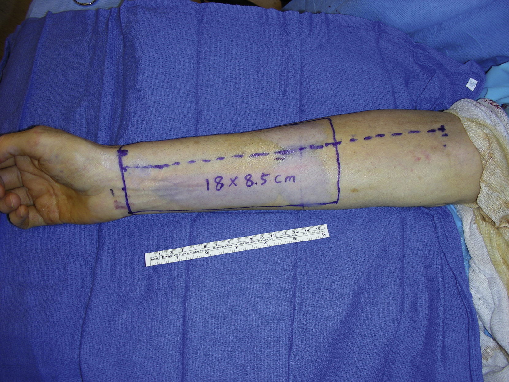
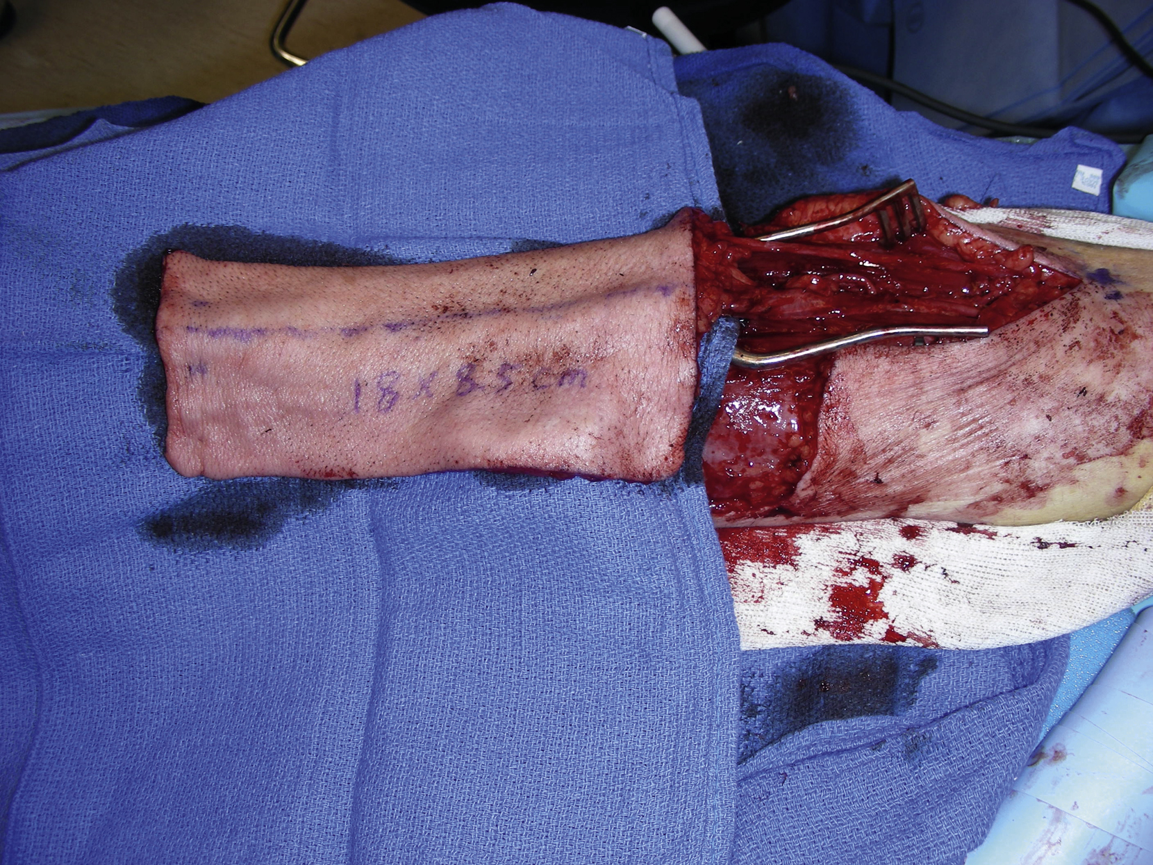
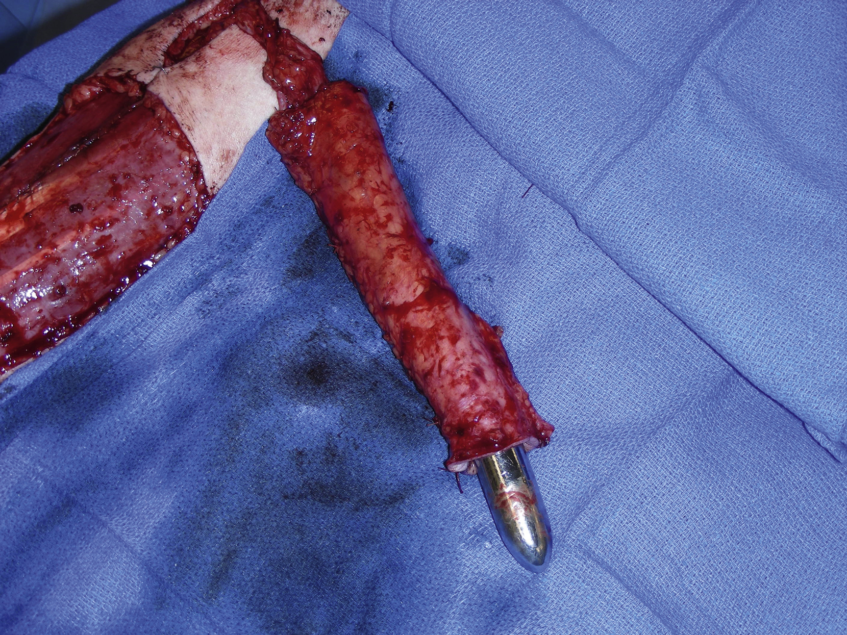
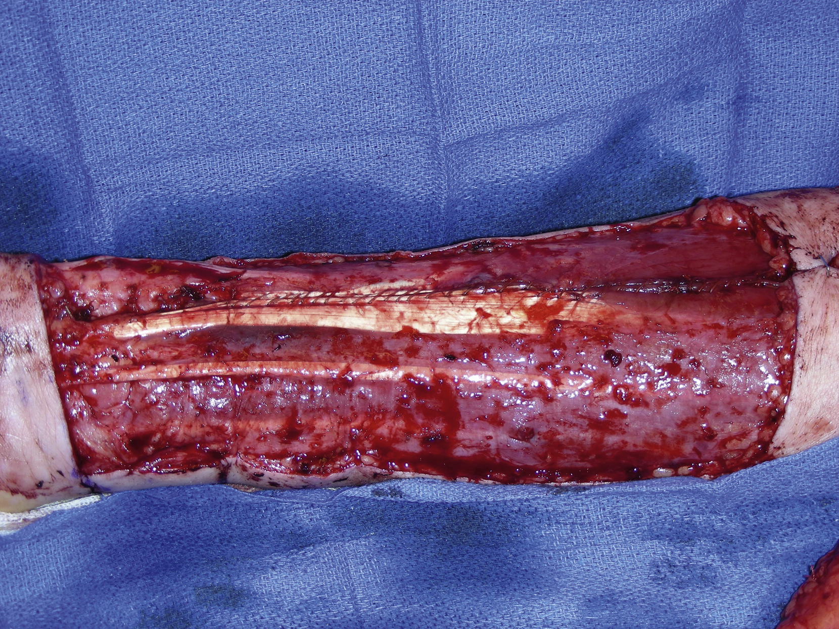
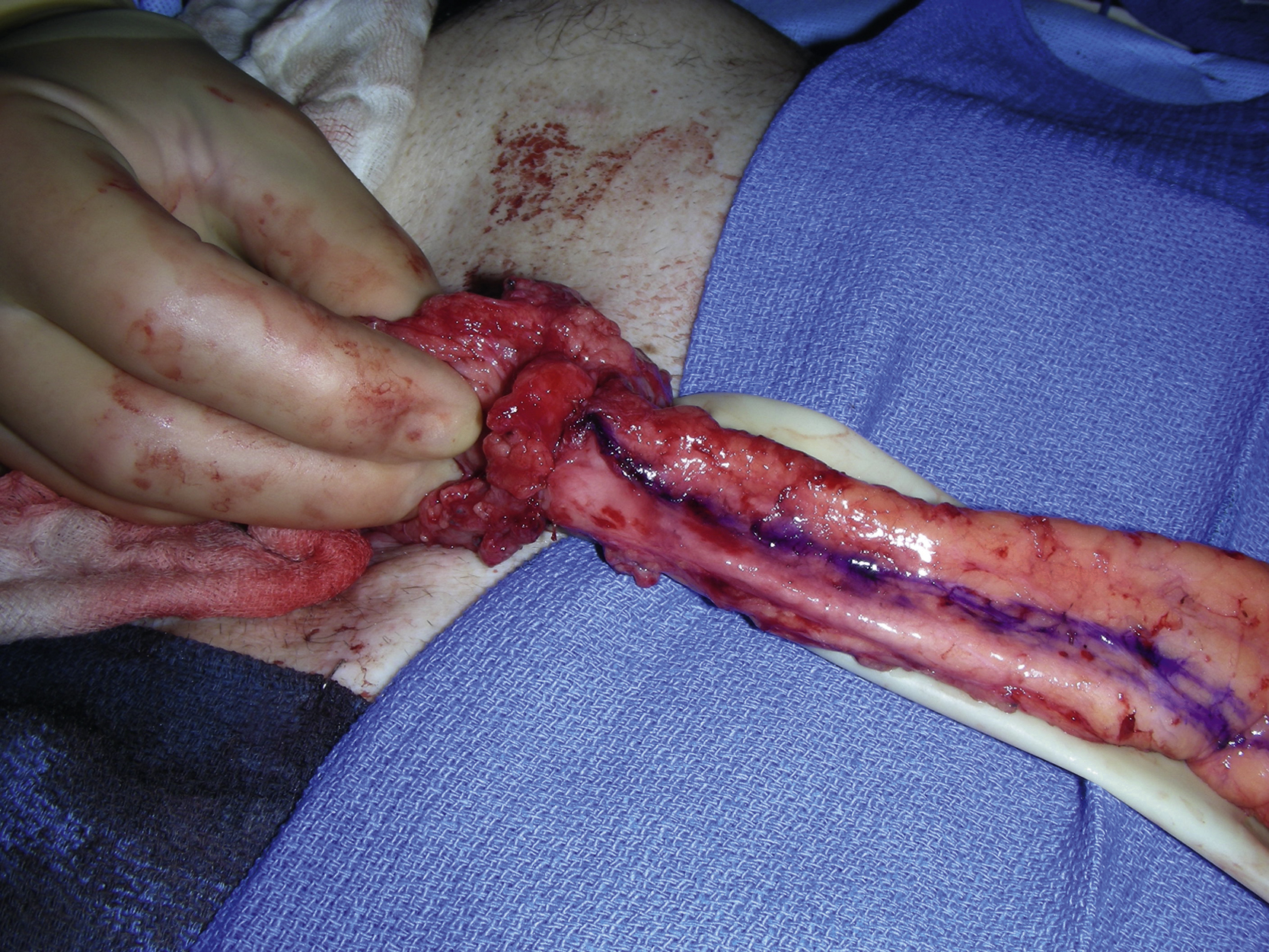
The right radial forearm flap was harvested by the plastic surgery service. The flap was marked out on the volar surface of the forearm and measured 18 × 8.5 cm ( Fig. 25.2 ). An intraoperative handheld Doppler was done to confirm pulses in the arch and the thumb with occlusion of the radial artery. The flap dissection was initially performed to the subcutaneous tissue. The suprafascial dissection was then performed and the cephalic vein, the radial artery, and its venae comitantes were identified and divided at the level of the wrist crease. The flap was raised from the ulnar to radial forearm and the septocutaneous perforators from the radial artery were preserved. This part of the flap dissection was performed under tourniquet control. The flap was raised from distal to proximal and the radial artery was dissected free to the level of the bifurcation of the brachial artery ( Fig. 25.3 ). The flap was then tubularized over a Hegar dilator, putting the skin at the internal lining of the flap and constructing this with a running dermal 3-0 Vicryl suture for a watertight seal. The tube was approximately 3 cm in diameter and 18 cm long ( Fig. 25.4 ).
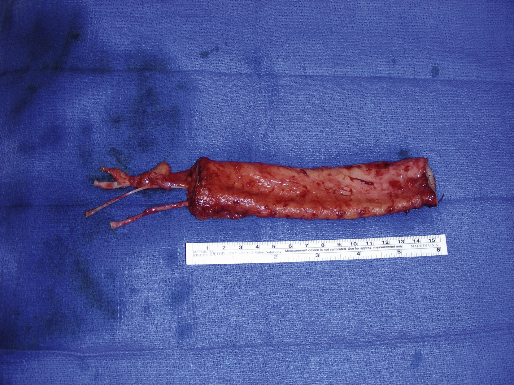
Once the colon and cervical esophagus had been prepared by the surgical oncology service for an anastomosis, the pedicle vessels of the radial forearm flap were divided proximally as far as possible ( Fig. 25.5 ). While waitfing for the flap inset and microvascular anastomoses, the fascial edges of the flexor carpi radialis and the brachial radialis were approximated with running 3-0 Vicryl suture and the loose skin around the periphery of the wound was fixed with 3-0 Vicryl suture, reducing the overall size of the wound to 17 × 7 cm for a skin graft placement ( Fig. 25.6 ). A split-thickness skin graft at 0.015 thickness was harvested from the left thigh with a dermatome. It was placed on the forearm wound as a sheet graft and stapled to the periphery of the wound edge. A volar forearm splint was placed for immobilization.
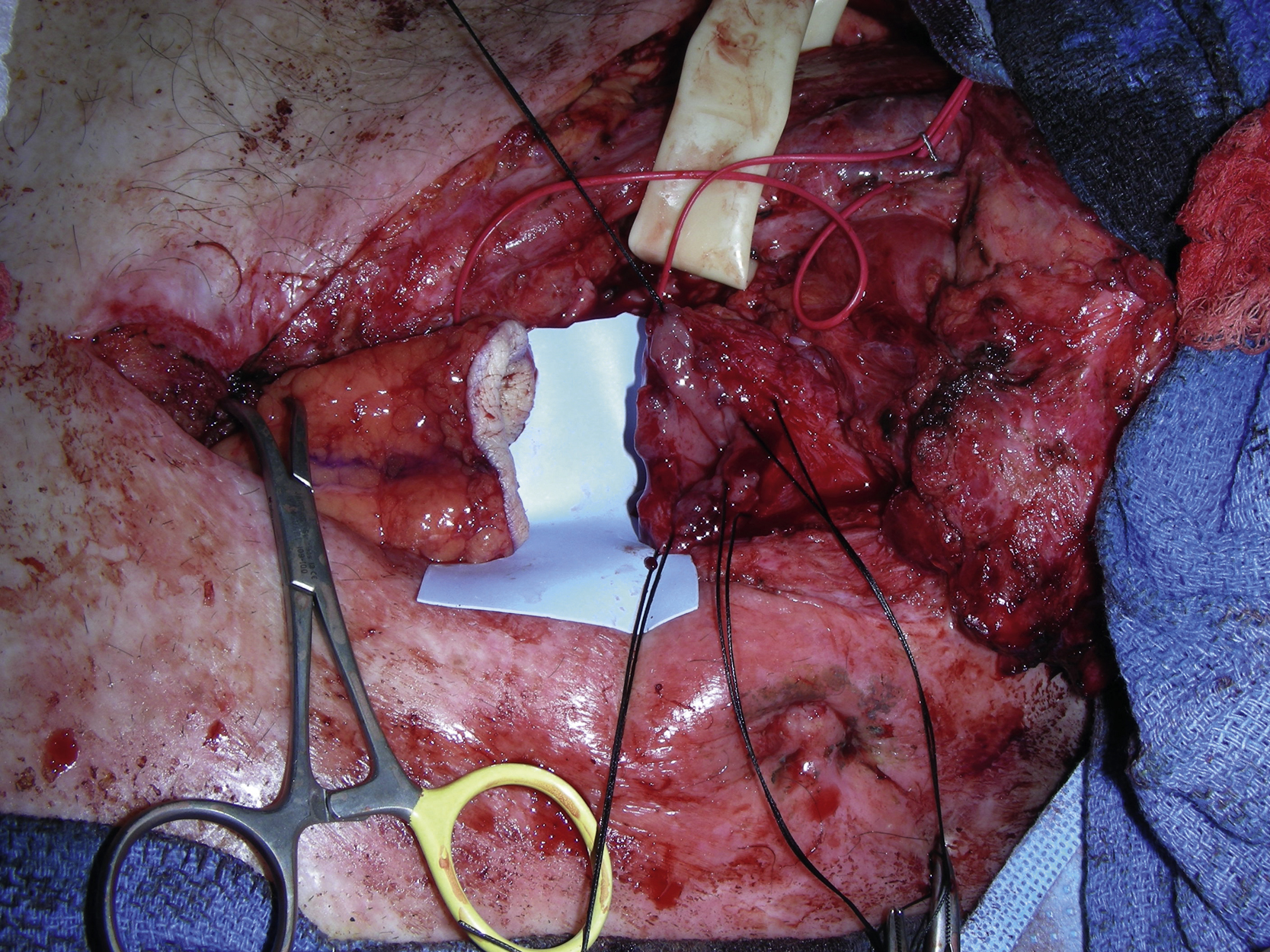
The colonic stump to the flap was anastomosed by the surgical oncology service ( Fig. 25.7 ). The flap was then tunneled under the sternum by the plastic surgery service and the flap to the cervical esophagus anastomosis was again performed by the surgical oncology service ( Fig. 25.8 ). The right transverse cervical artery was dissected out. A branch of the right external jugular was also dissected out. Both vessels were prepared for microvascular anastomoses. However, the right transverse cervical artery was found to have poor blood flow and the right inferior thyroid artery was therefore selected instead and dissected out for microvascular anastomosis. The artery was also tunneled under the right external jugular vein. The arterial microanastomosis was performed with interrupted 8-0 nylons in an end-to-end fashion. The venous anastomosis was performed with a 3-mm coupler device in end-to-end fashion between the donor cephalic vein and a branch of the right external jugular vein ( Fig. 25.9 ).
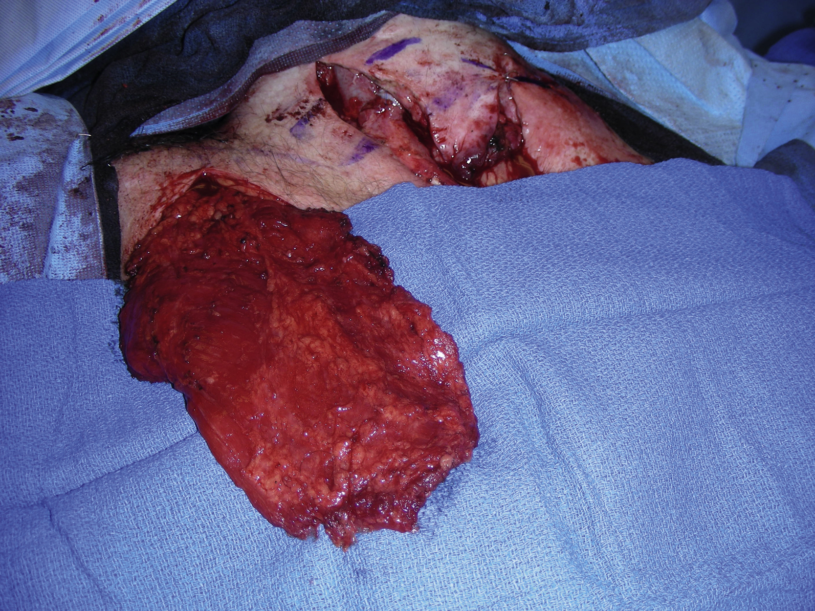
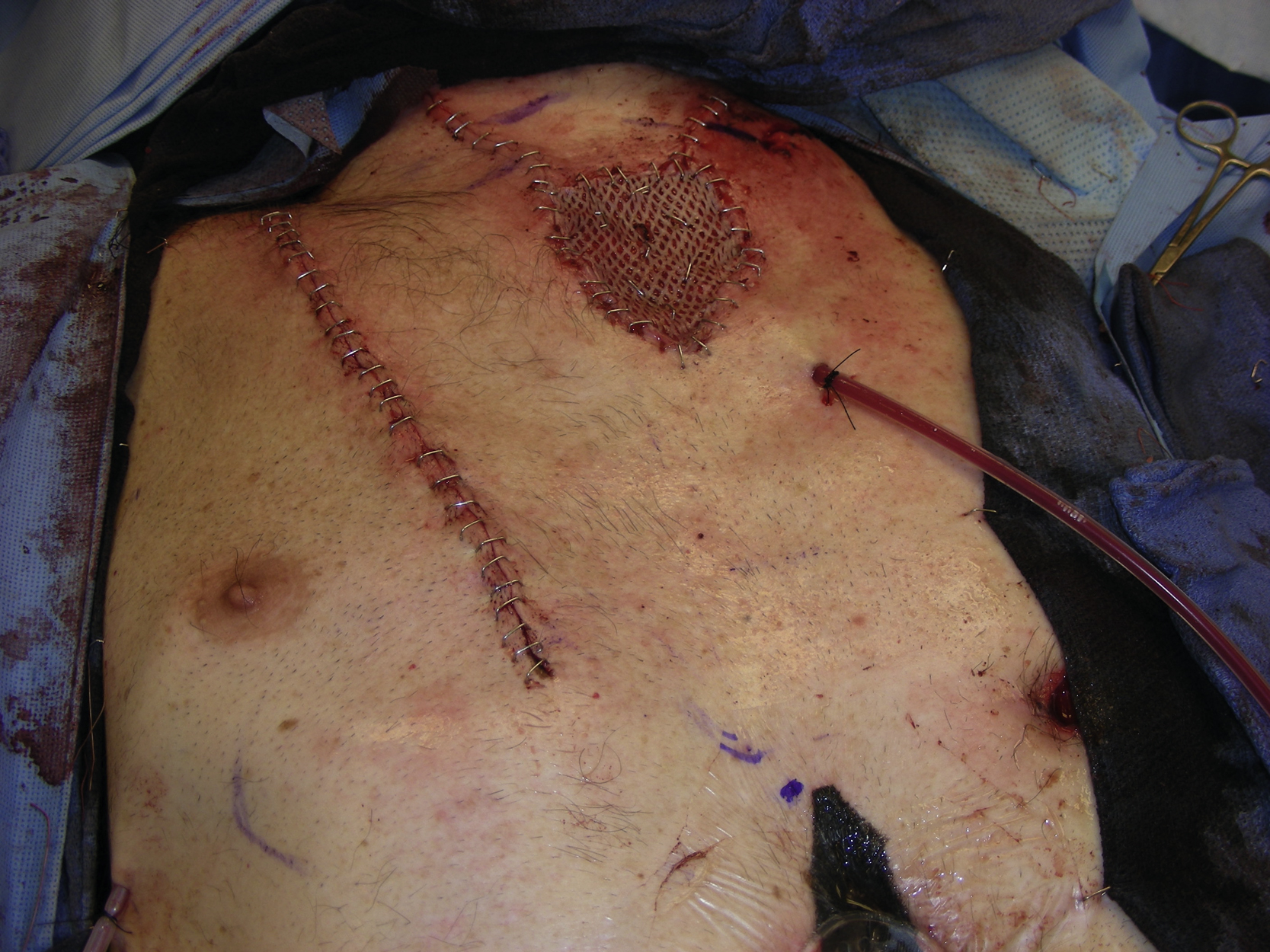
The abdominal wound was closed and a feeding tube was inserted by the surgical oncology team. The right pectoral muscle flap was elevaeted to cover the exposed the radial forearm flap and the neck wound. An oblique incision was made in the right chest wall medial to the nipple extending from the level of the xiphoid to the level of the clavicle. The flap dissection was done in a suprafascial plane from medial to lateral. Once all of its attachments had been divided, the pedicle of the flap was confirmed by a handheld Doppler and marked ( Fig. 25.10 ). The humeral attachment of the pectoralis major muscle was divided carefully. The flap was then completely mobilized on its pedicle and was advanced medially through a tunnel under the skin bridge to the neck wound. The flap was inset into the chest skin defect and approximated with interrupted 3-0 Vicryl sutures. A 15-Fr Blake drain was brought out through a stab incision in the right axilla. Additionally, a 10-Fr flat drain was placed between the muscle flap and the radial forearm flap in the neck wound. A 6 × 7 cm split-thickness skin graft, meshed 1 to 1.5 ratio at 0.015-inch thickness, was harvested from the left thigh with a dermatome and was placed over the muscle flap and secured with staples. The pectoral muscle donor site was closed in layers with 3-0 Monocryl suture for the dermal closure and staples for the skin closure. The neck wounds were closed in a similar fashion ( Fig. 25.11 ).
Management of Complications
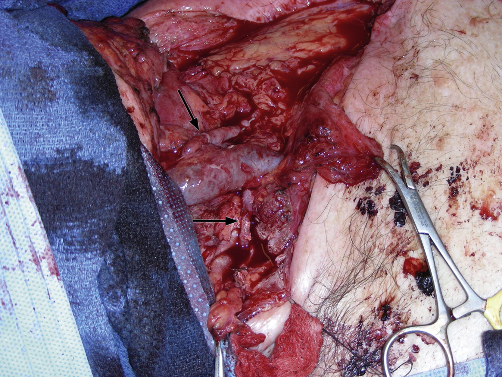
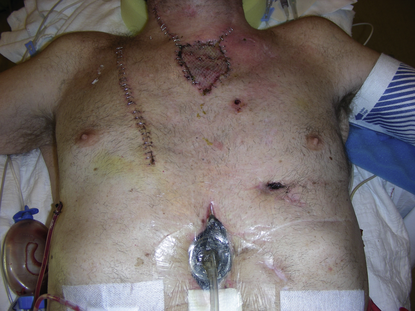
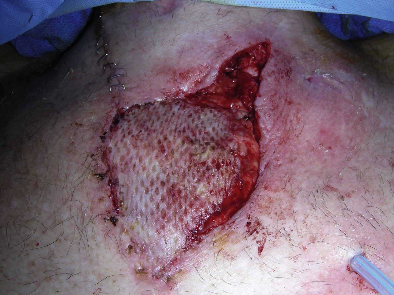
The patient did well postoperatively and tolerated this long procedure well. While he was recovering on postoperative day 11, he developed excessive air from the drain and persistent cellulitis around the left side of his neck. Amylase from the drain fluid was approximately 12,000, which was consistent with a leak at the cervical anastomosis ( Fig. 25.12 ). During reexploration in the operating room, a 1-cm anastomotic dehiscence with two 3-mm holes between the flap and the cervical esophageal stump were identified. The area had minimal local inflammation but a moderate degree of edema ( Fig. 25.13 ). The distal edge of the radial forearm flap and remnant cervical esophagus were viable and did not require debridement. The buried radial forearm flap was completely viable and both arterial and venous microvascular anastomoses were patent.
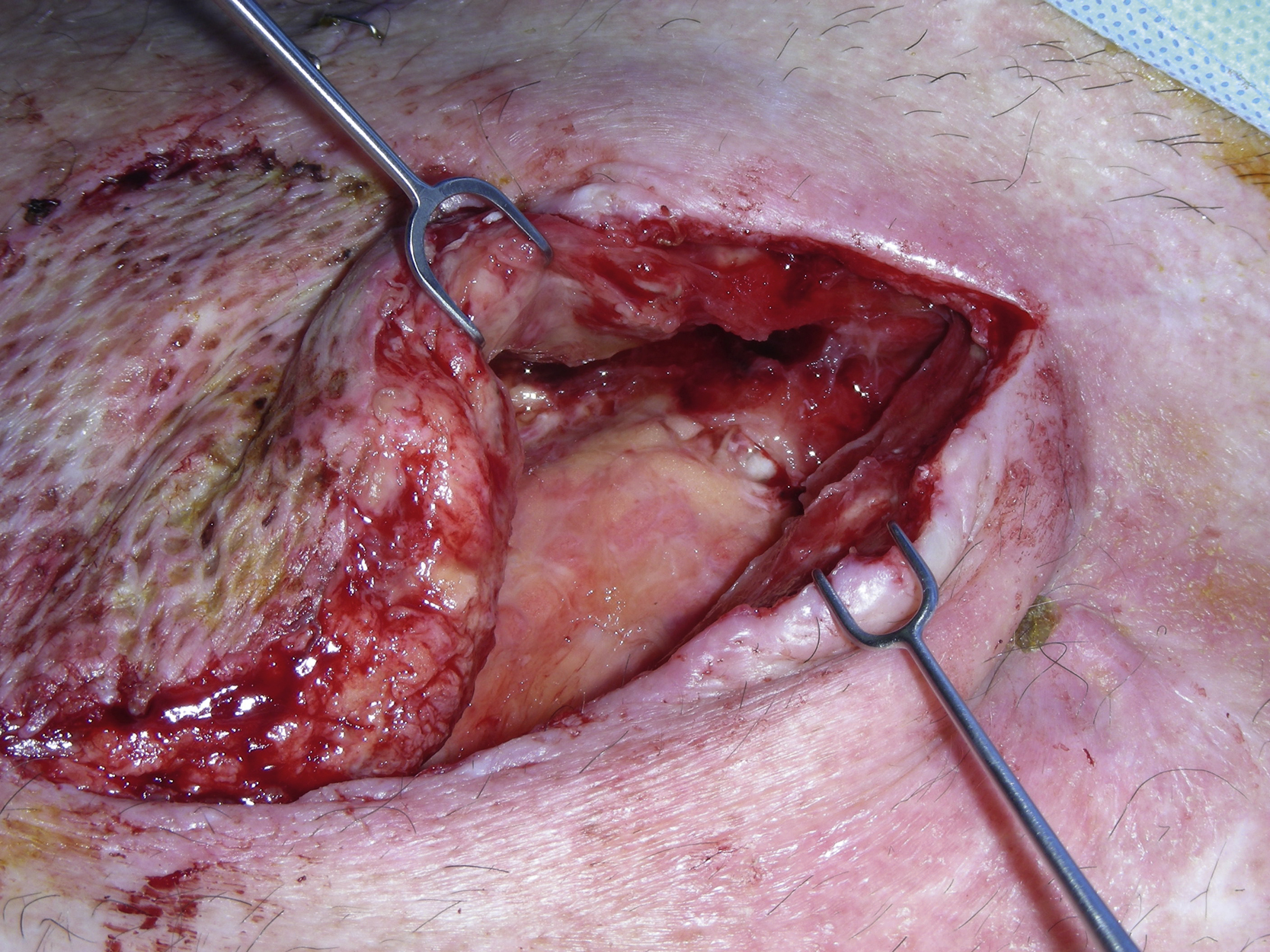
Reanastomosis of the flap to the cervical esophageal stump was not performed because of the condition of the tissue near the anastomosis. It was strongly felt that any attempted reclosure of the anastomotic dehiscence would fail because of the poor condition of tissues at the cervical esophageal site. Instead, the adjacent lower sternocleidomastoid muscle was partially mobilized and sutured to the superior portion of the readvanced right pectoralis major muscle flap with interrupted 3-0 PDS sutures. Approximation of the adjacent muscles allowed complete coverage of the cervical anastomotic leak site with well-vascularized tissue. In addition, a 10-mm closed-suction drain was placed under the muscle flaps but away from the anastomotic leak site to provide adequate drainage ( Fig. 25.14 ).
Follow-Up Results
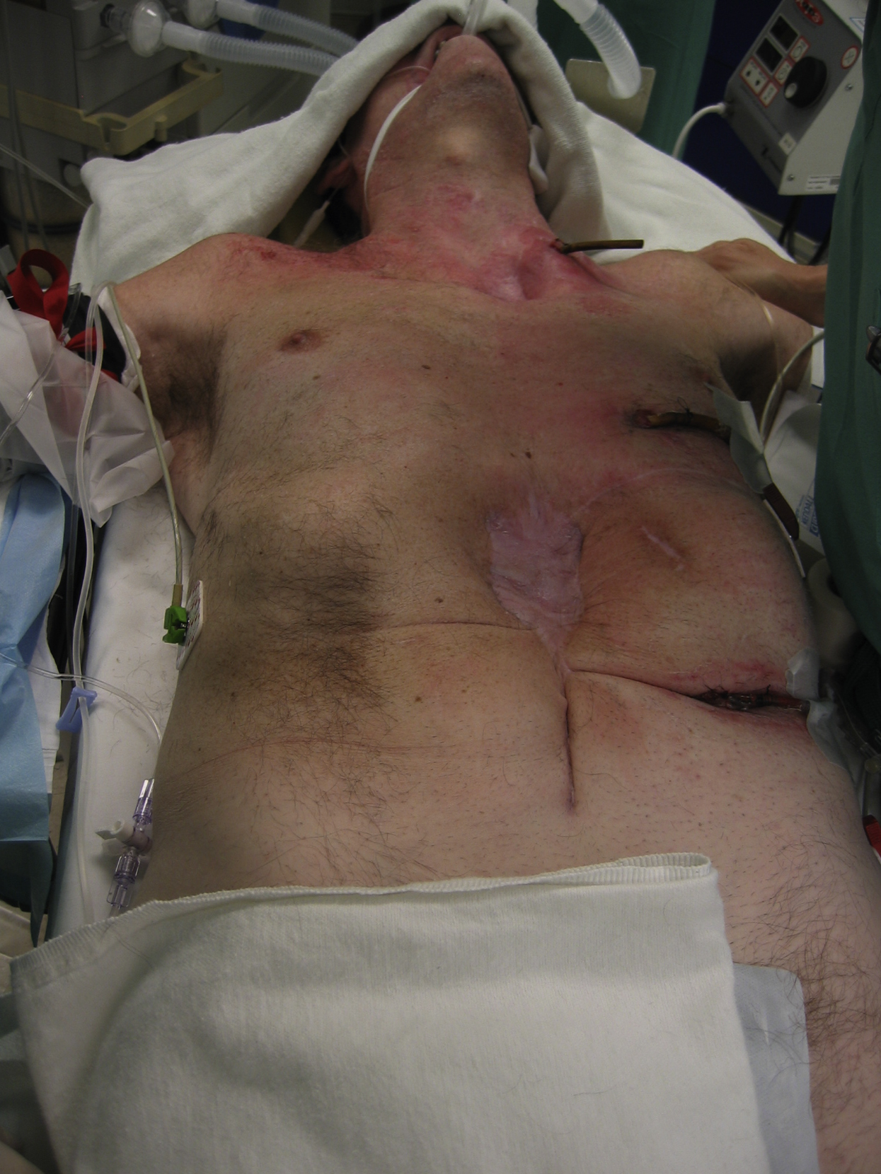
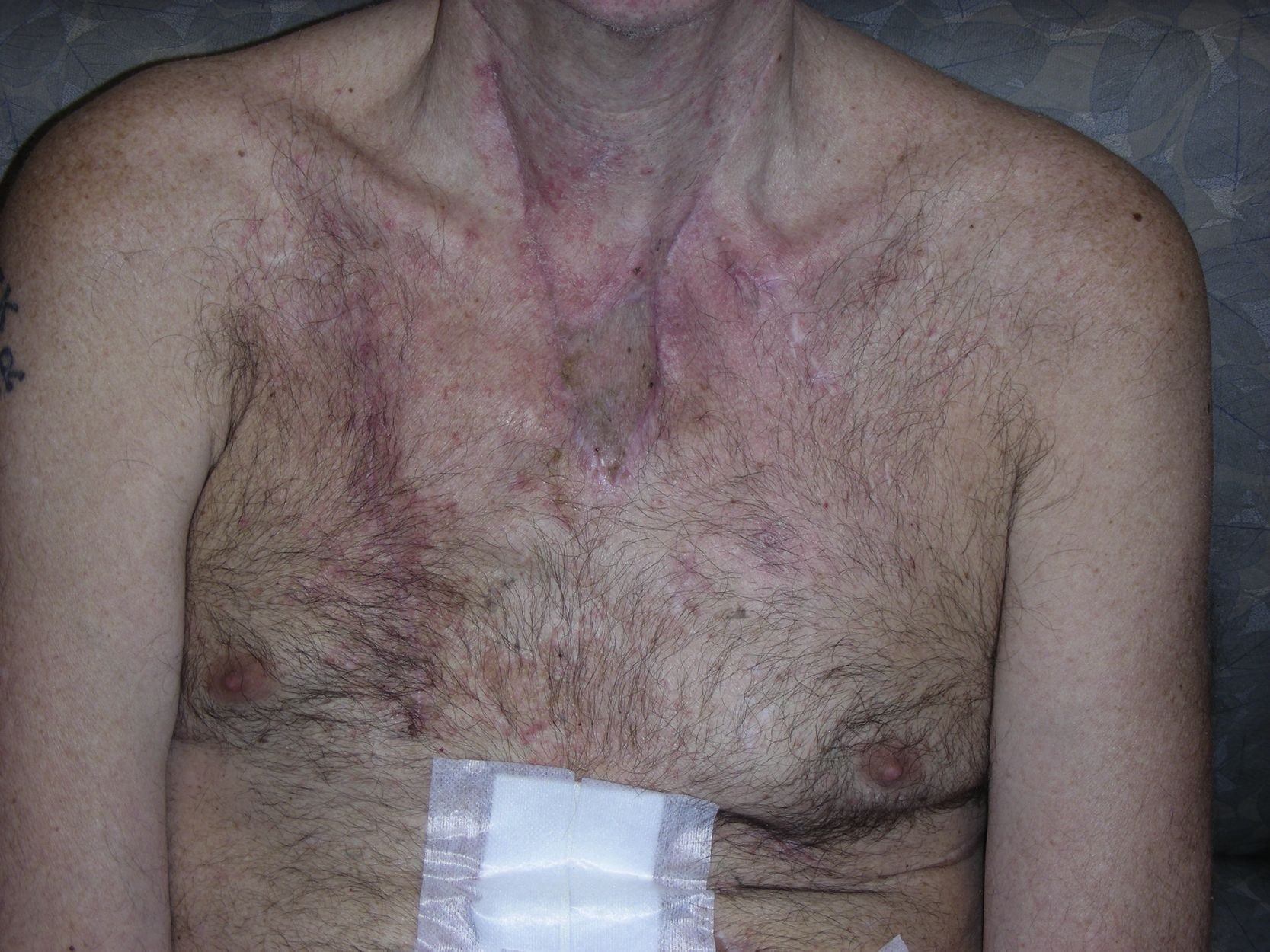
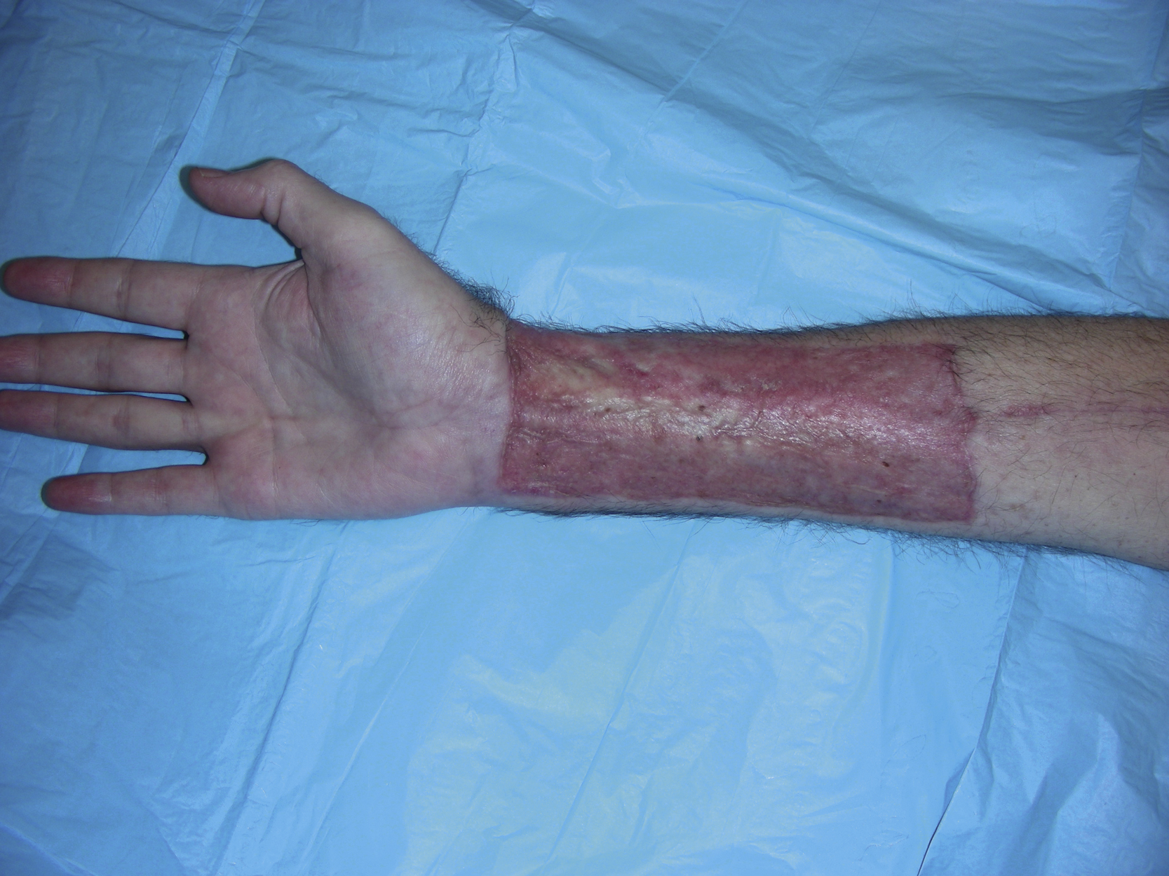
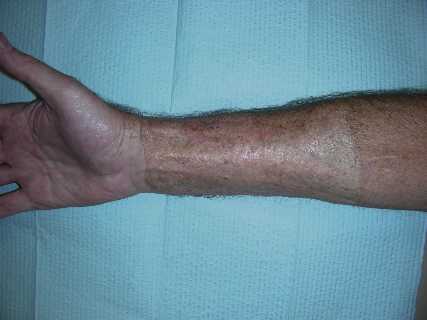
The patient did well after reoperation without any complications related to the salvage procedure. He showed no signs of an anastomotic leak of the proximal anastomosis between the cervical esophageal stump and the radial forearm flap. He had a barium swallow test, which showed the patent anastomosis with no signs of a leak. He had no further issues related to his additional postoperative care. He underwent dilatation of his reconstructed esophagus and started to eat and drink 5 years after his esophagectomy for cancer. The neck and abdominal incisions healed well ( Fig. 25.15 ), as did the right forearm flap donor site ( Fig. 25.16 ).
Final Outcome
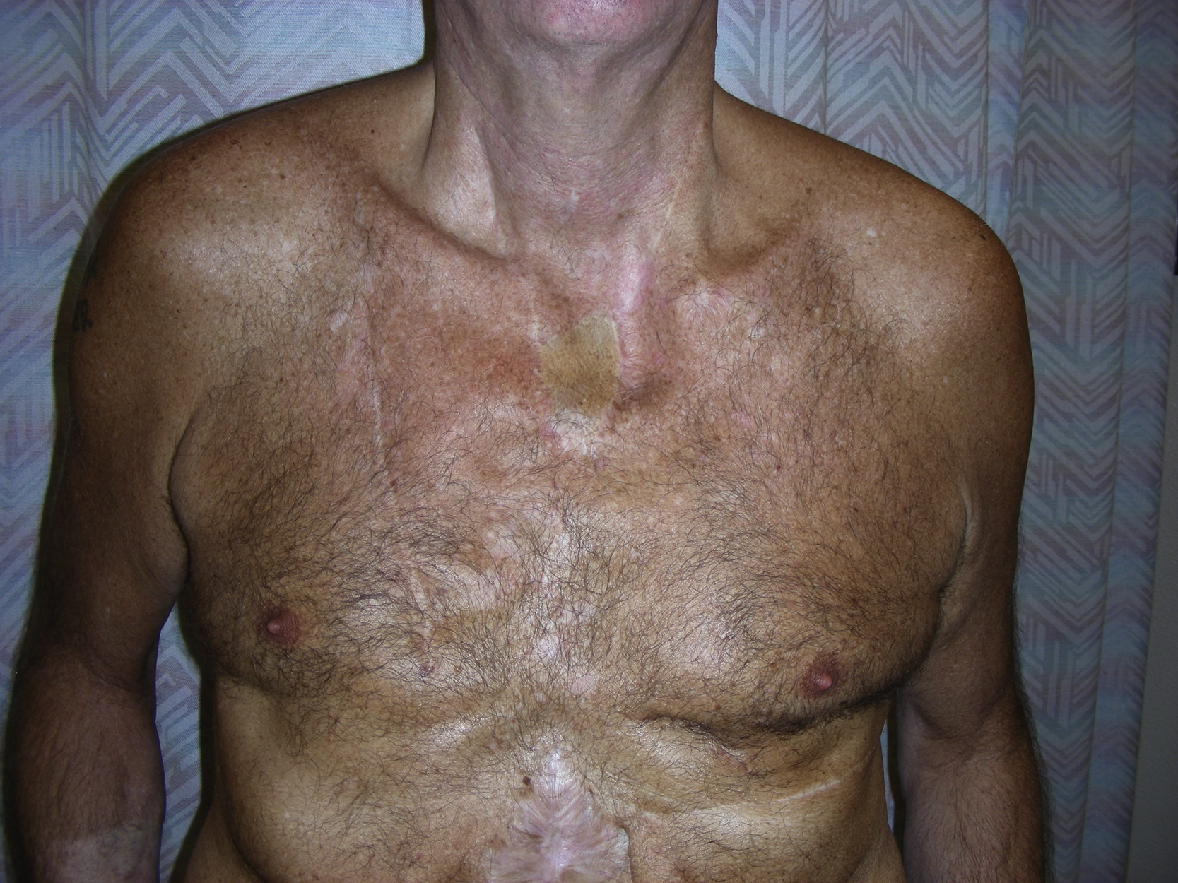
The patient underwent several dilatations of the reconstructed esophagus, especially the proximal anastomosis between the cervical esophageal stump and the radial forearm flap. He has maintained the ability to eat and drink freely. Most importantly, he has also remained cancer free. The neck and abdominal incisions healed well with minimal scarring ( Fig. 25.17 ). The right forearm flap donor site also healed well with minimal scarring ( Fig. 25.18 ). He has resumed his normal activities and has been followed by the surgical oncology service for routine follow-up.
Pearls for Success
The free radial forearm flap has been used to reconstruct noncircumferential or short circumferential segments of the esophagus because it can provide a reliable well-vascularized conduit for esophageal reconstruction. As demonstrated in this case, an 18-cm long-tubed radial forearm flap can be used successfully to reconstruct a substernal esophagus. The flap can be harvested suprafascially for a better donor site closure after a skin graft and a long tube can be made while the flap is still connected with its pedicle after a two-layer, water-tight longitudinal closure. Microvascular anastomoses are usually not difficult to perform because of the flap’s long pedicle and good caliber of the pedicle vessels. The cephalic vein should be selected as a primary pedicle vein.
The majority of cervical leaks after esophageal reconstruction result in local wounds with transient salivary fistula. However, the morbidity of cervical leaks should not be underestimated. Instead of causing more distal flap ischemia by attempting reanastomosis for the leak site, well-vascularized muscle flaps should be used to seal the leak and a closed suction drain should be placed to evacuate the contamination. The technique itself is relatively simple and reliable and works well in this clinical setting. This simple method potentially provides a quick solution for this complex clinical problem because of its sound reconstructive principle—the utilization of well-vascularized tissue to improve healing in a relatively ischemic environment. Simply covering a leak with vascularized tissue will avoid additional trauma and may result in fewer strictures because stricture formation is more likely from the greater tissue trauma during secondary revision and repair.
Case 2
Clinical Presentation
A 68-year-old White male underwent total esophagectomy and end-esophagostomy (spit fistula) secondary to gangrenous paraesophageal hernia and strongly desired to have esophageal reconstruction to restore his esophageal continuity. The thoracic surgery service proposed a supercharged jejunal flap for total esophageal reconstruction. This procedure would need combined efforts from the thoracic surgery, general surgery, and plastic surgery services. The plastic surgery service was asked to perform microvascular anastomosis for a supercharged jejunal flap.
Operative Plan and Special Considerations
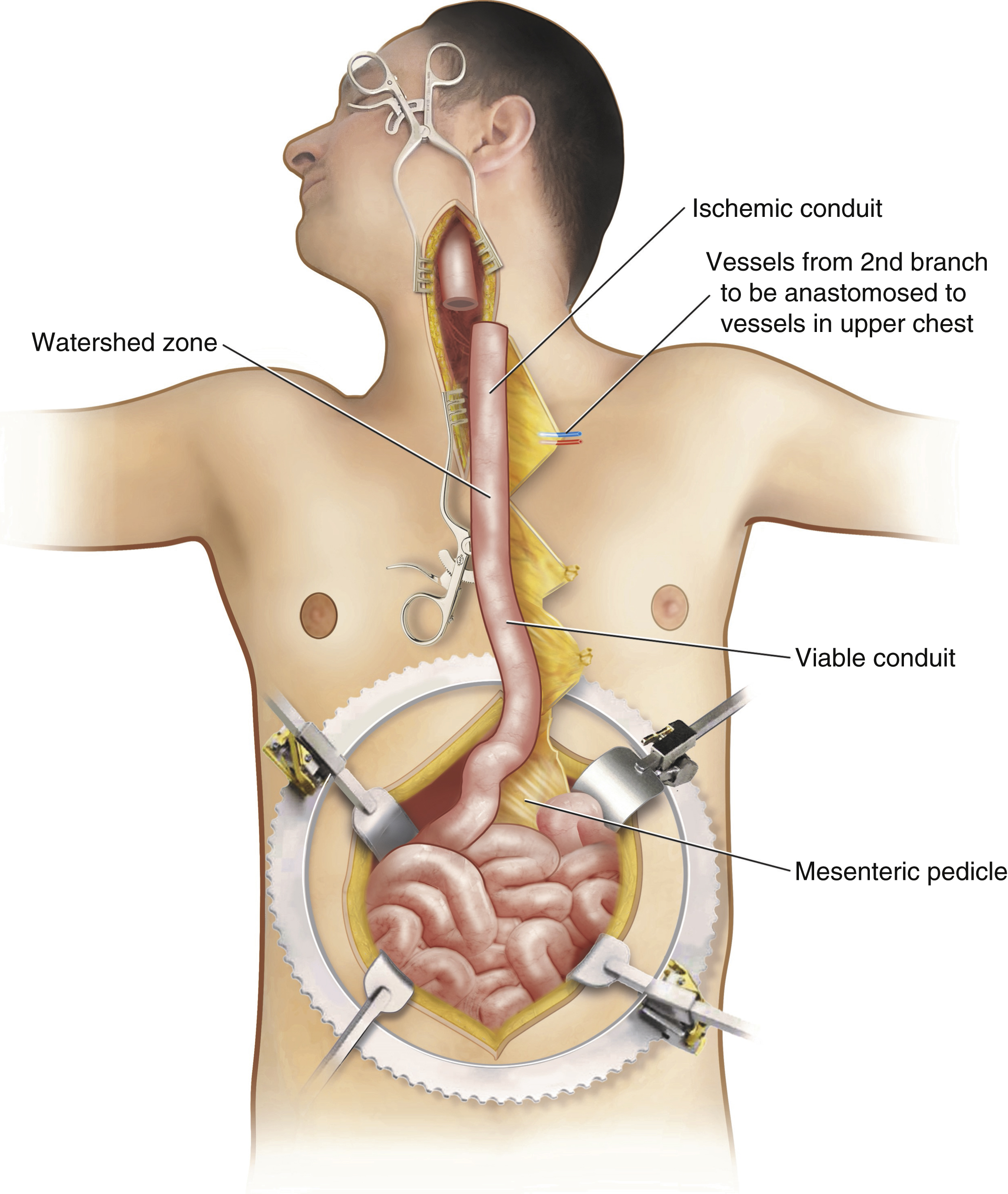
Pedicled jejunum has been used for esophageal reconstruction. However, it is generally unsuitable for reconstruction of the esophagus in the upper chest or neck because of the relatively short length of the mesentery and the lack of longitudinal vascular arcades. In addition, the curvature of the jejunum secondary to the fan-like foreshortening of its mesentery results in a sigmoidal conduit. Division of the mesentery to the mesenteric border of the jejunum allows the jejunum to unfurl, thus straightening the conduit and adding significant length, but this would create a devascularized segment of the jejunum proximally. However, the arterial and venous supply of this segment can be enhanced using microvascular techniques, anastomosing to vessels in the upper chest or neck. This results in a supercharged jejunal flap that can comfortably reach to the hypopharynx and more closely approximates the size of the native esophagus, retains peristalsis, and limits reflux ( Fig. 25.19 ).