Clinical Presentation
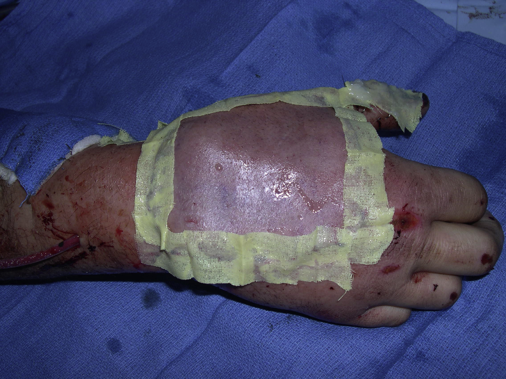
A 41-year-old White male had a degloving injury of his right hand as a result of a motor vehicle accident. He sustained a large soft tissue open wound, measuring 9 × 8 cm, over his dorsal hand with the underlying bones and extensor tendons exposed. He was managed initially by the trauma service and the plastic surgery service was consulted for soft tissue reconstruction of the dorsal hand wound ( Fig. 19.1 ). Preoperative evaluation using the Allen test confirmed an adequate blood supply to the right hand.
Operative Plan and Special Considerations
Based on the size and location of the soft tissue defect over the dorsal hand, a reversed radial forearm fasciocutaneous flap can be a good option. The flap is reliable and has a long pedicle for easy flap inset. It can provide durable soft tissue coverage to a dorsal hand wound. The Allen test should be performed preoperatively to evaluate the ulnar artery system to ensure there is an adequate blood supply to the hand when the radial artery is sacrificed after the flap elevation. A suprafascial flap dissection can be performed to improve donor site cosmesis after a skin graft procedure for the donor site closure.
Operative Procedures
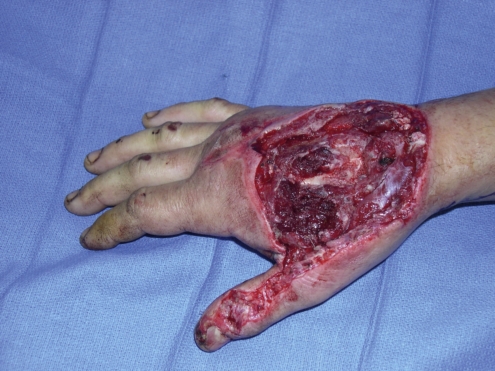
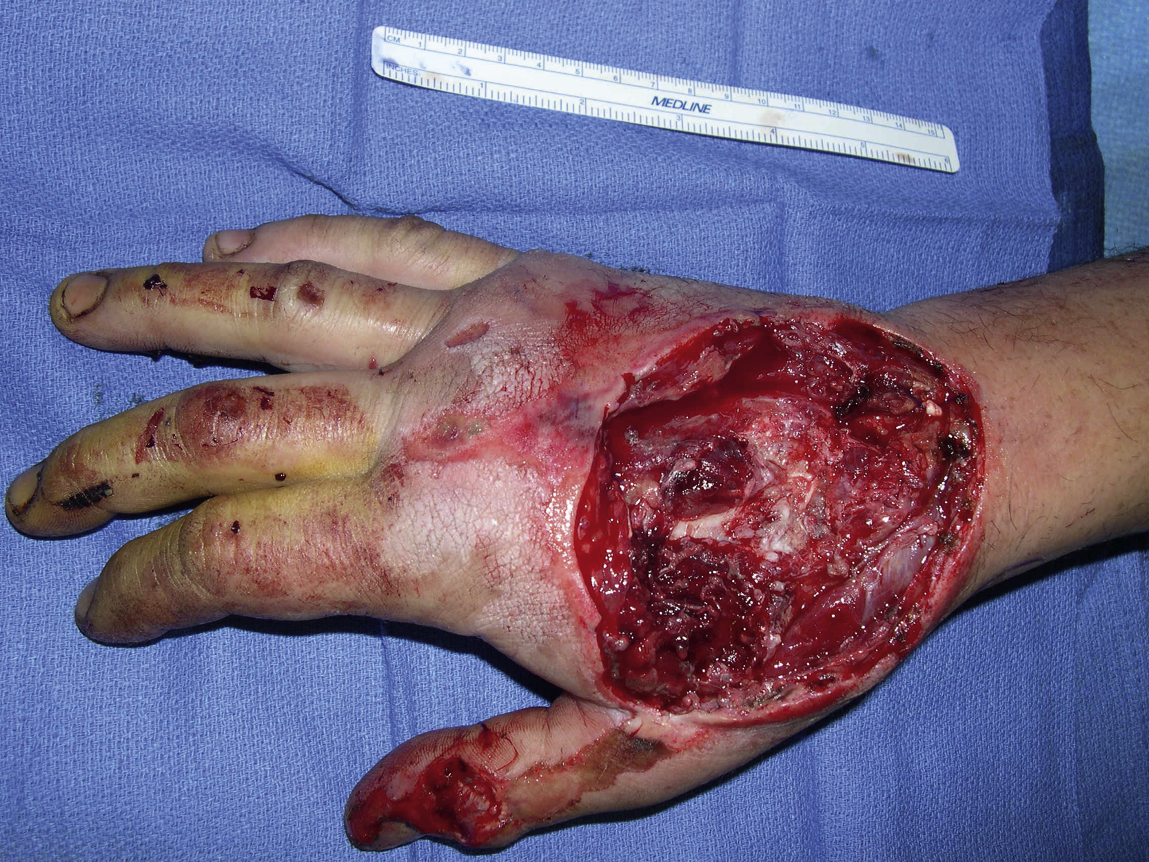
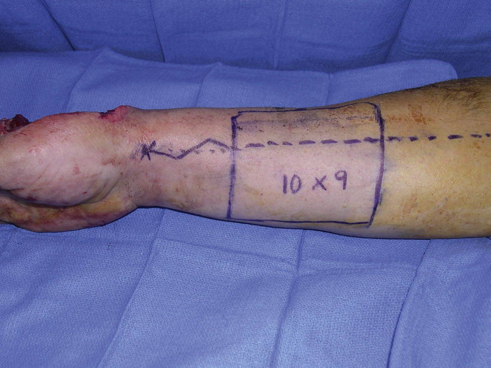
Under general anesthesia, the dorsal hand wound was debrided ( Fig. 19.2 ). A reversed radial forearm flap was designed and oriented longitudinally and a 10 × 9 cm skin paddle was marked. The proximal incision was determined based on the flap’s arc of turnover to cover the dorsal hand wound ( Fig. 19.3 ). The flap dissection was performed under a tourniquet control.
Once the skin incision had been made to the fascia, suprafascial dissection was performed for elevation of the skin paddle. When the dissection was about 1 cm from the pedicle vessels in both medial and lateral directions, subfascial dissection was performed to free the pedicle vessels. Once the proximal radial artery and its venae comitantes and the cephalic vein had been divided with hemoclips, the skin paddle was elevated freely. With a zigzag incision distal to the skin paddle, the dissection of the pedicle in the distal forearm was made between the flexor carpi radialis and brachioradialis to the wrist line. The flap was turned over, based on the radial vessels but in a reverse fashion, through a skin tunnel between the dorsal hand defect and the distal forearm. It was easily inserted into the dorsal hand wound in a reverse fashion without any tension. The flap was approximated to the adjacent skin with interrupted 3-0 Monocryl in half-buried horizontal mattress fashion. A drain was placed under the flap before the final closure ( Fig. 19.4 ).