Cutaneous Diseases Associated with Human Immunodeficiency Virus
Crystal Thomas MD
Antoanella Calame MD
Clay Cockerell MD
SAUER’S NOTES
1. Dermatologists have been at the forefront in diagnosis and therapy during the HIV pandemic.
2. The skin can be used as a marker for early diagnosis and follow-up for disease regression or progression.
3. Morbidity and occasionally mortality from skin diseases underline the importance of dermatology in HIV.
Worldwide, there are an estimated 33 million individuals infected with human immunodeficiency virus (HIV) and up to 3 million cases diagnosed annually. Cutaneous diseases are frequently an initial manifestation of HIV, and knowledge of the most common presentations may aid in the diagnosis of immunodeficiency. Over the course of the disease, more than 90% of HIV-positive patients will present with one or more dermatologic disorders. The spectrum of HIV-associated cutaneous manifestations is broad and includes infectious diseases, inflammatory disorders, neoplastic conditions, and hypersensitivity reactions, some of which may be related to antiretroviral therapies. The following is an outline of the most commonly encountered skin disorders in the HIV-infected population and the array of cutaneous manifestations associated with each entity.
Infectious Diseases
Viral Infections
Acute Retroviral Syndrome
Acute HIV infection, also termed acute retroviral syndrome (ARS), is the period from primary infection with HIV to complete seroconversion and is often subclinical. When symptomatic, ARS resembles an acute, transient, nonspecific viral infection with fever, malaise, myalgias, headache, pharyngitis, and lymphadenopathy. It has been shown that early identification and treatment of patients with ARS may help preserve immune function. Cutaneous manifestations may be seen in up to 75% of symptomatic patients with ARS and usually appear several days after the onset of symptoms. The most common presentation is a morbilliform eruption with erythematous macules and papules measuring up to 1 cm in diameter. The trunk is most commonly involved, followed by the face and extremities. Vesicles, pustules, urticarial lesions, alopecia, and desquamation of the palms and soles have also been reported. In addition, approximately 25% of patients with ARS exhibit painful erosions and shallow ulcerations of the mucous membranes, most commonly in the oral cavity.
Molluscum Contagiosum
Molluscum contagiosum (MC) is a viral infection present in 5% to 20% of HIV-positive patients. The causative virus is a member of the family Poxviridae and is passed by direct skin-to-skin contact. In adults the lesions are often sexually transmitted and occur most commonly in the genital region, lower abdomen, and thighs as solitary or multiple, skin-colored, umbilicated papules or nodules measuring 1 to 10 mm in diameter. Severely immunocompromised patients, particularly those with CD4 cell counts below 200/mm3, may exhibit unusual morphologies and growth patterns such as giant (>1 cm) lesions, clusters of several hundred papules, and extensive facial involvement. Although most lesions are generally self-limited, MC in immunocompromised patients is characteristically difficult to treat and lesions rarely spontaneously regress. Treatment options include cryotherapy, electrocauterization, laser surgery, and topical tretinoin, intralesional interferon, or the antiviral agent cidofovir for refractory lesions.
Human Papillomavirus
Human papillomavirus (HPV) is a double-stranded DNA virus of the Papovavirus class that induces hyperproliferative lesions of the skin and mucous membranes and is transmitted by direct skin-to-skin contact. More than 150 types of HPV have been identified, and several strains have been
shown to play a role in oncogenesis, particularly HPV types 16 and 18. HPV infections can be subdivided clinically into three categories: cutaneous lesions (e.g., verruca vulgaris), anogenital/mucosal lesions (e.g., condyloma acuminata), and epidermodysplasia verruciformis (EDV). EDV is believed to be related to an autosomal recessive impairment of cell-mediated immunity. HIV is associated with an increased incidence of high-grade anogenital intraepithelial dysplasias and invasive squamous cell carcinomas (SCCs), and therefore, these patients should undergo frequent surveillance. Treatment modalities for cutaneous lesions include cryotherapy, podophyllin, salicylic acid, trichloroacetic acid, laser therapy, topical imiquimod, and surgical techniques.
shown to play a role in oncogenesis, particularly HPV types 16 and 18. HPV infections can be subdivided clinically into three categories: cutaneous lesions (e.g., verruca vulgaris), anogenital/mucosal lesions (e.g., condyloma acuminata), and epidermodysplasia verruciformis (EDV). EDV is believed to be related to an autosomal recessive impairment of cell-mediated immunity. HIV is associated with an increased incidence of high-grade anogenital intraepithelial dysplasias and invasive squamous cell carcinomas (SCCs), and therefore, these patients should undergo frequent surveillance. Treatment modalities for cutaneous lesions include cryotherapy, podophyllin, salicylic acid, trichloroacetic acid, laser therapy, topical imiquimod, and surgical techniques.
Herpes Simplex Virus
Herpes simplex virus (HSV) infections are seen in 2% to 27% of HIV-positive patients and demonstrate an increased incidence when CD4 cell counts decrease to 100/mm3 or less. HSV-1 typically causes oral lesions, while HSV-2 is generally localized to the anogenital region, although significant crossover infection is present. Transmission occurs through the exchange of bodily fluids, as well as direct contact with vesicular fluid of active herpetic lesions. In the early stages of HIV infection, the clinical presentation of HSV infections is similar to that seen in immunocompetent individuals (Fig. 24-1). However, with advanced immunosuppression, lesions of herpes simplex often are atypical and can generally be divided into three clinical forms: chronic ulcerative herpes simplex, generalized acute mucocutaneous herpes simplex, and systemic herpes simplex. Chronic ulcerative herpes simplex exhibits recalcitrant ulcerations, most often of the anogenital and perioral regions, that peripherally expand, deepen, and become confluent. Generalized acute mucocutaneous herpes simplex is characterized by disseminated vesicular skin and mucous membrane lesions accompanied by high fever and other systemic symptoms. Systemic herpes simplex infection is relatively rare and most commonly involves the lungs, liver, adrenal glands, pericardium, and brain with an associated high mortality. Acyclovir remains the intravenous therapy of choice for severe local disease and disseminated cutaneous or systemic infections. However, acyclovir resistance has a prevalence of 6.4% and is encountered most commonly among AIDS patients in whom prolonged, low-dose administration is used for suppressive therapy. Foscarnet (vidarabine) has been shown to be effective against some acyclovir-resistant strains.
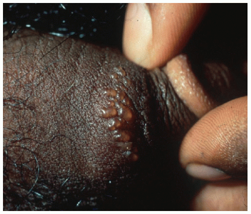 FIGURE 24-1 ▪ Grouped vesicles in a posterior auricular location in an AIDS patient. There is nothing characteristic about this as differing from an HIV(−) patient. |
Varicella-Zoster Virus
Varicella-zoster virus (VZV) infection, transmitted via aerosol droplets, initially manifests as varicella (chickenpox), whereas a reoccurrence of the disease is termed herpes zoster (shingles). When occurring in immunocompromised patients, VZV infection is often severe and can generally be categorized clinically as severe and persistent varicella, dermatomal zoster, recurrent zoster, chronic zoster, and disseminated zoster with or without precedent dermatomal lesions. Recurrent episodes of herpes zoster have an incidence of 10% to 23% in immunocompromised patients and serves as an indicator of progression of HIV-related immunosuppression. Chronic herpes zoster manifests as hyperkeratotic nodules, verrucous lesions, or ulcerations, most commonly affecting the buttock and lower extremities, with resolution requiring weeks to months. Disseminated zoster is defined as more than 20 lesions outside the originally affected dermatome, greater than 3 contiguous dermatomes involved, or systemic infections such as pneumonitis, hepatitis, or encephalitis. When HIV-positive individuals are exposed to VZV, varicella-zoster immune globulin (VZIG) should be given as soon as possible to prevent disease or alleviate its severity. If VZV infection occurs, acyclovir is the drug of choice, albeit administered at higher dosages than used for the treatment of HSV.
Bacterial Infections
Bacillary Angiomatosis
Bacillary angiomatosis is a rare cutaneous disease caused by the Bartonella species B. henselae and B. quintana and presents most commonly in HIV-positive patients, particularly those with CD4 cell counts of less than 200/mm3. Cutaneous lesions may manifest as tender violaceous papules resembling pyogenic granuloma, ulcerated or crusted nodules similar to Kaposi’s sarcoma, subcutaneous nodules, or widespread erythematous plaques (Fig. 24-2). While cutaneous manifestations are most common, visceral involvement may also occur and most commonly presents as peliosis hepatis of the liver. Lymph node, soft tissue, and lytic bone lesions have also been described. Erythromycin and doxycycline have been shown to be effective in eradicating the infection. Some patients may experience a disseminated inflammatory reaction after antibiotic administration, which may be alleviated by the use of an anti-inflammatory agent.
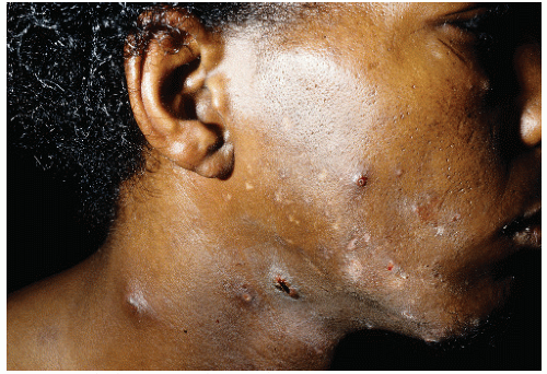 FIGURE 24-2 ▪ Bacillary angiomatosis in an AIDS patient showing crusted, pyogenic granuloma like, papules on side of the face and neck. |
Syphilis
Syphilis is caused by the spirochete Treponema pallidum and has a higher prevalence among HIV-positive populations. T. pallidum is sexually transmitted and initially manifests as a nontender, clean-based ulceration, or chancre, which is the hallmark of primary syphilis. Within 4 to 10 weeks, secondary syphilis develops and presents as numerous erythematous macules and papules presenting diffusely on the face, trunk, and genital region. Mucous membrane and palmoplantar involvement may also be noted. Latent syphilis, the asymptomatic period following the resolution of secondary syphilis, may continue indefinitely. However, if untreated, one third of patients develop tertiary syphilis within 15 years. The cutaneous manifestations of tertiary disease include granulomas, psoriasiform plaques, and gummas (painless, indurated nodules which may ulcerate and become locally destructive). Unusual features may be seen in immunocompromised hosts and include extensive chancres, lues maligna (syphilis with vasculitis), and a rapid progression to neurosyphilis despite appropriate treatment. In general, the treatment of syphilis in HIV-positive patients is similar to those without HIV, that is, parenteral penicillin G. HIV-positive patients should be followed closely for clinical and serologic evidence of treatment failure and if confirmed, treated appropriately.
Mycobacteria
Cutaneous mycobacterial disease, most commonly caused by Mycobacterium tuberculosis


Related posts:


Stay updated, free articles. Join our Telegram channel
Full access? Get Clinical Tree
