Clinical Presentation
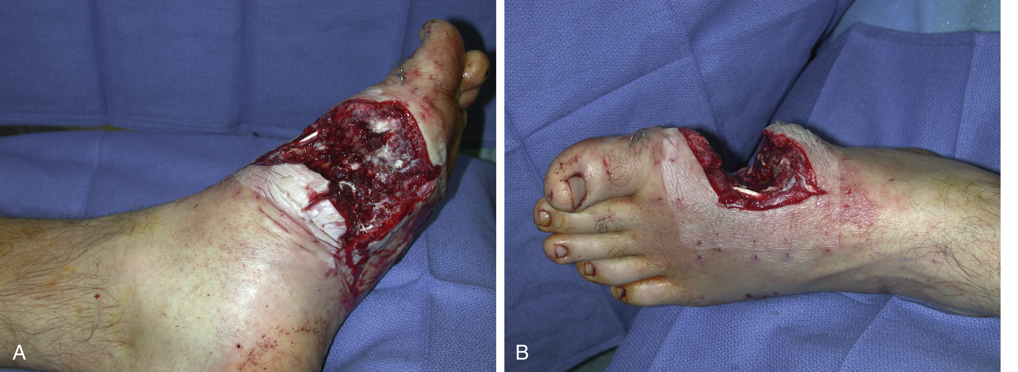
A 35-year-old White male sustained an accidental gunshot wound (GSW) to his left foot. The GSW (12 × 6 × 4 cm) was through-and-through to the medial aspect of the left midfoot with significant soft tissue loss. He also had a comminuted fracture of the first metatarsal with a 5-cm bony defect and complete destruction of the first metatarsophalangeal joint. The extensor hallucis longus tendon was completely destroyed. After initial wound debridement by the orthopedic trauma service, he was taken back to the operating room 2 days later for a second wound debridement by the orthopedic foot and ankle service. The bony stabilization of the first metatarsal was performed with K-wires at that time and the GSW of the left foot open fracture wound was filled with antibiotic beads. The plastic surgery service was consulted for soft tissue coverage of this complex foot wound with composite tissue loss ( Fig. 52.1 A and B).
Operative Plan and Special Considerations
For this relatively small but through-and-through composite defect of the foot, a free gracilis muscle flap can be an excellent choice for soft tissue reconstruction. The gracilis muscle is a type II flap. It is a narrow strap-like muscle (24 × 5 × 1.5 cm) with a pedicle length averaging 6 cm. It has become the author’s first choice to cover a small or medium-sized defect of the distal third tibial wound or foot. With many advances in surgical technique for flap dissection, the gracilis muscle can be used as a free muscle flap for various reconstructive needs. Its flap dissection can be relatively straightforward and pedicle length and size can be improved by dividing the branches of the medial circumflex femoral vessels to the adductor longus and brevis muscles. In this case, the proximal dorsalis pedis vessels could be dissected and used as recipient vessels for a comfortable and relatively easy end-to-end microvascular anastomosis. About 6 weeks after a free gracilis muscle flap transfer once the foot GSW healed, bone grafts could be performed to achieve fracture union of the first metatarsal.
Operative Procedures
Six days after the initial injury, the patient underwent a free gracilis muscle flap transfer to the left foot wound by the plastic surgery service. Under general anesthesia, with the patient in the supine position, the left foot open fracture wound was debrided. All colonized tissues were removed and the skin edge was freshened with a blade. The wound was irrigated with Pulsavac and looked clean and fresh after definitive wound debridement.
The proximal dorsalis pedis vessels were exposed through a zigzag incision. They were easily dissected free and would act as the recipient vessels. Both the artery and the vein were identified and dissected free under loupe magnification. Each vessel was then wrapped with a vessel loop.
A 20-cm longitudinal incision was made in the medial left thigh along a line between the pubic tubercle and the medial femoral condyle. The skin and subcutaneous tissue were incised and the fascia was opened. Once the anterior board of the gracilis muscle had been identified and dissected free, the adductor longus muscle was retracted laterally to expose the main pedicle vessels. After identifying the main pedicle of the gracilis muscle, all minor pedicles in the distal portion of the muscle were divided. The distal muscle was also divided near the origin. Proximally, the muscle insertion to the pubic tubercle was divided and the muscle dissection was completed. The pedicle dissection was performed under loupe magnification and during its dissection, all small branches were divided with microclips. The branches of the medial circumflex femoral vessels to the adductor longus and brevis were divided, which produced an additional 2 cm of pedicle length with a possible vessel diameter greater than 2.0 mm of the pedicle size. The pedicle vessels were divided with hemoclips from the profunda, ready for free tissue transfer.
The pedicle of the flap was prepared under loupe magnification. The artery and vein were then identified and separated. The artery was flushed with heparinized saline solution. One artery and two veins were prepared for microvascular anastomoses. After further preparation of the pedicle vessels under an operating microscope, the flap was temporarily inset into the through-and-through medial foot wound. Under an operating microscope, an end-to-end arterial microanastomosis was performed between the pedicle and the dorsalis pedis artery with an interrupted 8-0 nylon suture. The venous microanastomosis was performed with a 2.0-mm coupler device in an end-to-end fashion. Once all clamps had been released, the flap appeared to be well perfused with a strong Doppler signal.
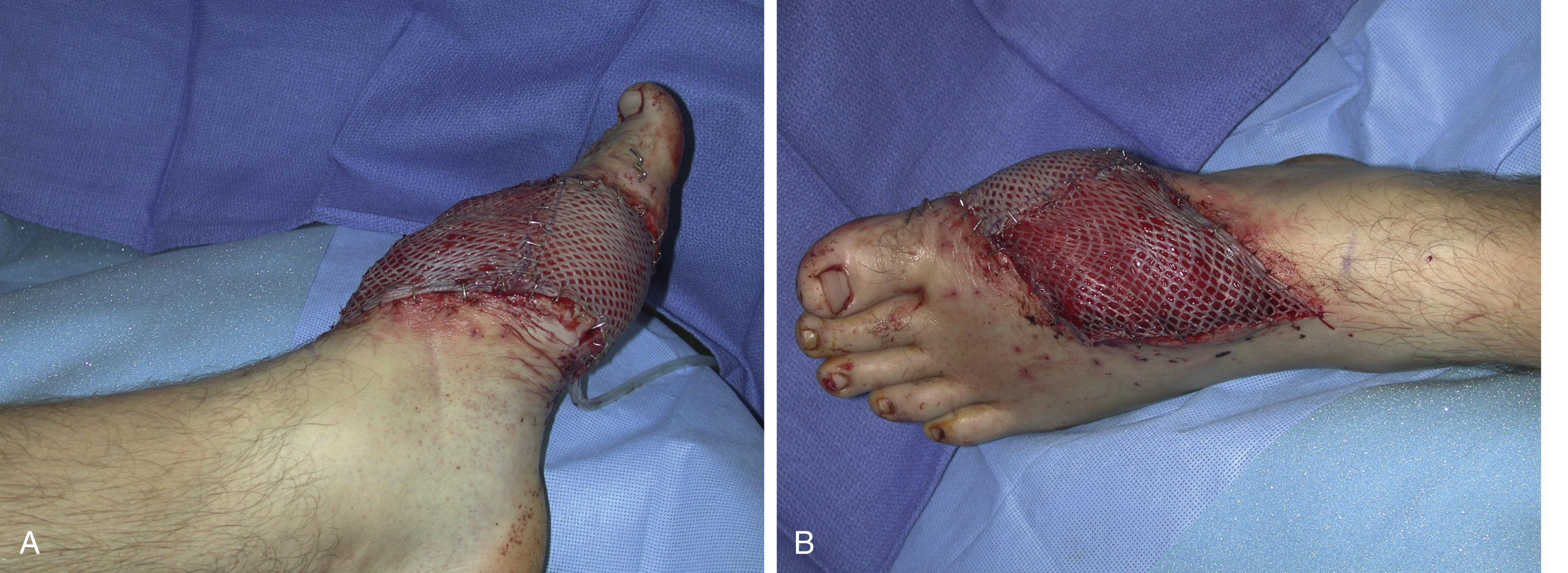
The final inset of the flap was performed. A 10-mm flat JP was inserted into the wound under the flap. The muscle flap was placed inside the through-and-through wound and sutured to the adjacent skin edge with interrupted 3-0 Monocryl sutures in a half-buried horizontal mattress fashion. Both microvascular anastomoses were also covered with the flap. A split-thickness skin graft was harvested from the left lateral thigh. The skin graft was meshed to 1:1.5 ratio, placed over the muscle flap, and secured to the adjacent skin with skin staples ( Fig. 52.2 A and B).