Common Disorders of the Nail Apparatus
Chao Li
Human nails are both aesthetic and functional. Not only do they complete the healthy appearance of hands and feet, they also protect the distal phalanges, allow interaction with small objects, and serve as a natural weapon and grooming tool. Additionally, their appearance, composition, and state of health can hold a wealth of diagnostic information to the careful clinical observer. This chapter endeavors to address common diseases that afflict the nails as well as illuminate the various signs of systemic illnesses that can be seen in the nails.
Normal Nail Biology
The nail apparatus is comprised of the nail plate and four types of supporting soft tissue: the nail matrix, the proximal nail fold, the nail bed, and the hyponychium. The various anatomic regions of the nail are displayed in Figure 25-1. The nail plate is the hard, protective product of the nail apparatus; it derives strength from matrix proteins high in sulfur and its shape relates to the shape of the underlying bone. The nail matrix contains a proliferating layer of basal cells that produce keratinocytes that differentiate, harden, die, and become the building blocks of the nail plate. Nail matrix melanocytes are also found in the lower layers of the nail matrix and remain quiescent unless activated by certain pathologic conditions in which case they can cause nail pigmentation. The proximal nail fold is a fold of skin that closely adheres to and covers one-fourth of the nail plate. It also forms the cuticle, which prevents separation of the nail plate from the nail fold and protects the region from foreign infection. The dermis of the proximal nail fold contains many longitudinally oriented capillaries that run parallel to the skin surface; the organization of these capillaries are disrupted or become more prominent (see Fig. 25-21) in connective tissue diseases such as lupus or dermatomyositis. The lunula region (Fig. 25-2) underlies the proximal nail fold and is the most distal region of the nail matrix; it normally appears white. The nail bed contains a network of elastic fibers, fat cells, lymphatics, and blood vessels, which contribute to its normal pink appearance through the translucent nail plate. The hyponychium describes the space from the point of separation between the nail plate and the nail bed to the distal edge of the nail plate. Fingernails have a mean growth rate of 3 mm/month and toenails have a mean growth rate of 1 mm/month; this rate varies between individual as well as between different digits of the same individual. Total regeneration of fingernails can take between 3 to 6 months and toenails require 12 to 18 months. Conditions that
slow growth rate include infection, malnutrition, peripheral vascular and neurologic diseases, systemic diseases, and treatment with antimitotic drugs. Pregnancy, trauma, psoriasis, and antifungal drugs can also lead to accelerated nail growth.
slow growth rate include infection, malnutrition, peripheral vascular and neurologic diseases, systemic diseases, and treatment with antimitotic drugs. Pregnancy, trauma, psoriasis, and antifungal drugs can also lead to accelerated nail growth.
 Figure 25-1 Anatomic terms for the nail. Asset provided by Anatomical Chart Co. |
 Figure 25-2 The white lunula region, which is the most distal region of the nail matrix. |
Nomenclature of Nail Abnormalities | ||||||||||||||
|---|---|---|---|---|---|---|---|---|---|---|---|---|---|---|
|
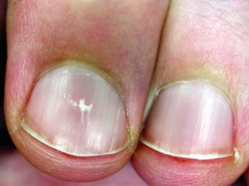 Figure 25-3 Leukonychia. |
 Figure 25-4 Melanonychia. |
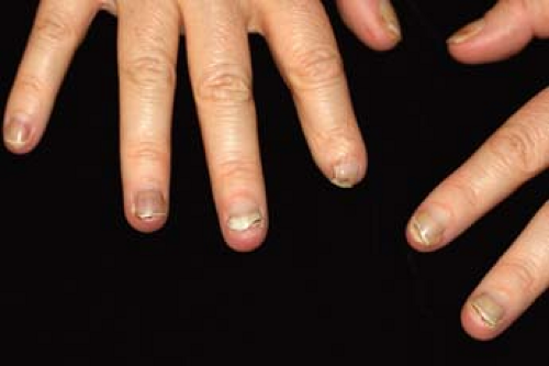 Figure 25-5 Onycholysis: separation of the nail plate from the nailbed. |
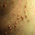
 Figure 25-6 Onychomadesis secondary to chemotherapy with docetaxel. |
 Figure 25-7 Nail pitting from psoriasis. From Goodheart HP. Goodheart’s Photoguide of Common Skin Disorders, 3rd ed. Philadelphia: Lippincott Williams & Wilkins, 2009. |
Nail Infections
 |
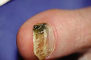
A 75-year-old man presents to your clinic complaining of yellowing and thickening of his toenails on both feet (Fig. 25-8), as well as an itchy and scaly rash in the interdigital spaces of the same foot. He works out daily at his local gym and uses communal showers, though he relates having slowly thickening toenails “since I was overseas in military service.” What is the most likely diagnosis? Is this condition curable?
Fungal Nail Infections: Onychomycosis
Onychomycosis is a broad term that describes an infection of the nail apparatus typically caused by dermatophyte fungi, less frequently by nondermatophyte fungi and yeasts (Fig. 25-8). Onychomycosis is the most common disease of the nail, and its incidence increases with age. Fungal infections of the nail are transmitted by fomite or direct contact, commonly among family members. Therefore, it is believed that tight shoes and use of communal locker rooms are risk factors. Individuals who suffer from atopy, diabetes mellitus, immunosuppression, or human immunodeficiency virus (HIV) disease are also at an increased risk. Onychomycosis is discussed in detail in Chapter 9.
Key Features
Onychomycosis is the most common disease of the nail; incidence increases with age.
It commonly presents with areas of nail opacification and discoloration with intermittent paronychia with periungual tenderness, erythema, and pus. Toenails are more commonly involved, with associated tinea pedis.
The diagnosis can be made by light microscopy of nail scrapings in a potassium hydroxide solution, nail biopsy, and fungal culture. Confirmation of diagnosis is recommended before initiating oral antifungal agents.
A combination of topical and oral antifungal agents is most effective.
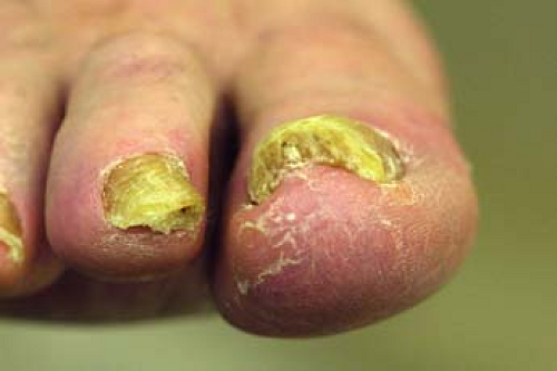 Figure 25-8 Total dystrophic onychomycosis. |
Bacterial nail Infections
 |
A 48-year-old attorney who swims in a communal pool for exercise four to five times weekly presents with black-green discoloration of the right great toe (Fig. 25-9). He has a history of chronic onycholysis and onychomycosis of that toe. He is worried that he might have a melanoma. What is the most likely diagnosis? How is this condition treated?
Green Nail Syndrome (Chloronychia)
Background
Bacteria are not typically able to invade or infect under the nail plate. However, patients with chronic onycholysis or paronychia of the fingernails/toenails who endure prolonged immersion of the affected hand or feet in fresh water may develop a secondary P. aeruginosa infection of diseased nail plate.
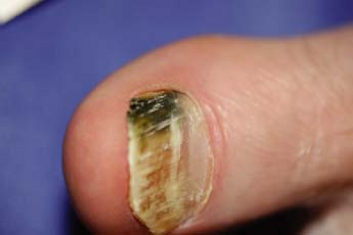 Figure 25-9 Subungual Pseudomonas aeruginosa infection with characteristic green/black color. |
Pathogenesis
Green nail syndrome or chloronychia is a secondary infection of an onycholytic nail plate by incidental or chronic environmental exposure to P. aeruginosa-contaminated water. This organism produces a pyocyanin pigment, which adheres to the surfaces of the nail plate and stains it a green-black color.
Clinical Presentation
Patients typically present with green-black discoloration of one to two nail plates (Fig. 25-9). These nail changes often occur in nail plate that already suffer from chronic onycholysis, onychomycosis, or paronychia.
Key Features
Pseudomonas aeruginosa infection of the nail produces a green-black discoloration of the nail bed, associated with chronic onycholysis and exposure to water.
The affected nail can also suffer from concurrent fungal infection.
The diagnosis consists of visual observation, history of prolonged nail exposure to water, and exclusion of green dyes and fungal infection.
It is treated with topical bleach, vinegar, or chlorhexidine drops combined with reduced water exposure.
Diagnosis
Differential Diagnosis
Common: onychomycosis. Stain from lacquers, dyes, or paints. Uncommon: subungual melanoma.
Clinical diagnosis consists of observing a typical triad of: green discoloration of the nail plate, chronic proximal nail paronychia, and distolateral onycholysis. Rule out fungal infections by potassium hydroxide (KOH) exam and fungal culture of nail scrapings. In recalcitrant cases, subungual melanoma should be suspected.
Treatment
Patients should be counseled to avoid prolonged immersion of nails in water, even when gloves are worn. Care should be taken to dry the nail thoroughly after washing; a hair dryer can help. Topical therapy is usually successful. Oral antibiotics are not necessary or effective in most cases. Over-the-counter remedies such as vinegar (acetic acid 1%) or solutions of 2% sodium hypochlorite may help. Prescription chlorhexidine solution soaks twice daily for 3 to 4 weeks will suppress bacterial growth. Another topical option is ciprofloxacin 0.2% otic solution BID to the affected nail for 2 to 4 weeks. In some cases, it may take up to 12 months for clearance of the green-black pigmentation as normal nail grows out. Oral antibiotic treatment is not necessary.
“At a Glance” Treatment
OTC:
Acetic acid (vinegar) 1% or sodium hypochlorite 2% soaks
Prescription:
Chlorhexidine solution BID × 3 to 4 weeks
Ciprofloxacin 0.2% otic solution BID to the affected nail for 2 to 4 weeks
When nail discoloration does not resolve with treatment or for management of comorbid chronic paronychia and onycholysis; if there is any suspicion of subungual melanoma, the patient should be referred to a surgeon with the tools to perform a nail matrix biopsy (podiatry, dermatology, or general).
Subungual melanoma should be considered for all recalcitrant/spreading cases of chloronychia.
Course and Complications
In general, chloronychia is self-limited and resolves with therapy. Avoiding excess water immersion is essential. This condition can also present with a concomitant fungal infection of the same nail. In these cases, treatment of concurrent onychomycosis is indicated.
ICD9 Code
| 041.7 | Pseudomonas infection in conditions classified elsewhere and of unspecified site |
Pigmented Lesions
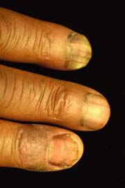 |
A 45-year-old African American man presents with multiple dark longitudinal bands in his thumb and fingernails, bilaterally (Fig. 25-10). Upon closer inspection of his fingernails, pigmented bands are found in the thumbnails bilaterally as well as the index and middle fingernails of his dominant hand. The bands are straight with distinct borders. He says that he remembers having the bands in his fingers for many years but he has grown more concerned because the other bands have appeared more recently, within the last few months. He also has had psoriasis for many years. What may be causing the recent development of additional longitudinal bands in his fingernails? What questions may help better evaluate his condition? Would you refer this patient to be evaluated by dermatology? Would you still refer this patient to dermatology if he says he began azidothymidine (AZT) for recently diagnosed HIV disease?
Longitudinal Melanonychia
Background
Pigmented lesions of the nail usually present as longitudinal pigmentation of the nail or longitudinal melanonychia. It appears as a brown to black band that runs longitudinally in the nail plate from the proximal nail fold to the free edge. These bands can be congenital or acquired later in life and they can be common in African American, Latino, or Asian patients. Many different conditions can cause this feature, including early subungual melanoma.
Pathogenesis
Longitudinal melanonychia is either caused by activation of normally dormant nail matrix melanocytes or from benign or malignant nail matrix melanocyte hyperplasia.
Clinical Presentation
Typically a brown or black longitudinal band in the nail plate; however, the width of the band is variable between nails (but uniform within a given band) (Fig. 25-10). There may be more than one band per nail plate. Full-width melanonychia presents as pigment involvement of the entire nail plate.
 Figure 25-10 Longitudinal melanonychia and nail dystrophy secondary to psoriasis. |
Diagnosis
Related posts:

Stay updated, free articles. Join our Telegram channel

Full access? Get Clinical Tree


