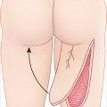
Chest Wall Defect
Description
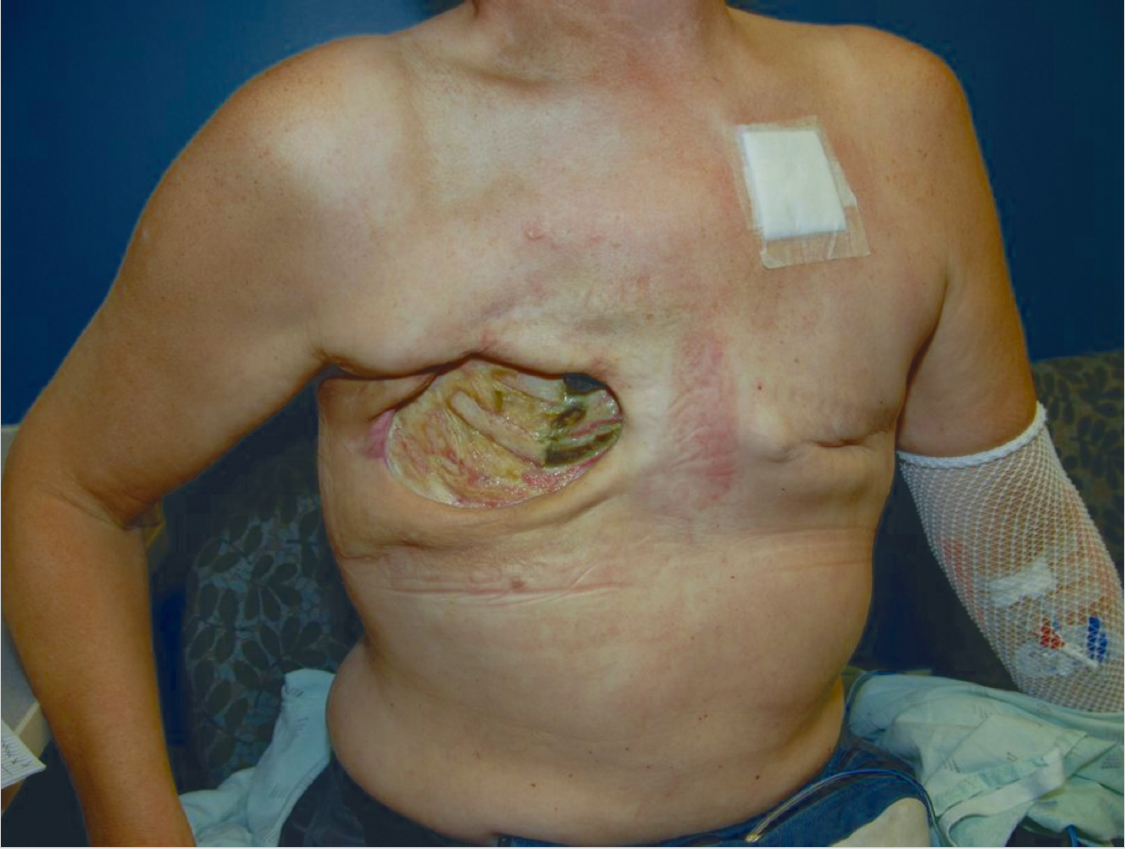
Large chest wall defect, including the inferior portion of the pectoralis major, lateral portion of the serratus, and the fourth and fifth ribs.
Clear evidence of osteomyelitis (with exposed, necrotic bone).
Healthy-appearing tissue in lateral wound.
Left mastectomy scar.
Work-up
History
Coronary artery disease
History of coronary artery bypass grafting: Possible absence of internal mammary artery.
Pulmonary disease (chronic obstructive pulmonary disease [COPD], asthma): Increased risk for respiratory compromise in the absence of chest wall skeletal reconstruction.
Previous history of chest, back, or abdominal surgery/trauma: Potential compromise of specific flaps.
Other comorbidities.
Tobacco use.
Nutritional status.
Etiology of chest wall wound/deformity
Traumatic, oncologic, infectious, radiation, congenital.
If oncologic, benign versus malignant: History of (or plan for) radiation therapy.
Physical examination
Define defect or mass: Location, depth, fixed or mobile.
Perform lymph node examination.
Assess muscle involvement in the chest: Is the pectoralis major involved?
Assess abdomen for hernias, diastasis recti.
Evaluate back musculature and soft-tissue laxity.
Assess for chest wall, back, or abdominal scars.
Related posts:

Stay updated, free articles. Join our Telegram channel

Full access? Get Clinical Tree


